Case Report : Non-surgical Treatment of Inferior Alveolar Nerve Injury as a Result of Overinstrumented Root Canal
Treatment
Kook-Jin Bae
1, D.D.S., Jong-Mo Ahn
1, D.D.S.,M.S.D.,Ph.D.
Chang-lyuk Yoon
1, D.D.S.,M.S.D.,Ph.D., Young-Gon Cho
2, D.D.S.,M.S.D.,Ph.D., Ji-won Ryu
1, D.D.S.,M.S.D.,
Department of Oral medicine
1, Department of Operative Dentistry
2, School of Dentistry, Chosun University
During root canal treatment, overinstrumentation with hand or mechanically driven files can perforate the mandibular canal, allowing the extrusion of endodontic sealers, dressing agents, and irrigant solutions out of the tooth and into the canal. The patient may report symptoms such as pain, hyperesthesia, hypoesthesia, anesthesia, dysesthesia and paresthesia.
Such problems must be resolved as quickly as possible to avoid irreversible sequelae caused by certain neurotoxic materials that form part of endodontic sealants. Although there have been no controlled trials of treatment protocols involving endodontically related injuries to the inferior alveolar nerve, the normal therapeutic sequence for this complication is the control of pain and inflammation and, whenever possible, the surgical elimination of the cause.
However, total resolution of pain and reduction in or disappearance of paraesthesia after a non-surgical management have been reported. Antiepileptic drugs such as gabapentin or pregabalin have been used for the treatment of neuropathic pain.
This article describes a case of inferior alveolar nerve(IAN) damage after endodontic treatment of a mandibular right second molar and the treatment with non-surgical approach using prednisone and gabapentin medication, monitoring the patient's condition with clinical neurosensory examination and current perception threshold test(Neurometer).
Key words : Current perception threshold, Endodontic complication, Endodontic sealer, Gabapentin, Inferior alveolar nerve injuries
Corresponding author : Ji-Won Ryu
Department of Oral Medicine, Chosun University 421, College of Dentisty
Seosuk-dong Dong-Gu, Gwang-Ju, 501-825 Tel: 062-220-3897
Fax: 062-234-2119
E-mail: [email protected] Received: 2011-08-03 Accepted: 2011-09-01
* This study was supported by research funds from Chosun University, 2010
Ⅰ. INTRODUCTION
The inferior alveolar and lingual nerve injury can occur after several types of dental treatment, including local anesthetic injections
1,2), endodontic treatment
3), implant placement
1), and dentoalveolar surgery, particularly involving the removal of mandibular third molars.
1,4)These dental treatments can cause permanent nerve damage resulting in anesthesia, paresthesia, and dysesthesia.
5,6)One of the potential iatrogenic causes of this
problem is the incorrect treatment of the root canals of a lower molar or premolar (overextension and/or overfilling). The mechanism by which such treatment can damage the inferior alveolar nerve(IAN) may be mechanical, thermal, or chemical.
7)There have been reports of IAN damage in up to 1% of cases when performing defective root canal treatment (overextended or overfilled) of a lower premolar.
8)The patient may report symptoms such as pain, hyperesthesia, hypoesthesia, anesthesia, dysesthesia and paresthesia.
9,10)Such problems must be resolved as quickly as possible to avoid irreversible sequelae caused by certain neurotoxic materials that form part of endodontic sealants.
11)Although there have been no controlled trials of treatment protocols involving endodontically related injuries to the inferior alveolar nerve, the normal therapeutic sequence for this complication is the control of pain and inflammation and, whenever possible, the surgical elimination of the cause.
3)However, total resolution of pain and reduction in or disappearance of paraesthesia after a non-surgical management have been reported.
10,12-14)
Antiepileptic drugs such as gabapentin or pregabalin have been used for the treatment of neuropathic pain.
This article describes a case of IAN damage after endodontic treatment of a mandibular right second molar and the treatment with non-surgical approach using prednisone and gabapentin medication, monitoring the patient’s condition with clinical neurosensory examination and current perception threshold test(Neurometer).
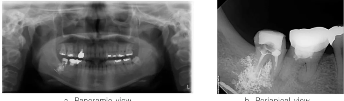
a. Panoramic view b. Periapical view
Fig. 1. The results of the radiographic examination at the first visit
Ⅱ. CASE REPORT
A 32-year-old woman was referred to the department of oral medicine, Chosun University Dental Hospital(CUDH) for pain and numbness of the right chin, mandibular gingiva and buccal mucosa. 1 day before, in a local clinic, she had undergone root canal treatment of the madibular right second molar. During the treatment, she felt a sudden, severe pain in the tooth being treated. The treatment was immediately terminated and the endodontic sealer, Vitapex
®, was injected into the root canal. The pain continued and was so intense the patient could not sleep that night. After 1 day, the pain was reduced because she took the analgesics. However the numbness in the right chin, lower lip, buccal mucosa and gingiva continued.
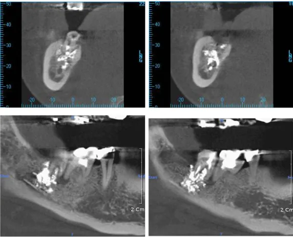
Clinical and radiographic examinations were taken at CUDH to assess her symptoms. Panoramic and periapical radiography showed radiopaque material in the right mandibualr canal(Fig. 1.a,b). We tentatively diagnosed her symptoms as inferior alveolar nerve injury owing to the result of radiographic examination. To confirm this diagnosis, we ordered the Cone Beam-Computed Tomography (CBCT) test for assessment of the relationship between the endodontic sealer(Vitapex
®) and the mandibular canal. The result was the same as the panoramic examination, radiopaque material was noted in the right mandibular canal(Fig. 2).
Treatment started with an anti-inflammatory
regimen including prednisolone(Solondo
®5 mg),
tapering from 30 mg/day to 5 mg/day over 6 days.
Fig. 2. The result of the Come-Beam Computered tomography(CBCT) at the first visit
1 week later, the patient revisited and reported that the pain and numbness were slightly relieved. After discussing about treatment options, she preferred the non-surgical treatment to surgical debridment and decompression of the inferior alveolar canal.
Gabapentin(Neurontin
®) was prescribed, increasing the dosage from 600 mg/day to 1200 mg/day, according to the pain reported in the regular monthly visit. At follow-up appointments, we evaluated the symptoms using three methods; mapping, Clinical Neurosensory Examination(CNE), and the Current Perception Threshold(CPT) test. The affected lesion was mapped for any notable changes in size or position. The CNE evaluated mechanoceptive and nociceptive function on the affected side versus the non-affected side. For CNE, the pressure, touch, pin prick sensation were tested. Sensation was quantified on a scale of 0 to 100, where 0 is complete anesthesia and 100 is normal sensation compared to
the non-affected site. CPT was assessed by the Neurometer(Neurotron Inc., Baltimore, MD). A noninvasive transcutaneous electrical stimulus was delivered to the skin overlying the chin. The site included the affected side and non-affected contralateral side. Each site received 3 stimulus frequencies, 2,000, 250, and 5Hz, which are known to stimulate A-beta, A-delta, and C fibers, respectively. CNE and Neurometer examination were not done on the same day, except on the first visit, due to the lack of time. Instead, on every follow up visit, only one test was carried out in the order of CNE and Neurometer. The results of the tests follows(Fig. 3., Fig. 4., Table 1.).
After six months of the medication, the patient
reported that the sensation had recovered almost
completely, but the patient occasionally felt tingling
sensation in the right chin area. Finally, the
medication was stopped and the patient was referred
a. the first visit b. 2 months later c. 4 months later Fig. 3. Comparison of affected lesion
Fig. 4. Comparison of the result of clinical neurosensory evaluation(CNE)
to the department of Operative Dentistry for completion of the root canal treatment of the mandibular right second molar. A final periodic follow up at the end of the dental treatment was planned, but the patient did not return to our clinic.
CPT Measures and Analysis Summary
side Frequency First visit 3 months 5 months
Rt.
2000 Hz 484 112 52
250 Hz 264 16 5
5 Hz 163 3 2
Lt.
2000 Hz 132 72 120
250 Hz 20 10 31
5 Hz 7 7 9
Table 1. Comparison of the current perception threshold(CPT) test
Ⅲ. DISCUSSION
During root canal treatment, overinstrumentation with hand or mechanically driven files can perforate the mandibular canal, allowing the extrusion of endodontic sealers, dressing agents, and irrigant solutions out of the tooth and into the canal.
15)Ideally, during the root canal treatment, the filling material should be confined to the root canal.
9)Overextension and/or overfilling of mandibular molar and premolar is a potential iatrogenic cause of inferior alveolar nerve injury.
6)Severe endodontic pain after endodontic sealer extrusion requires early diagnosis and prompt management to reduce the risk of permanent nerve damage.
14)Meaningful symptoms can be easily obtained by a medical examination and interview with patient.
The patient can easily express what had happened
since the event is definite, that is root canal
treatment. In this case, the patient reported severe pain during the root canal treatment, and after taking analgesics, the pain was relieved but the numbness in the affected area persisted.
The radiographic examinations(panoramic and periapical radiograph) revealed a radiopaque material around the madibular canal.(Fig. 1.a,b) In most case articles, a radiopaque material was found in the panoramic radiograph or periapical radiograph.
6,9,10)In this case, Cone Beam Computed Tomograph(CBCT) was taken additionally.(Fig .2.) The conventional radiographic image of the mandibular canal is dark linear shadow with thin radiopaque superior and inferior borders cast by the lamella of bone that binds the canal. Furthermore, the mandibular canal can be superimposed over the apex of a molar.
16)Recently, cone beam computed tomography(CBCT) has been proposed as an effective radiographic diagnostic device when endodontic related inferior alveolar nerve or mental foramen paraesthesia is suspected.
17)We considered that CBCT has more advantage in the diagnosis of such case over the conventional radiograph. By using CBCT, the limitation of conventional radiograph can be overcome. (생략 CBCT has been proposed as an effective radiograpic diagnostic device when endodontic related inferior alveolar nerve or mental nerve damage is suspected.)
Studies have shown that all root canal sealants are neurotoxic to some degree.
3)Even root canal sealants that are believed to be more benign, such as zinc oxide and eugenol and calcium hydroxide (owing to its high pH)
18), have been shown to be neurotoxic in vitro
19-24)and are almost certainly neurotoxic in vivo.
25)In this case, the patient reported the sharp, severe pain when the doctor injected Vitapex
®into the root canal of the right mandibular second molar. Vitapex
®which is a premixed Calcium hydroxide with iodoform paste may gain entry into the mandibular canal, and it can damage the nerve owing to its high pH. Pogrel and Thamby
3)suggested that root canal sealants' have neurotoxic properties only when they come into direct contact with the individual fascicles, and as
long as they are outside the epineurium, they are safe. In this case, the symptoms of the patient recovered in a short period of time, assessed by clinical neurosensory evaluation(CNE) and the current perception threshold(CPT) test(Fig.4., Table 1), however, the size of the affected lesion was almost the same in the mapping test.(Fig. 3.) It was postulated that Vitapex
®might have compressed the inferior alveolar nerve, mechanically, but as it did not enter through the perineurium, therefore, there might be no chemical damage in the manibular canal.
During treatment, we followed the symptoms up with the clinical neurosensory evaluation(CNE) combined with the current perception threshold (CPT) test(Neurometer;Neurotron Inc., Baltimore, MD), this was the uniqueness of this article, as far as we know. Neurometer provides reliable measurement of sensory nerve function for large and small myelinated as well unmyelinated nerve fibers. The stimulator delivers sine wave stimuli at frequencies of 2,000, 250 and 5 Hz, which have been shown to selectively stimulate large myelinated Aβ, small myelinated Aδ and small unmyelinated C fibers, respectively.
16)Ziccardi VB et al.
26)suggested that CNE combined with CPT may be used to aid in the assessment of the IAN injuries.
Treatment of this endodontic complication remains controversial, varying from a wait and-see approach, including anti-inflammatory drugs and periodic follow-up
10,12-14), to surgical debridement of the inferior alveolar nerve involving bone removal of the vestibular cortical plate
16)or sagittal mandibulotomy.
27)In the present case, the patient refused the surgical approach, but agreed to attend frequent follow-up appointments. Therefore, non-surgical management was agreed upon including anti-inflammatory treatment with prednisone and analgesic treatment with gabapentin.
Steroid was prescribed to reduce the edema and
inflammatory response. Morse DR
25)advise
immediate steroid administration, though there is no
agreement regarding the type, dosage or duration of
steroid treatment. The antiepileptic medication,
gabapentin(Neurontin
®) was selected. Antiepileptic drugs such as pregabalin and gabapentin have been used for the treatment of neuropathic pain.
9)This binds to the α
2δ subunit of calcium channel, however, the precise mechanism by which it produces analgesia in not well understood.
28)Finally, we suggest that the non-surgical approach with combination of prednisone and gabapentin was a good option in the management of inferior alveolar nerve damage subsequent to overinstrumentation. Immediate management and periodic follow-up is necessary for obtaining a predictable prognosis
REFERENCES
1. Pogrel MA, Thamby S. The etiology of altered sensation in the inferior alveolar, lingual, and mental nerves as a result of dental treatment. Journal of the California Dental Association 1999;27(7):531,534-538.
2. Pogrel MA, Thamby S. Permanent nerve involvement resulting from inferior alveolar nerve blocks. Journal of the American Dental Association 2000;131(7):
901-907.
3. Pogrel MA. Damage to the inferior alveolar nerve as the result of root canal therapy. Journal of American Dental Association 2007;138:65-69
4. Robert RC, Bacchetti P, Pogrel MA. Frequency of trigeminal nerve injuries following third molar removal. Journal of Oral and Maxillofacial Surgery 2005;63(6):732-736.
5. Pogrel MA. Complications of Third Molar Surgery.
Oral Maxillofac Surg Clin North Am 1990;2:441.
6. Escoda-Francoli J, Canalda-Sahli C, Soler A, Figueiredo R, Gay-Escoda C. Inferior alveolar nerve damage because of overextended endodontic material:
a problem of sealer cement biocompatibility? Journal of Endodontics 2007;33(12)1484-1489.
7. Fanibunda K, Whitworth J, Steele J. The management of thermomechanically compacted gutta percha extrusion in the inferior dental canal. British Dental Journal 1998;184(7):330-332.
8. Knowles KI, Jergenson MA, Howard JH. Paresthesia associated with endodontic treatment of mandibular premolars. Journal of Endodontics 2003;29(11):768- 770.
9. López-López J, Estrugo-Devesa A, Jané-Salas E, Segura-Egea JJ. Inferior Alveolar Nerve Injury
Resulting From Overextension of an Endodontic Sealer: Non-surgical Management Using the GABA Analogue Pregabalin. International Endodontic Journal 2011;Aug 23;44(10):1-7.
10. González-Martín M, Torres-Lagares D, Gutiérrez- Pérez JL, Segura-Egea JJ. Inferior Alveolar Nerve Paresthesia After Overfilling of Endodontic sealar into the mandibular canal. Journal of Endodontics 2010;36(8):1419-1421.
11. Kothari P, Hanson N, Cannell H. Bilateral mandibular nerve damage following root canal therapy. British Dental Journal 1996;180(5):189-190.
12. Blanas N, Kienle F, Sándor GK. Inferior alveolar nerve injury caused by thermoplastic guttapercha overextension. Journal of the Canadian Dental Association 2004;70(6):384–387.
13. Poveda R, Bagán JV, Fernández JM, Sanchis JM.
Mental nerve paresthesia associated with endodontic paste within the mandibular canal: report of a case.
Oral Surgery, Oral Medicine, Oral Pathology, Oral Radiology, and Endodontics 2006;102(5):e46–49.
14. Froes FG, Miranda AM, Abad Eda C, Riche FN, Pires FR. Non-surgical management of paraesthesia and pain associated with endodontic sealer extrusion into the mandibular canal. Australian Endodontic Journal 2009;35(3);183–186.
15. Köseoğlu BG, Tanrikulu S, Sübay RK, Sencer S.
Anesthesia following overfilling of a root canal sealer into the mandibular canal: a case report. Oral Surgery, Oral Medicine, Oral Pathology, Oral Radiology, and Endodontics 2006;101(6):803–806.
16. Stuart C. White, Michael J. Pharoah, Oral Radiology:
principles and interpretation, Fifth edition, St. Louis, 2004, Mosby Inc., pp.184.
17. Gambarini G, Plotino G, Grande NM et al . Differential diagnosis of endodontic-related inferior alveolar nerve paraesthesia with cone beam computed tomography: a case report. International Endodontic Journal 2011;44(2):176–181.
18. Conrad SM. Neurosensory disturbances as a result of chemical injury to the inferior alveolar nerve. J Oral Maxillofac Surg Clin North Am 2001;13(2):255-263.
19. Pogrel MA. The results of microneurosurgery of the inferior alveolar and lingual nerve. Journal of Oral and Maxillofacial Surgery 2002;60(5):485-489.
20. Asgari S, Janahmadi M, Khalilkhani H. Comparison of
neurotoxicity of root canal sealers on spontaneous
bioelectrical activity in identified Helix neurones using
an intracellular recording technique. International
Endodontic Journal 2003;36(12):891-897.
21. Asrari M, Lobner D. In vitro neurotoxic evaluation of root-end filling materials. Journal of Endodontics 2003;29(11):743-746.
22. Hume WR. An analysis of the release and the diffusion through dentin of eugenol from zinc oxide-eugenol mixtures. Journal of Dental Research 1984;63(6):881- 884.
23. Hume WR. The pharmacologic and toxicological properties of zinc oxide-eugenol. Journal of the American Dental Association 1986;113(5):789-791.
24. Hume WR. In vitro studies on the local pharmaco- dynamics, pharmacology and toxicology of eugenol and zinc oxide-eugenol. International Endodontic Journal 1988;21(2):130-134.
25. Morse DR. Endodontic-related inferior alveolar nerve and mental foramen paresthesia. Compendium of Continuing Education in Dentistry 1997;18(10):963- 968.
국문초록
근관 충전제의 과충전에 의한 하치조 신경손상에 관한 비수술적 치료 증례