The first case of a bifid mandibular condyle(BMC) was presented by Hrdlička,1 who identified it in the skull spe
cimen of a cadaver in 1941. A total of 103 cases of BMC have been reported from 1987 to 2011.2 The paucity of the reported cases indicates that BMC is an extremely un
usual morphological variation. The morphological details of BMC have been reported to vary. The second condylar head has been reported to usually be located in the anteri
or or medial site of the original condylar head. Other cas
es of BMC showed variation in the depth of the groove dividing the mandibular condyle into two sections sym
metrically in the sagittal plane. The extremely rare forma
tion of a trifid mandibular condyle, with medial, central, and lateral divisions, has also been reported.3
Several authors have proposed etiologies of BMC;311 but no consensus has emerged. Szentpétery et al.4 found only 7 cases of BMC in 1882 skulls(0.48%). From 2008 to 2010, Menezes et al.5 and Miloglu et al.6 found that the prevalence of BMC was 0.018% and 0.3%, respectively, using panoramic radiographs. Sahman et al.9,10 reported that the prevalence of BMC was 0.52% and 1.82% using
panoramic radiographs and computed tomography(CT), respectively. In Cho and Jung’s retrospective study using conebeam computed tomography(CBCT),11 the preva
lence of BMC was 0.5% in 7424 patients. Moreover, the femaletomale ratio ranged from 1.1:1 to 3.5:1 in these studies.3,5,6,911 Some authors4,7 have suggested that trauma at an early age may be a crucial factor in the development of BMC, whereas other authors3,6,8 reported no age pre
dilection. These disagreements in statistical analyses of BMC were caused by the lack of adequate data. Further
more, its uncertain pathogenesis and etiology make BMC a complicated subject to study.
This report describes a case of posttraumatic BMC with radiographic findings. Additionally, we summarize the pathogenic features of BMC in a review of the litera
ture.
Case Report
A 9yearold female patient was referred to the Depart
ment of Oral and Maxillofacial Surgery at Sanggye Paik Hospital with complaints of pain and trismus in the right temporomandibular joint(TMJ) after a bicycle accident.
Spontaneous pain in the right TMJ, mandibular deviation to the right side, restricted mouth opening, malocclusion, concussion of the upper anterior teeth, and an upper lip
Post-traumatic bifid mandibular condyle: A case report and literature review
MinHo Woo1, KyuHo Yoon1, KwanSoo Park1,*, JaeAn Park1
1Department of Oral and Maxillofacial Surgery, Sanggye Paik Hospital, College of Medicine, Inje University, Seoul, Republic of Korea
AbstRACt
Bifid mandibular condyle(BMC) is an uncommon morphological variant of the mandibular condyle. Although authors have proposed various etiologies for BMC, no consensus has emerged. In addition, varying findings have been reported regarding the epidemiological parameters of BMC(e.g., prevalence, gender ratio, and age), possibly due to its low incidence. BMC is occasionally associated with symptoms of the temporomandibular joint, such as ankylosis, pain, and trismus; however, it is difficult to detect this condition on conventional radiographs. This study reports a case of BMC with radiographic findings, and reviews the literature on the epidemiology of BMC.(Imaging Sci Dent 2016; 46: 217-22)
Key woRds: Mandibular Condyle; Anatomic Variation; Congenital Abnormalities; Prevalence; Epidemiology
Copyright ⓒ 2016 by Korean Academy of Oral and Maxillofacial Radiology
This is an Open Access article distributed under the terms of the Creative Commons Attribution NonCommercial License(http://creativecommons.org/licenses/bync/3.0) which permits unrestricted noncommercial use, distribution, and reproduction in any medium, provided the original work is properly cited.
Imaging Science in Dentistry·pISSN 22337822 eISSN 22337830 Received May 16, 2016; Revised June 14, 2016; Accepted July 2, 2016
*Correspondence to : Prof. KwanSoo Park
Department of Oral and Maxillofacial Surgery, Sanggye Paik Hospital, College of Medicine, Inje University, Dongilro 1342, Nowongu, Seoul 01757, Korea Tel) 8229501167, Fax) 8229501167, Email) [email protected]
laceration were detected on the clinical examination(Fig.
1). A maximum jaw opening of 10mm was achieved pas
sively. Computed tomography(CT) showed a fracture of the right mandibular condyle(Fig. 2). Nonsurgical re
duction with a gunning splint was recommended while the patient remained juvenile. Circumzygomatic and cir
cummandibular wiring were performed under general anesthesia since the patient had primary teeth, which did not allow a toothretaining reduction technique such as an arch bar. Active physiotherapy was conducted begin
ning 7 days postoperatively. A maximum jaw opening of 10mm was obtained. The elastic intermaxillary fixa
tion was removed, and mouth opening exercises using a tongue blade were initiated 1 month postoperatively. The gunning splint was removed and a jaw opening of 26mm was obtained at a 2month followup visit. A mouth open
ing of 38mm was achieved 4 months postoperatively, and the patient was instructed to continue mouth opening
exercises to achieve a normal range of motion. The pa
tient achieved a maximum mouth opening of 45mm at a 6month followup visit and did not have any subjective symptoms. She displayed no clinical signs such as click
ing sounds and tenderness of the TMJ, trismus, maloc
clusion, and pain; a 4mm rightward deviation of the jaw upon protrusive movement was observed. Clinical and radiological examinations were performed regularly. A se
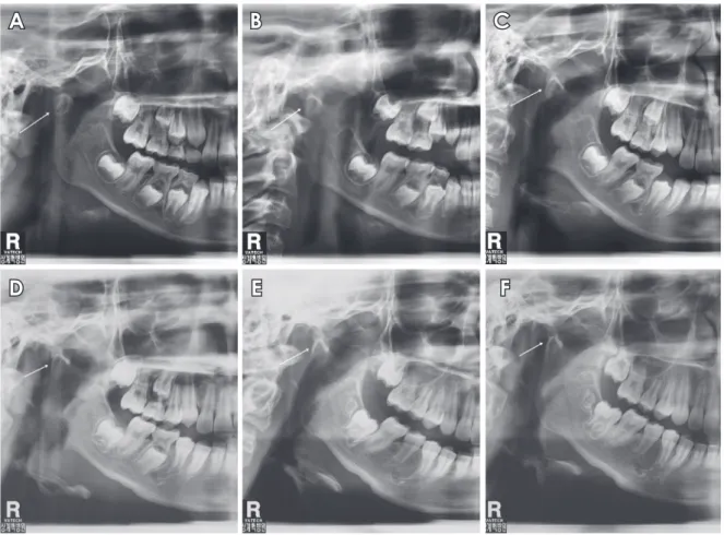
ries of panoramic radiographs showed the fractured con
dylar head, indicating a gradual healing process(Fig. 3).
At a routine 3year routine visit after the operation, CBCT with Uni3D S(Vatech, Hwaseong, Korea) was conduct
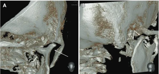
ed to examine the condition of the TMJ for orthodontic treatment planning. Bifidity of the condylar head was found incidentally on CBCT radiographs(Fig. 4). Another medially placed condylar head, which had been bent by the fracture, was discovered on the 3dimensional recon
structed image(Fig. 5). The patient exhibited no clinical symptoms or signs. The jaw deviation on protrusion was also corrected.
discussion
BMC is a unique anomaly consisting of a doubleheaded condyle. Its specific pathogenesis and etiology remain un
known due to its rarity. Some authors have suggested that most BMCs are asymptomatic.2,3,5,6,8 GarcíaGonzález et al.12 argued that treatment is not required for an asymptom
atic BMC, even if longterm followup is necessary due to the possibility of delayed symptoms. However, symptom
atic BMCs have also been described.24,7,1216 Bifid forma
tion with a temporomandibular disorder(TMD) such as a clicking sound, restricted mandibular movement, pain, swelling, ankylosis, and facial asymmetry were noted in
Fig. 1. Intraoral photograph of the patient after trauma. The image shows malocclusion and a sutured upper lip.
Fig. 2. Medially deviated right man
dibular condyle. A. Axial computed tomography(CT) section. B. Coro
nal CT section. The arrowhead indi
cates the low position of the fracture site of the condylar neck.
A B
these studies. Antoniades et al.3 treated patients suffering from restricted jaw movement with nonsteroidal antiin
flammatory drugs, muscle relaxants, occlusal splints, and wooden tongue spatulas. GarcíaGonzález et al.12 also suggested that patients who had an internal derangement of the TMJ disc should receive medical treatment with splints and arthroscopic surgery. If facial asymmetry, man
dibular hypoplasia, restricted mandibular movement, and other TMDs accompany a BMC with TMJ ankylosis, it should be treated surgically, through procedures such as gap arthroplasty, interpositional arthroplasty, condylecto
my, and joint reconstruction.12,1416
Two major causes of BMC have been suggested. The possibility that developmental factors may be involved in the development of BMC has been discussed by sev
eral authors. Blackwood17 proposed the idea of a retained wellvascularized fibrous septum embedded on the man
dibular condyle in the anteroposterior direction. This sep
tum was proposed to divide the condylar head into 2 parts mediolaterally during the developmental process. This
could explain how a forceps delivery causing hematoma around the condylar head led to BMC formation.15,17 Sz
entpétery et al.4 supported the retained septum theory, and insisted that a doubleheaded condyle with anteroposte
riorly positioned morphology was associated with early childhood fractures, whereas those with a mediolaterally positioned morphology were caused by the persistence of a septum in the connective tissue. The growthcenter the
ory regarding condylar head development was suggested by Antoniades et al.3 and Kahl et al.18 These researchers argued that minor trauma during puberty or childhood could result in a bifid condyle, as well as facial asymme
try, because the mandibular condyle is an important cen
ter of facial growth.3 However, Gundlach et al.19 found a BMC that did not contain fibrous septa. They induced a BMC using a teratogenic substance in a rat experiment, which caused maldirection of the muscle affecting the condylar head position. Infection, irradiation, nutritional disorders, genetic, endocrinological, and pharmacological factors have been postulated to account for this phenom
Fig. 3. Panoramic radiographs reveal the remodeling process of the fractured right condylar head at 6month intervals. A. The white arrow indicates a deviated right condylar head. B and C. A cortical radiopaque line is seen. D. The morphology of the fractured condylar head has changed due to remodeling. E and F. Another round condylar head is seen.
A B C
D E F
enon. Cho and Jung11 suggested that developmentally originated BMCs were not associated with TMD in a ret
rospective study. Some authors have reported that the fre
quency of nontraumatic origin was much higher than that of traumatic origin.2,3,5,6,8 The prevalence of nontraumatic BMC was reported to be 64.07% by Almasan et al.2 and
Fig. 4. Conebeam computed tomo graphy images demonstrate the ap pearance of the bifid mandibular condyle. A. Coronal image. B. Sagittal image. C. Axial image. D. Threedimensional reconstruction image.
A B
C D
Fig. 5. Threedimensional reconstructed images show bifid mandibular condyle(BMC) formation. A. The appearance of the BMC in a pos
terior view. B. The appearance of the BMC in an oblique posterior view.
A B
75% by Antoniades et al.3
Another important cause of BMC has been argued to be trauma related to the condylar neck or a sagittal fracture.
Poswillo20 induced a BMC by condylectomy in Macaca monkeys. This study revealed that the resection site of the condylar head was able to generate a new condylar head. Hotz21 also reported formation of a new condyle in the fractured mandibular condyle of a living person. Hotz observed radiographs taken during the healing process of a fractured condylar head; an entirely new condyle had grown at the original position and the fractured condyle underwent partial resorption due to insufficient remodel
ing capacity. As mentioned above, Szentpétery et al.4 in
sisted that anteroposteriorly arranged bifid condyles must be the result of early childhood fractures. Although most reported cases of posttraumatic BMC follow the hypoth
esis of Szentpétery et al., Li et al.7 noted a case of BMC that was due to a sagittal fracture of the condylar head ar
ranged in a mediolateral position.
Thorough evaluation is needed to explain these con
flicting results related to the etiology of BMC. Various ar
guments have been adduced in favor of a developmental cause of BMC, but no consensus has emerged regarding the most likely explanation. However, it is apparent that BMC caused by developmental factors tends to form me
diolaterally on the frontal plane and appears as a symmet
rical form. These BMCs exhibit variation in the depth of the groove that separates the condylar head mediolaterally into 2 symmetric parts.
Posttraumatic BMC involves relatively specific factors that cause a doubleheaded mandibular condyle. Szent
pétery et al.4 insisted that the emergence of a BMC and its symptoms were influenced by the type of injury(direct or indirect, high or low fracture), the extent of damage to the joint structures(disc, capsule, and articular surfaces), the presence or absence of inflammation, hemarthrosis, and the patient’s age. Sahm and Witt22 found that a high condylar head fracture underwent highgrade remodel
ing, but a low condylar head fracture did not show a good remodeling result. Moreover, BMC formation occurred in some cases. Li et al.7 reported a series of 4 cases of a BMC caused by fracture, and classified the BMC appear
ance based on trauma severity, the fracture site, and rela
tionship to the insertion of the lateral pterygoid muscle.
Dahlström et al.23 also suggested that remodeling of the fractured condylar head was affected by the demands of function and growth. In our review of the literature, the direction of the fractured condylar head, site, severity, and its remodeling capacity were found to be the key factors
involved in the formation of a BMC from the fractured state.
The lateral pterygoid muscle, which affects the direc
tion of the fractured condylar fragment, is an important factor in BMC formation.4,7,16,22 If the muscle force is ad
equate to dislocate the condylar head, it meets the mini
mum requirement for the formation of a BMC. Moreover, the ability to create a new condylar head at the original site and insufficient remodeling capacity of the fractured condylar head must occur simultaneously. It is not cer
tain whether remodeling ability is related with age, sex, or race. The vector of the fractured condyle is also im
portant because it regulates the arrangement of the BMC.
Although Szentpétery et al.4 argued that the anteroposteri
orly arranged BMC resulted from trauma, this dichotomy did not successfully explain variation in the morphology of BMCs. Rather, it seems that the arrangement of the BMC depends on the vector of the fractured condylar head and its relationship with the lateral pterygoid muscle force. Variation in BMC morphology might be also regu
lated by the extent of trauma. The anteroposterior groove or notch in the middle of doubleheaded condyle is main
ly seen in sagittally(vertically) fractured or slightly de
viated condylar heads. Immediately apparent Yshaped condylar heads, however, are found in cases of severe de
viation.
BMCs are difficult to detect in conventional radiographs.
Most of the reported cases were found with considerable difficulty due to a lack of symptoms and the superimposi
tion of nearby anatomic structures. A panoramic radiogra
ph is the first choice of diagnostic tool for detecting BMC due to its easy accessibility and low cost.12 However, it has a substantial limitation because the 2dimensional plane only allows the view to be shown on one side of the plane. Thus, midsagittal grooves of the condylar head were not seen on panoramic radiographs. In addition, apparent BMCs can be confused with other abnormal an
atomic structures due to the presence of overlapping near
by structures or inherent radiographic distortion. There
fore, several authors have suggested that CT or CBCT is the goldstandard diagnostic tool for detecting the true prevalence of BMCs.5,6,11,16,18 These developments in di
agnostic imagery have resulted in an increasing number of reports of BMCs in recent decades.
In this study, a young patient showed bifidity of the condylar head on the right side after a condylar neck frac
ture healed. The condylar head was arranged in the me
diolateral position, which is the opposite of what would be expected according to the hypothesis of Szentpétery et
al.4 This led us to conclude that the direction and appear
ance of the BMC are affected by multifactorial causes.
Additionally, patients such as ours may present with no symptoms and a normal range of mouth opening with deflection to the right side. Although further investiga
tion including more cases is needed, we speculate that BMC formation may be associated with the fracture site, deviation extent and direction, and remodeling capacity with age. The degree of symmetry of the BMC may indi
cate whether the origin was traumatic or developmental.
A symmetric appearance usually occurs in BMCs with a developmental cause, and an asymmetric appearance is associated with a traumatic origin. In spite of these ten
dencies, the precise pathophysiology of BMC formation remains unresolved. Recently, advancements in imaging devices have helped to further characterize the prevalence of BMCs. Since BMC cases have been reported with in
creasing frequency, further study is required to ascertain the etiology of the bifidity of the condylar head.
References
1. Hrdlička A. Lower jaw: double condyles. Am J Phys Anthro
pol 1941; 28: 7589.
2. Almasan OC, Hedesiu M, Baciut G, Baciut M, Bran S, Jacobs R. Nontraumatic bilateral bifid condyle and intermittent joint lock: a case report and literature review. J Oral Maxillofac Surg 2011; 69: e297303.
3. Antoniades K, Hadjipetrou L, Antoniades V, Paraskevopoulos K. Bilateral bifid mandibular condyle. Oral Surg Oral Med Oral Pathol Oral Radiol Endod 2004; 97: 5358.
4. Szentpétery A, Kocsis G, Marcsik A. The problem of the bifid mandibular condyle. J Oral Maxillofac Surg 1990; 48: 12547.
5. Menezes AV, de Moraes Ramos FM, de VasconcelosFilho JO, Kurita LM, de Almeida SM, HaiterNeto F. The prevalence of bifid mandibular condyle detected in a Brazilian population.
Dentomaxillofac Radiol 2008; 37: 2203.
6. Miloglu O, Yalcin E, Buyukkurt MC, Yilmaz AB, Harorli A.
The frequency of bifid mandibular condyle in a Turkish pa
tient population. Dentomaxillofac Radiol 2010; 39: 426.
7. Li Z, Djae KA, Li ZB. Posttraumatic bifid condyle: the patho
genesis analysis. Dent Traumatol 2011; 27: 4524.
8. Loh FC, Yeo JF. Bifid mandibular condyle. Oral Surg Oral Med Oral Pathol 1990; 69: 247.
9. Sahman H, Sekerci AE, Ertas ET, Etoz M, Sisman Y. Preva
lence of bifid mandibular condyle in a Turkish population. J Oral Sci 2011; 53: 4337.
10. Sahman H, Sisman Y, Sekerci AE, TarimErtas E, Tokmak T, Tuna IS. Detection of bifid mandibular condyle using com
puted tomography. Med Oral Patol Oral Cir Bucal 2012; 17:
e9304.
11. Cho BH, Jung YH. Nontraumatic bifid mandibular condyles in asymptomatic and symptomatic temporomandibular joint subjects. Imaging Sci Dent 2013; 43: 2530.
12. GarcíaGonzález D, MartínGranizo R, López P. Imaging quiz case 4. Bifid mandibular condyle. Arch Otolaryngol Head Neck Surg 2000; 126: 7959.
13. Quayle AA, Adams JE. Supplemental mandibular condyle. Br J Oral Maxillofac Surg 1986; 24: 34956.
14. To EW. Mandibular ankylosis associated with a bifid condyle.
J Craniomaxillofac Surg 1989; 17: 3268.
15. Stadnicki G. Congenital double condyle of the mandible caus
ing temporomandibular joint ankylosis: report of case. J Oral Surg 1971; 29: 20811.
16. Daniels JS, Ali I. Posttraumatic bifid condyle associated with temporomandibular joint ankylosis: report of a case and review of the literature. Oral Surg Oral Med Oral Pathol Oral Radiol Endod 2005; 99: 6828.
17. Blackwood HJ. The doubleheaded mandibular condyle. Am J Phys Anthropol 1957; 15: 18.
18. Kahl B, Fischbach R, Gerlach KL. Temporomandibular joint morphology in children after treatment of condylar fractures with functional appliance therapy: a followup study us com
puted tomography. Dentomaxillofac Radiol 1995; 24: 3745.
19. Gundlach KK, Fuhrmann A, BeckmannVan der Ven G. The doubleheaded mandibular condyle. Oral Surg Oral Med Oral Pathol 1987; 64: 24953.
20. Poswillo DE. The late effects of mandibular condylectomy.
Oral Surg Oral Med Oral Pathol 1972; 33: 50012.
21. Hotz RP. Functional jaw orthopedics in the treatment of con
dylar fractures. Am J Orthod 1978; 73: 36577.
22. Sahm G, Witt E. Longterm results after childhood condylar fractures. A computertomographic study. Eur J Orthod 1989;
11: 15460.
23. Dahlström L, Kahnberg KE, Lindahl L. 15 years followup on condylar fractures. Int J Oral Maxillofac Surg 1989; 18: 1823.