1)서 론
방사성의약품(radiopharmaceuticals)은 방사성핵종(radionuclide) 또는 특정 수용체에 결합하는 화합물을 방사성핵종에 표지한 물질로서 질병의 진단이나 치료를 위해 사용되는 제제이다.1) 진단용 방사성의약품을 인체에 투여하면 특정 장기에 집적되
∙ Received: September 16, 2019 Accepted: October 27, 2019
∙ Corresponding author: Sang-Hyeong Kil
∙ Department of Nuclear Medicine, Pusan National University Yangsan Hospital, 20, Geumo-ro, Mulgeum-eup, Yangsan-si, Gyeongsangnam-do, 50612, Korea
Tel: +82-55-360-1912, Fax: +82-55-360-1915 E-mail: rlf1007@naver.com
며, 그 부위에서 방출하는 방사선을 섬광카메라(scintillation camera)나 양전자방출전산화단층촬영(positron emission tomography/computed tomography, PET/CT) 장치로 수집하 여 관심장기의 형태 및 기능을 평가 할 수 있다. 섬광카메라로 영상을 획득할 때 사용되는 여러 방사성핵종 중 Tc-99m은 다 른 방사성동위원소들에 비해 Mo-99/Tc-99m 발생기로부터 편리하게 용출하여 사용할 수 있기 때문에 체내 영상용 방사 성핵종으로 가장 많이 이용되고 있다.2) 방사성의약품 Tc-99m HMPAO (Tc-99m Hexamethyl Propylene Amine Oxime)는 지 용성으로 뇌혈류장벽(blood brain barrier, BBB)을 통과한 후 뇌혈류에 비례하여 장시간 고정적인 국소분포를 유지하기 때 Sang-Hyeong Kil1, Yung-Hyun Lim2, Gwang-Yeol Park3 and Seong-Mook Cho4
Department of Nuclear Medicine, Pusan National University Yangsan Hospital, Yangsan-si, Korea
Purpose Tc-99m HMPAO is widely used radiopharmaceutical for brain perfusion SPECT. Tc-99m HMPAO is chemically unstable and is liable to show deterioration of labeling efficiency due to high incidence of secondary Tc-99m HMPAO complex, free pertechnetate and reduced-hydrolyzed Tc-99m. In this study, we investigated whether sialogogues administration could reduce the impurities of Tc-99m HMPAO.
Materials and Methods In thirty subjects(20 male and 10 female, age range 19~89 years, mean age 60.7±14.5 years), brain perfusion SPECT were performed at basal and citric acid stimulation states consecutively after injection of 555 MBq of Tc-99m HMPAO. In the salivary glands, the uptake coefficient was calculated using Siemens processing program. Statistical comparison between before and after the citric acid stimulation performed paired t-test. P value less than 0.05 was regarded as statistically significant.
Results Salivary glands uptake was 12900±3101 counts in basal and 10677±2742 counts in citric acid stimulation states.
Unnecessary impurities in the body is much decreased after citric acid administration(t=10.78, P<0.05). The image quality was much improved after administration of citric acid and the regional cerebral perfusion was clearly from demarcated the background.
Conclusion The impurity is distributed throughout the body particularly in the salivary glands and nasal mucosa when Tc-99m HMPAO brain perfusion SPECT is performed. If this impurities is not removed, the quality of the image may deteriorate, resulting in errors in visual evaluation. The use of sialogogues could be helpful for decreasing unnecessary impurities in the body.
Key Words Brain perfusion SPECT, Tc-99m HMPAO, Sialogogues, Salivary glands uptake coefficient
문에 뇌졸중(stroke), 간질(epilepsy), 치매(dementia) 및 기타 뇌혈관질환에서 변형된 뇌관류 평가에 널리 이용되고 있 다.3,4) 그러나 Tc-99m HMPAO는 표지 과정에서 주석 이온의 양과 방사분해에 의해 발생하는 중간산물 때문에 화학적으로 불안정하고, secondary Tc-99m HMPAO complex, free pertechnetate, reduced-hydrolyzed Tc-99m 등 방사화학적 불 순물 생성이 많아 표지효율 저하가 나타나기 쉽다.5,6) 또한 과 산화테크네슘(free pertechnetate)은 뇌혈류장벽을 통과하지 못하고 침샘과 구강 점막에 집적되며, 제거되지 않은 불순물 로 인하여 영상의 품질이 저하되어 뇌혈관질환 평가에 오류가 발생할 수 있다. 따라서 본 연구는 Tc-99m HMPAO를 이용한 뇌혈류 단일광자방출단층촬영(brain perfusion single photon emission computed tomography, brain perfusion SPECT)에서 타액분비촉진제(sialogogues) 구연산(citric acid) 투여가 침샘 및 구강 점막에 집적된 불순물을 줄여 영상의 품질 향상과 뇌 혈관질환 진단 평가에 유용한지 알아보고자 하였다.
실험재료 및 방법 1. 연구 대상
본 연구는 2016년 7월부터 12월까지 뇌혈류 단일광자방출 단층촬영(brain perfusion SPECT)을 시행한 환자 중 연구에
동의한 30명(나이 분포 19∼89세, 평균 연령 60.7±14.5세, 남 성 20명, 여성 10명)을 대상으로 하였다.
2. 방사화학적 순도 측정
Mo-99/Tc-99m 발생기(Ultra-TechneKow FM Generator, Mallinckrot Medical B.V., Netherlands)에서 용출한 과테크네 슘산나트륨(99mTcO4-) 주사액을 30분 이내 동결 건조된 HMPAO kit(Stabilised Ceretec TM, GE Healthcare Limited, UK)에 표지 시킨 후 박층 크로마토그래피(Thin Layer Chromatography, TLC) 측정기(Bioscan AR-2000, Eckert &
Ziegler Medical, USA)를 이용하여 방사화학적 순도 (radiochemical purity)를 측정하였다.
박층 크로마토그래피용 실리카켈(Instant Thin Layer Chromatography Silica Gel, ITLC-SG) strips 2개를 준비 한 후 하단 2.5 ㎝ 부분에 과테크네슘산나트륨과 표지된 HMPAO를 점적 하였다. ITLC-SG strip 하나는 메틸에틸케톤(Methyl Ethyl Ketone, MEK)에 다른 하나는 생리식염수(0.9%
aqueous sodium chloride)에 넣고 15분 동안 전개하여 꺼낸 후 말린 다음 AR-2000 radio-TLC imaging scanner로 각각 방사 능 분포를 계수하였다. 메틸에틸케톤을 사용하여 secondary Tc-99m HMPAO complex와 reduced-hydrolyzed Tc-99m의 방사능 비율(A%)을, 생리식염수를 사용하여 pertechnetate의 Fig. 1. Flow chart of radiochemical purity of measurement. Tc-99m HMPAO radiochemical purity analyzed using combination of 2 chromatographic systems.
방사능 비율(B%)을 각각 계산 하였다(Fig. 1). 또한 Tc-99m HMPAO의 방사화학적 순도는 식 (1)을 이용하여 계산하였다 (Eq. 1).
Radiochemical purity = 100 - (A% + B%) ··· (Eq. 1) A%: secondary Tc-99m HMPAO complex + reduced- hydrolyzed Tc-99m
B%: pertechnetate
3. 영상 획득
방사성의약품 Tc-99m HMPAO의 뇌혈류 분포는 신경정신 기능 활성도에 따라 변화한다. 따라서 재현성 있는 영상 획득 과 시각 활성자극을 피하기 위해 안정실의 환경은 조광기를 이용하여 조명을 낮추었으며 조용한 상태로 유지 시켰다. 영 상 획득 장치는 저에너지 고해상력(Low Energy High Resolution, LEHR) 조준기를 장착한 단일광자방출단층촬영 장치(Symbia E, Siemens, USA)를 이용하였다. 방사성의약품 투여 시 연구 대상이 통증을 느끼지 않도록 편안하게 누운 상 태에서 오른쪽 팔의 전주정맥(antecubital vein)에 약물투여경 로(intravenous route)를 미리 확보하고, 20분 정도 안정을 취 한 다음 방사성의약품 Tc-99m HMPAO 555 MBq을 투여하였
다. Tc-99m HMPAO 투여 10분 경과 후 환자를 배뇨 시킨 다음 테이블에 눕히고 두·경부를 검출기 시야 중심부에 위치시켰 으며, 안와외이공선(Orbitomeatal Line, OML)이 단일광자방 출단층촬영장치 회전축에 수직이 되도록 머리를 고정하였다.
먼저 3분 동안 기저(basal) 두·경부 전면 영상(head & neck anterior image)을 획득하였으며, 0˚ 에서 시계방향으로 180˚
‘step and shoot circular' 방식으로 검출기를 회전시켜 64 프레 임(frame) 기저(basal) 뇌혈류 투사상(projection view)을 획득 하였다. 또한 기저영상(basal image)을 획득 한 후 침샘 및 구강 점막에 집적된 불순물을 줄이고자 타액분비촉진제 구연산 (citric acid) 1 ㏄를 1분간 입에 머금게 하여 침샘을 자극시킨 다음 삼키게 하고, 기저영상 획득 방법과 동일하게 침샘 자극 후 영상(post-citric acid image)을 얻었다(Fig. 2).
4. 침샘 섭취계수 계산
침샘 섭취계수(uptake coefficient)는 Siemens사의 영상처 리 프로그램을 사용하여 침샘 자극 전·후 획득한 두·경부 전면 영상(head & neck anterior image)에서 그림 3과 같이 좌·우 침 샘부위에 관심영역(Regions of Interest, ROI)를 설정한 다음 관심영역의 계수 값(salivary glands counts of ROI)을 식 (2)에 대입하여 계산하였다(Eq. 2).
(A) Basal head & neck anterior image (B) Post-citric acid head & neck anterior image Fig. 3. Salivary glands uptake coefficient was calculated using Siemens processing program.
Uptake coefficient of salivary glands = (ROIrs + ROIls) / 2
··· (Eq. 2) ROIrs: right salivary glands frame counts of ROI ROIls: left salivary glands frame counts of ROI
5. 뇌혈류 영상 비교
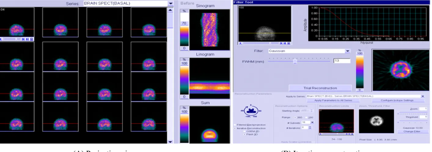
단일광자방출단층촬영장치를 이용하여 획득한 뇌혈류 투 사상을 시네모드(cine mode)로 환자의 움직임 및 인공산물 유 무를 확인하였다. 또한 투사상을 여과기(gaussian filter)를 사 용하여 여과하였으며, 반복적 재구성(iterative reconstruction) 방법으로 영상을 재구성하였다(Fig. 4). 횡단면(transverse)은 OML에 평행하게 얻고, 뇌의 크기와 및 형태에 따라 기울어진 영상을 보정하여 횡단면에 수직인 시상면(saggital plane)과 관상면(coronal plane)을 각각 얻었으며, 타액분비촉진제 구 연산(citric acid) 투여 전·후 뇌혈류 영상 비교는 횡단면, 시상 면, 관상면을 동시에 보면서 시행하였다.
6. 통계분석
자료 분석은 SPSS 21.0(IBM-SPSS Inc., Chicago, Il, USA) 을 이용하였으며, 타액분비촉진제 구연산(citric acid) 투여 전 (basal image)·후(post-citric acid image) 침샘 섭취계수의 평 균비교는 대응표본 T검증(paired t-test)을 하였다. P값이 0.05 미만인 경우 통계적으로 유의하다고 판단하였다.
결 과 1. 방사화학적 순도
조제된 방사성의약품 Tc-99m HMPAO는 secondary Tc-99m HMPAO complex, free pertechnetate, reduced- hydrolyzed Tc-99m 등 불순물이 존재할 수 있다. 따라서 AR-2000 radio-TLC imaging scanner를 이용하여 방사화학적 순도를 측정한 결과 평균과 표준편차는 93.6±0.85이었으며, 90% 이상의 표지율을 보였다(Table 1).
2. 침샘 섭취계수 비교
타액분비촉진제 구연산(citric acid) 투여 전(basal image)과 후(citric acid stimulations image)에 획득 한 두·경부 전면 영상 의 침샘 섭취계수(uptake coefficient)를 비교 분석한 결과 타 액분비촉진제 투여 전 침샘 섭취계수의 평균과 표준편차는 12900±3101, 투여 후 침샘 섭취계수의 평균과 표준편차는 10677±2742였다(Fig. 5). 타액분비촉진제 구연산(citric acid) 투여 후 불순물이 많이 감소되었으며 통계적으로 유의하게 나 타났다(Table 2).
Fig. 5. Salivary glands uptake coefficient was 12900±3101 in basal and 10677±2742 in citric acid stimulation states.
(A) Projection view (B) Iterative reconstruction
Fig. 4. These data acquired from SPECT system are reconstructed OSEM 3D(ordered-subsets expectation maximization) method called by Flash 3D from Siemens with Subsets 16, Iteration 8 and FWHM 13.
3. 뇌혈류 단일광자방출단층촬영 영상 비교
타액분비촉진제 구연산(citric acid) 투여 전·후 뇌혈류 단일 광자방출단층촬영 영상을 비교 한 결과 침샘과 구강 점막에 집적되었던 불순물이 제거되어 뇌-연부조직의 방사능비가 좋 아졌으며, 기억·집중·사고·언어 등의 중요 기능을 담당하고 있는 대뇌피질(cerebral cortex)과 운동·반복적 업무 수행·강 박행동 등과 관련된 부위인 미상핵(caudate nucleus), 담창구 (globus pallidus), 피각(putamen) 등 심부 뇌 구조(deep brain structure)가 선명하게 나타났다(Fig. 6).
고 찰
이상적인 뇌혈류 영상용 방사성의약품은 뇌혈류장벽을 자 유롭게 이동할 수 있으면서 가능한 한 혈액내에서 단백질과 결합이 적어야 한다. 또한 체내 주사 후 최초 순환 뇌 조직 추출 률이 높아야 하며, 뇌 조직에 섭취되지 않는 화합물은 배후 조 (A) Basal image
(B) Citric acid stimulation image
Fig. 6. The image quality was improved after administration of citric acid and the basal ganglia perfusion was clearly from demarcated the background.
Table 2. Result of statistical comparison between before and after the citric acid stimulation
Salivary gland uptake(counts) t P*
Mean±SD Basal image Citric acid stimulation image
10.78 0.001
12900±3101 10677±2742
*P : P value by paired t-test
Tc-99m HMPAO8 P20,P21 3.7% 96.3% 97.1% 2.9% 93.4%
Tc-99m HMPAO9 P22,P23,P24 4.1% 95.9% 96.8% 3.2% 92.7%
Tc-99m HMPAO10 P25,P26 2.8% 97.2% 97.8% 2.2% 95.0%
Tc-99m HMPAO11 P27,P28 2.8% 97.2% 97.6% 2.4% 94.7%
Tc-99m HMPAO12 P29,P30 3.5% 96.5% 97.1% 2.9% 93.6%
Rf1 : secondary Tc-99m HMPAO complex + reduced-hydrolyzed Tc-99m Rf2 : lipophilic Tc-99m HMPAO complex + pertechnetate
Rf3 : lipophilic Tc-99m HMPAO complex + secondary Tc-99m HMPAO complex + reduced-hydrolyzed Tc-99m Rf4 : pertechnetate
직에서 빨리 제거되어야 한다.7,8) 본 연구에 사용된 방사성의 약품 Tc-99m HMPAO는 주사 후 1분 이내에 최대섭취에 도달 하며, 뇌 조직에 안정되게 섭취되므로 이상적인 뇌 혈류 영상 제제이다.1,9) 그러나 99mTc-d,l-HMPAO 복합체는 불안정하기 때문에 체내에서 수용성 유도체로 분해되어 이차 복합체를 생 성하며, 지용성이 낮은 이차 복합체들은 뇌혈류장벽을 통과 할 수 없게 된다. 뇌로 들어가지 못한 과산화테크네슘 (pertechnetate)과 같은 불순물은 침샘 및 구강 점막, 전신에 걸 쳐 근육과 연조직에 광범위하게 분포하며 배후 방사능은 천천 히 제거된다.2,10) 침샘 및 구강 점막 부위에 방사능 집적은 인접 배후 방사능으로부터 야기되는 방사선 때문에 뇌-연부조직의 방사능비가 떨어져 영상 품질이 저하되고, 시각적 평가에 오 류가 발생할 수 있다. 본 연구에 사용된 방사성의약품 Tc-99m HMPAO는 박층 크로마토그래피 측정기로 화학적 순도를 측 정한 결과 표지율이 평균 93% 이상 이었으며, 유럽핵의학학 회(European Association of Nuclear Medicine, EANM)와 제 조사에서 권고하고 있는 표지율(radiochemical purity of >
80%)보다 높게 나타났다.4) 그러나 7% 정도의 불순물은 침샘 에서 활동적으로 섭취, 농축, 분비되므로 영상의 질을 저하시 킬 수 있을 것이다. 또한 뇌혈류 단일광자방출단층촬영 영상 을 시각적으로 분석하면, 판독자간의 일치도 차이가 발생 할 수 있으며 뇌 혈류 분포차이를 정량적으로 나타내기 어렵
다.11,12) 따라서 향후 Tc-99m HMPAO를 이용한 뇌혈류 영상
평가에 있어서 시각적 영상비교뿐만 아니라 확률뇌지도분석 법(Statistical Probabilistic Anatomical Mapping, SPAM)과 같 은 지표를 사용하여 국소 뇌혈류 변화 정도를 정량화하면 진 단적 가치를 더 높일 수 있을 것으로 생각된다.13-15)
결 론
뇌혈류 단일광자방출단층촬영에서 타액분비촉진제 구연 산(citric acid)을 투여하면 침샘 및 구강 점막에 집적된 불순물 이 제거되어 뇌-연부조직의 방사능비가 향상되었다. 또한 생 리적 섭취에 따른 영상 품질저하를 줄이고, 대뇌피질(cerebral cortex)과 심부 뇌 구조(deep brain structure)를 선명하게 나타 낼 수 있어 이미지 품질이 크게 증가되었으며, 뇌혈류 감소 부 위 국소화가 가능하여 정확한 뇌관류 평가에 도움을 줄 수 있 을 것이다.
요 약
뇌혈류 영상용 방사성의약품 Tc-99m HMPAO는 방사분해
에 의해 발생하는 중간산물 때문에 화학적으로 불안정하고, 불순물 생성이 많아 표지효율 저하가 나타나기 쉽다. 과산화 테크네슘(free pertechnetate)은 뇌혈류장벽을 통과하지 못하 고 침샘과 구강 점막에 집적되며, 불순물이 제거되지 않으면 영상 품질이 저하되어 시각적 평가에 오류가 발생할 수 있다.
따라서 본 연구는 Tc-99m HMPAO를 이용한 뇌혈류 단일광 자방출단층촬영(brain perfusion SPECT)에서 타액분비촉진 제(sialogogues) 투여가 침샘 및 구강 점막에 집적된 불순물을 줄여 영상 품질 향상에 유용한지 알아보고자 하였다. 뇌혈류 단일광자방출단층촬영을 시행한 환자 중 연구에 동의한 30명 을 대상으로 침샘 섭취계수(uptake coefficient)와 뇌혈류 영상 을 비교 분석 하였다. 방사성의약품 Tc-99m HMPAO 555 MBq을 투여 후 기저(basal) 두·경부 전면 영상과 뇌혈류 투사 상(projection view)을 획득하였다. 타액분비촉진제 구연산 (citric acid)을 사용하여 침샘을 자극시킨 후 기저영상 획득 방 법과 동일하게 침샘 자극 후 영상(post-citric acid image)을 얻 었다. Siemens사의 영상처리 프로그램을 사용하여 좌·우 침샘 부위에 관심영역을 설정한 다음 타액분비촉진제 투여 전·후 침샘 섭취계수의 평균차이를 분석(paired t-test)하였으며, 뇌 혈류 영상 비교는 획득한 투사상을 반복적재구성(iterative reconstruction) 방법으로 영상을 재구성하여 비교 분석하였 다. 두·경부 전면 영상의 침샘 섭취계수를 비교 분석한 결과 구 연산 투여 전 섭취계수의 평균값은 12900±3101, 투여 후 섭취 계수의 평균값은 10677±2742였다. 타액분비촉진제 투여 후 불순물이 많이 감소되었으며 통계적으로 유의하게 나타났다 (t = 10.78, P < 0.05). 뇌혈류 영상을 비교 한 결과 침샘과 구강 점막에 집적되었던 불순물이 제거되어 이미지 품질이 크게 향 상되었다. 타액분비촉진제를 투여하면 침샘 및 구강 점막에 집적된 불필요한 불순물이 제거되어, 생리적 섭취에 따른 영 상 품질저하를 줄일 수 있어 정확한 뇌혈류 평가에 도움이 될 것이다.
REFERENCE
1. Saha GB. Fundamentals of nuclear pharmacy. 7rd ed. New York: Springer; 2017. p. 93-121
2. 정준기, 이명철. 핵의학. 제3판. 고려의학; 2008. p. 395-465 3. Martinez, Teresa, et al. "Influence of radiolytic‐produced
hydrogen peroxide on the stability of commercial kit 99mtechnetium‐exametazime preparation." J. Label. Compd.
Radiopharm. 2014;57(1):49-52
flow tracer in focal cerebral ischaemia." Eur J Nucl Med.
1994;21(9):913-923
7. Lear, James L. "Quantitative local cerebral blood flow measurements with technetium-99m HM-PAO: evaluation using multiple radionuclide digital quantitative autoradiography."
J Nucl Med. 1988;29(8):1387-1392
8. Podreka, I., et al. "Initial experience with technetium-99m HM-PAO brain SPECT." J Nucl Med. 1987;28(11):
1657-1666
9. Shen, Yeh‐You, et al. "Regional cerebral blood flow in patients with systemic lupus erythematosus." J Neuroimaging . 1999;9(3):160-164
10. Ziessman, Harvey A., Janis P. O'Malley, and James H.
Thrall. Nuclear Medicine: The Requisites E-Book. 4rd ed. Elsevier Health Sciences, 2013. p. 350-377
al. "Efficacy assessment of cerebral arterial bypass surgery usingstatistical parametric mapping and probabilistic brain atlas on basal/Acetazolimide brain perfusion SPECT." J Nucl Med. 2004;45(2):202-206 14. Eo JS, Oh CW, Kim Yu, Park EK, Lee WW, Kim SE, et
al. "Hemodynamic outcome of successful bypass surgery in patients with atherosclerotic cerebrovascular disease: a study with Acetazolamide and 99mTc-ECD SPECT." J Nucl Med. 2006;47(1):293-301
15. Mazziotta, John, et al. "A probabilistic atlas and reference system for the human brain: International Consortium for Brain Mapping (ICBM)." Philos Trans R Soc Lond B Biol Sci. 2001; 356(1412):1293-1322