∙ Received: August 4, 2010. Accepted: August 24, 2010.
∙ Corresponding author: Seung Jeong Kim
Department of Nuclear Medicine, Seoul National University Hospital, 101 Daehang-ro, Jongno-gu, Seoul, 110-744, Korea
Tel: +82-2-2072-2535, Fax: +82-2-766-9083 E-mail: [email protected]
Original Article
유방암 환자의 Positron EmissionMammography에서의 유용성에 관한 연구
서울대학교병원 핵의학과
김승정⋅김재일⋅김진의⋅김현주
A Study on Availability about Positron Emission Mammography of Breast Cancer
Seung Jeong Kim, Jae Il Kim, Jin Eui Kim and Hyun Joo Kim
Department of Nuclear Medicine, Seoul National University Hospital, Seoul, Korea
Purpose: Until now the general study for breast cancer patient has been mammography, breast sonography for anatomic diagnostics and
18F-FDG whole body PET for functional one. But the PEM (Positron Emission Mammography) was developed to increase sensitivity, specificity and accuracy to improve the disadvantage of each study. Therefore this present study aims to describe availability of PEM for improving diagnosis of breast cancer. Materials and Methods: During 3 months from January in 2010, PEM was performed on 100 patients who had history of breast cancer. Using Naviscan’s PEM Flex Solo Ⅱ scanner, PEM images of breast were acquired. And then we evaluated sensitivity, specificity and accuracy of PEM data by comparing results of PEM images with postoperative pathologic finding. Results: On the 100 patients, it could reveal 89 true positive, 9 false positive, 7 false negative and 87 true negative. Thus the results of sensitivity, specificity and accuracy for PEM was evaluated 92.7%, 90.6% and 91.7%. Conclusion: The sensitivity, specificity and accuracy have been reported 83.7%, 68.5% and 77.1% in mammography, and that was 89.1%, 79.1% and 83.4% in breast sonography. However the results of PEM were more increased than others. Therefore the results of this study will be available that PEM used diagnosis with breast cancer, used preoperative operation plan and helps to discovery of a part recurrence. (Korean J Nucl Med Technol 2010;14(2):60-64)
Key Words : PEM, breast PET, breast cancer imaging, Sensitivity, Specificity, Accuracy
서 론
유방암은 우리나라에서 가장 흔한 여성암 중 하나이고 연 령이 증가할수록 발병률이 증가하고 있다. 특히 40세 여성 의 250명 중 1명이 매년 유방암으로 진단받고 있다.1)
유방암의 진단을 위해 단순유방 X-선 촬영과 유방초음파 검사가 대부분 함께 이루어지고 있다. 그 중 단순유방 X-선 촬영은 가장 많이 하는 검사로 높은 예민도를 나타내지만 특
이도와 양성 예측률은 유방초음파검사에 비해 낮은 편이다.
그리고 특히 유방의 조직밀도가 증가되어 있는 경우 병변의 구별이 어려울 때가 많아 50세 미만의 환자에 있어서 유방암 의 진단이 늦어지는 경우가 많다.2)
이러한 단점을 보완해 주기 위해 다양한 방사성동위원소를 이용한 핵의학 검사가 병행되는데, 최근 유방영상평가에 사용 하기 위한 고해상도 PEM (Positron Emission Mammography) 이 개발되었다. 더불어 PEM을 전신 18F-FDG PET 검사와 함께 시행함으로 보다 유방암의 진단률을 높이고 병변의 위 치 정보를 정확하게 알 수 있게 되었다.
본 연구에서는 유방암의 진단에 있어서 단순유방 X-선 촬 영과 유방초음파검사에 대한 PEM의 유용성을 알아보고자 한다.
ⓒ Naviscan PET Systems, Inc.
Fig. 1. The PEM Flex Solo Ⅱ scanner has dual detector, com-
pression and support ones.Fig. 2. PEM imaging displays the 3D breast image with 12
tomographic slices.Fig. 3. Decision Matrices consist of 4 cell, TP, FP, FN and
TN.실험재료 및 방법
1. 연구 대상 및 검사 방법
2010년 1월부터 3월까지 본원 유방외과에서 유방암 진단 을 받은 환자 100명(연령: 30~73세)을 대상으로 양쪽 유방에 대한 PEM을 시행하였다. 장비는 Naviscan사의 PEM Flex Solo Ⅱ scanner (Naviscan PET Systems, Inc., San Diego, CA) 를 사용하였다.
PEM은 gantry 내 360° 방향에 검출기가 고정된 일반 PET 과는 다르게 감마카메라와 같이 2개의 검출기가 양방향으로 위치해 있다. 각 검출기 크기는 5.6×16.3 cm2이고 스캔 시 이 동하여 16×23 cm2의 작은 FOV (Field of View)를 만든다 (Fig. 1). 스캔이 완료되면 재구성을 통해 축방향의 단층영상 12개가 만들어진다(Fig. 2).4-5)
사용된 방사성의약품은 18F-FDG이고 PEM 검사를 위해 환자의 체중에 비례하여 5.18 MBg/kg (0.14 mCi/kg)을 병소 가 있는 반대편 팔에 정맥 주사하고 30분 후 검사를 시작하 였다. 환자는 앉은 자세로 상하 방향(Cranio-caudal view), 내 외 사 방향(Medio-lateral Oblique view) 촬영을 시행하였고, 각각 좌우 4분씩 검사하였다.
2. 분석 방법
검사를 시행한 후 결과를 평가하기 위해 간단한 2×2 행렬 을 만든다. PEM 검사의 결과를 양성과 음성으로 분류하여
행으로 표시하고 수술 후 병리결과를 통한 실제 질병상태의 유, 무를 분류하여 열로 표시한다. 이 행렬을 판정행렬(Decision Matrices)이라고 한다. 행렬의 각 항목에는 해당 검사결과와 질병상태에 해당하는 환자의 수를 기입한다(Fig. 3).
검사결과가 양성(positive)이고 질병이 있는 경우는 진양성 (true positive, TP), 검사결과가 양성인데 질병이 없는 경우는 위양성(false postivie, FP), 검사결과가 음성(negative)이고 질 병이 없는 경우는 진음성(true negative, TN)이라 하며, 검사 결과가 음성인데 질병이 있는 경우는 위음성(false negative, FN)으로 구분한다. 이렇게 구분된 각각의 검사결과를 토대 로 예민도, 특이도, 정확도를 산출하였다(Table 1).
예민도(sensitivity)
특이도(specificity)
위음성률(false-negative ratio, FNR)
위양성률(false-positive ratio, FPR)
정확도(accuracy)
Table 1. Ratio
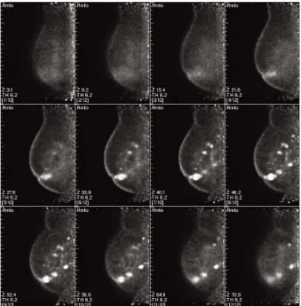
Fig. 4. This picture shows the true positive result.
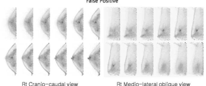
Fig. 5. This picture shows the false positive result.
Fig. 6. This picture shows the false negative result.
Fig. 7. This picture shows the true negative result.
예민도(sensitivity)는 진양성 결과수를 질병이 있는 총 환 자 수로 나눈 값이고, 진양성률(true positive ratio, TPR)이라 고도 한다. 특이도(specificity)는 진음성 결과수를 질병이 없 는 총 환자수로 나눈 값이며, 진음성률(true negative ratio, TNR)이라고도 한다. 정확도(accuracy)는 진양성 결과수와 진음성 결과수의 합을 총 검사대상수로 나눈 지표를 말한 다.6)
이러한 지표를 통해 PEM 결과를 단순유방촬영, 유방초음 파검사와 비교하였다.
결 과
1. PEM 영상결과와 수술 후 병리결과 비교
환자 100명의 양쪽 유방의 PEM 영상결과에서는 18F-FDG 섭취가 보이는 병변은 98, 섭취가 보이지 않은 곳은 96으로 나타났고 수술 후 병리결과에서는 종양이라고 확진된 곳은 94, 종양이 아니라고 확진된 곳은 96으로 나타났다.
이 중 PEM 영상과 수술 후 병리결과를 통해 알아본 결과 로 18F-FDG 섭취가 보이고 종양이라고 확진된 결과수인 진 양성은 89(Fig. 4), 18F-FDG 섭취가 보이지만 종양이 아니라 고 확진된 결과수인 위양성은 9(Fig. 5), 18F-FDG 섭취가 보 이지 않지만 종양이라고 확진된 결과수인 위음성은 7(Fig. 6),
18F-FDG 섭취도 보이지 않고 질환도 없는 결과수인 진음성 은 87(Fig. 7)로 나타났다.
2. 분석 결과
PEM 영상결과에서 18F-FDG 섭취를 보이는 병변이 있고, 수술 후 병리결과에서도 종양으로 나타난 환자 수인 진양성 결과수를 질병이 있는 총 환자 수로 나누어 예민도를 구하였 다. 질병이 있는 총 환자 수는 진양성과 위음성 결과수를 합
한 값으로 96에 대한 진양성 결과수 89를 백분율화 하면 92.7%로 나타난다.
PEM 영상결과에서 18F-FDG 섭취를 보이는 병변이 없고 수술 후 병리 결과에서도 종양으로 나타나지 않는 환자 수인 진음성 결과수를 질병이 없는 총 환자수로 나누어 특이도를 구하였다. 질병이 없는 총 환자 수는 진음성과 위양성 결과 수를 합한 값으로 96에 대한 진음성 결과수 87을 백분율화 하면 90.6%로 나타난다.
정확도는 진양성과 진음성 결과수의 합을 총 검사대상수 로 나누어 구하였다. 총 검사대상수는 진양성, 위양성, 위음 성, 진음성 결과수를 합한 값으로 192에 대한 진양성과 진음 성 결과수의 합인 176을 백분율화 하면 91.7%로 나타난다.
결 론
유방암의 진단에 있어서 단순유방촬영과 유방초음파검사 는 예민도가 높은 검사 중의 하나로 정기적 건강검진의 필수 이다. 그러나 단순유방촬영의 양성 예측률이 비교적 낮은 특 이도를 나타내어 이로 인해 생검의 대상이 되는 환자를 가려 내기 위한 새로운 보조적 진단 방법이 필요하게 되었다. 유 방초음파검사는 방사선피폭의 위험이 없으며 비침습적 검사 로서 고형 종괴와 낭성 종괴의 구분이 유용하다. 또한 젊은 여성이나 단순유방촬영 상 유방이 치밀 유방이거나 유선우 세음영을 보이는 경우 유방실질 내에 숨어있는 종괴의 발 견에 효과적이고 단순유방촬영보다 예민도와 특이도 모두 높다.2)
단순유방촬영의 예민도는 86.2%, 특이도는 64.5%로 보고 되어 있고, 유방초음파검사의 예민도는 87.9%, 특이도는 76.7%로 알려져 있다.7) 본 연구에서 PEM의 예민도는 92.7%, 특이도는 90.6%, 정확도는 91.7%로 단순유방촬영과 유방초음파검사보다 높게 나타났다. 그러나 PEM 검사가 다 른 검사보다 예민도와 특이도가 높게 나타났다고 해서 모든 유방검사를 대체할 수는 없다. 일반적으로 18F-FDG PET과 PEM에서 유방암의 진단에 대한 음성 예측률이 낮다고 보고 되어 있어서8-9) 단순유방촬영, 유방초음파검사, 유방 MRI 검 사 등과 더불어 양성과 악성을 구분하여 진단해 내는데 도움 을 줄 것이라고 생각한다.
검사의 예민도는 질병이 있는 환자만 검사해도 나타낼 수 있으며, 질병이 없는 환자의 검사결과에 대해서는 아무것도 알 수가 없어서 예민도 단독으로는 정상과 질병상태를 구분 하는 검사의 능력을 반영하지는 못한다. 또한 검사의 특이도 는 질병이 없는 환자만 검사해도 나타낼 수 있으며, 예민도 와 마찬가지로 단독으로는 정상과 질병상태를 구분하는 검 사의 능력을 반영하지 못한다. 예민도와 특이도 중에서 어느 지표가 중요한가는 상황에 따라 그리고 양성 또는 음성 검사 결과가 가져오는 영향에 따라 달라진다.6) 검사의 정확도는 질병의 유병률에 영향을 많이 받으므로 검사의 정보내용을 잘 반영하지 못할 경우가 많아 본 연구에서는 PEM 검사의 정확도만 나타내었고 단순유방촬영과 유방초음파검사의 정
확도는 배제하였다.
최근 2.4 mm의 해상도를 가진 고 해상도 유방 전용 PET 장비인 PEM의 도입으로 인해 비교적 작은 크기의 병변의 발견이 가능하였다. 그리고 유방암에 대한 예민도와 특이도 가 높아 유방암 진단에 아주 유용한 검사가 될 것이다. 뿐만 아니라 전신 18F-FDG PET 검사를 위해 주입하는 18F-FDG 한 번의 주사만으로도 전신 PET 검사와 PEM 검사 모두를 시행할 수 있으므로 환자에게는 불필요한 피폭을 경감시킬 수 있는 이점도 있다.
따라서 PEM은 유방암을 진단하고 수술하기 전의 더 나은 수술 계획에 사용될 수 있으며 국소 부위 재발견에 도움이 될 것으로 사료된다.
요 약
유방암의 진단을 위해 단순유방촬영, 유방초음파검사,
18F-FDG PET 검사가 대부분 함께 이루어지고 있다. 그러나 이들 검사의 단점을 보완하고자 보다 더 높은 예민도, 특이 도, 정확도를 가진 PEM이 도입되었다. 본 연구에서는 유방 암의 진단을 향상시키는 PEM의 유용성을 알아보고자한다.
2010년 1월부터 3월까지 유방외과에서 유방암 진단을 받 은 환자 100명을 대상으로 PEM 검사를 시행하였다. 장비는 Naviscan사의 PEM Flex Solo Ⅱ scanner를 사용하였다.
PEM 영상결과와 수술 후 병리결과를 비교하여 PEM 검사의 예민도, 특이도, 정확도를 평가하였다.
환자 100명에서 PEM 영상과 수술 후 병리결과를 통해 알 아본 결과로 진양성은 89, 위양성은 9, 위음성은 7, 진음성은 87이었다. PEM 검사의 예민도, 특이도, 정확도는 각각 92.7%, 90.6%, 91.7%로 나타났다.
단순유방촬영에서 예민도 86.2%, 특이도 64.5%로 보고되 어 있고 유방초음파검사에서 예민도 87.9%, 특이도 76.7%로 알려져 있다. 본 연구에서 PEM은 예민도 92.7%, 특이도 90.6%로 다른 검사보다 높게 나타났다. 이로써 PEM은 유방 암을 진단하고 수술하기 전의 더 나은 수술 계획에 사용될 수 있으며 국소 부위 재발의 발견에 도움이 될 것으로 사료 된다.
REFERENCES
1. Joon-Kee Yoon. Clinical Application of 18F-FDG PET in Breast Cancer. Nucl Med Mol Imaging 2008;42:76-90
2. 이은주, 이한경, 장미선, 장경아, 차경호, 김종호. 유방암 진
단에 있어서 99mTc-MIBI 스캔의 유용성: 유방촬영술과 초음 파유방촬영술과의 비교.
대한방사선의학회지
2000;42:191-7 3. Kavita Murthy, Marianne Aznar, Christopher J. Thompson,Antoine Loutfi, Robert Lisbona and Jean H. Gagnon. Results of Preliminary Clinical Trials of the Positron Emission Mammography System PEM-Ⅰ: A Dedicated Breast Imaging System Producing Glucose Metabolic Images Using FDG. J Nucl Med 2000;41:
1851-8
4. Lawrence MacDonald, John Edwards, Thomas Lewellen, David Haseley, James Rogers and Paul Kinahan. Clinical Imaging Characteristics of the Positron Emission Mammography Camera:
PEM Flex Solo Ⅱ. J Nucl Med 2009;50:1666-75
5. David Beylin. Physics of Positron Emission Mammography (PEM):
Basic Principles of Operation. Naviscan PET Systems, Inc.
6. 고창순. 제3판 핵의학.
고려의학
2008;8-117. Sabine Malur, Susanne Wurdinger, Andreas Moritz, Wolfgang Michels and Achim Schneider. Comparison of written reports of mammography, sonography and magnetic resonance mammography for preoperative evaluation of breast lesions, with special emphasis on magnetic resonance mammography. Breast Cancer
Res 2001;3:55-60
8. Rakesh Kumar, Anil Chauhan, Hongming Zhuang, Prem Chandra, Mitchell Schnall and Abass Alavi. Clinicopathologic Factors associated with false negative FDG-PET in primary breast cancer. Breast Cancer Res Treat 2006;98:267-74 9. Eric L. Rosen, Timothy G. Turkington, Mary Scott Soo, Jay A.
Baker and R. Edward Coleman. Detection of Primary Breast Carcinoma with a Dedicated, Large-Field-of-Vies FDG PET Mammography Device: Initial Experience. Radiology 2005;234:
527-34