INTRODUCTION
The supraclavicular lymph nodes comprise a final common pathway of metastatic nodal involvement from various malig- nancies. These lymph nodes are easily accessible by palpation and their enlargement may be the first sign of a metastatic tu- mor, mostly from lung, head and neck, breast, esophageal, gastric, pancreatic, gynecologic, and prostate cancers (1).
Examination of the supraclavicular lymph node has tradi- tionally been performed by palpation; however, this method has been found to be unreliable in the literature (2-5). These investigators have suggested that noninvasive imaging tech-
niques such as CT and ultrasound (US) can be used to improve the detection of lymph node metastasis. Several studies have reported on CT and US methods for assessing supraclavicular lymphadenopathy (1, 2, 6-11).
18F-fluorodeoxyglucose positron emission tomography (18F- FDG PET) is a noninvasive method that plays an important role in the evaluation of lymph node metastasis in patients with various malignancies. Moreover, 18F-FDG PET is more accu- rate than CT for detecting or excluding nodal disease (12-16).
However, some limitations exist for the use of 18F-FDG PET alone. In combining functional PET data and morphologic CT data, 18F-FDG PET/CT studies have produced promising initial
J Korean Soc Radiol 2012;66(1):83-92
Received September 10, 2011; Accepted October 13, 2011 Corresponding author: Eun Bi Ryu, MD
Department of Radiology, Kosin University Gospel Hospital, Kosin University College of Medicine, 34 Amnam-dong, Seo-gu, Busan 602-072, Korea.
Tel. 82-51-990-6341 Fax. 82-51-255-2764 E-mail: [email protected]
Copyrights © 2012 The Korean Society of Radiology
Purpose: The purpose of this study is to compare the usefulness of 18F-fluorodeoxy- glucose positron emission tomography (18F-FDG PET)/CT, contrast-enhanced CT and ultrasound (US) for diagnosing metastatic supraclavicular lymph nodes.
Materials and Methods: This study included 53 supraclavicular lymph nodes of 48 consecutive patients with various malignancies observed on 18F-FDG PET/CT, contrast- enhanced CT and US. Detection of supraclavicular lymph nodes was determined by
18F-FDG PET/CT where uptake was greater than that of surrounding tissue and con- trast-enhanced CT with a node short-axis diameter of 5 mm or more. On US, we clas- sified the supraclavicular lymph node as benign or malignant by sonographic criteria.
The diagnostic values of these modalities were compared in the detection of meta- static supraclavicular lymph nodes.
Results: Metastatic supraclavicular lymph nodes were diagnosed cytologically in 44 (83%) of 53 lesions. In the detection of metastatic supraclavicular lymph nodes, the diagnostic accuracies of 18F-FDG PET/CT, contrast-enhanced CT, and US were 92%, 89%, and 91%, respectively. The specificity (67%) and negative predictive value (86%) of 18F-FDG PET/CT were higher than those of contrast-enhanced CT and US.
Conclusion: 18F-FDG PET/CT is more useful for detecting and characterizing supra- clavicular lymph nodes in patients with cancer, because of its high specificity and negative predictive value.
Index terms Lymph Node Metastasis
Positron Emission Tomography/CT Malignancy
Supraclavicular Lymph Node Metastasis from Various Malignancies:
Assessment with 18F-Fluorodeoxyglucose Positron Emission Tomography/CT, Contrast-Enhanced CT and Ultrasound
다양한 원발암으로부터의 쇄골상림프절 전이: 18F-Fluorodeoxyglucose Positron Emission Tomography/CT, 조영증강 CT 그리고 초음파 간의 평가
Eun Bi Ryu, MD, Kyung Seung Oh, MD, Kyung Soon Jeong, MD
Department of Radiology, Kosin University Gospel Hospital, Kosin University College of Medicine, Busan, Korea
from the head to the pelvic floor according to a standard pro- tocol with the following settings: 130 kVp; 30 mA; tube rota- tion time, 0.8 seconds per rotation; pitch, 6; section thickness, 5 mm to match the PET section thickness. Immediately after non-enhanced CT, PET was performed in the identical trans- verse field of view. PET data sets were obtained with an iterative reconstruction and an ordered subset expectation maximiza- tion algorithm was performed by the application of segment- ed attenuation correction (two iterations, 28 subsets) to the CT data. Co-registered scans were displayed with software that enabled image fusion and analysis.
18F-FDG PET/CT data sets were prospectively evaluated by one nuclear medicine physician. The physician was unaware of the CT findings or any clinical information except that the pa- tients had primary malignancy. For imaging interpretation, posi- tive FDG uptake in supraclavicular lymph nodes was considered when glucose uptake of the lesion was greater than the sur- rounding tissue. Further, the maximum standardized uptake val- ue (SUV) adjusted for the patient’s body weight, was recorded.
CECT Acquisition and Imaging Interpretation
CT scans were acquired using a helical technique, which was performed with a Somatom Plus-4 (Siemens Medical Solutions, Erlangen, Germany) or a Somatom Sensation 64 (Siemens Medical Solutions, Erlangen, Germany) scanner. Chest CT scanning was performed from the lower part of the neck to the middle portion of the kidneys. In neck CT, scanning was performed from the skull base to the level of the aortic arch.
All scanning was performed after IV administration of con- trast medium (100 mL of iopromide, Redisence 300, Accuzen, Seoul, Korea) at a rate of 2 mL/s with a power injector (Mallinck- rodt, Tyco and Vistron CT, Medrad, Pittsburgh, PA, USA).
The scanning parameters were as follows: 120 kVp; chest CT, 90 mA; neck CT, 150 mA; beam width, 2.5 mm; and a table speed of 15 mm per rotation in the chest CT and 13.8 mm per rotation in the neck CT. Data were interfaced directly to a pic- ture archiving and communication system, which displayed all imaging data on monitors (two monitors, 1,536 × 2,048 im- age matrices, 8-bit viewable gray scale, 60-foot-lambert lumi- nescence). Scans were viewed with both mediastinal (window width, 400 H; window level, 20 H) and lung (window width, 1,500 H; window level, -700 H) window settings.
oncologic imaging results (17-19). To our knowledge, the use- fulness of 18F-FDG PET/CT in the characterization and detec- tion of supraclavicular lymph node metastases from various malignancies has not yet been reported.
Accordingly, the purpose of this study is to compare the use- fulness of 18F-FDG PET/CT, contrast-enhanced CT (CECT), and US in the diagnosis of metastatic supraclavicular lymph nodes.
MATERIALS AND METHODS
Patients
From January 2008 to September 2009, 158 consecutive pa- tients with suspected or proven malignancy underwent US, CECT, and 18F-FDG PET/CT examinations. Our institutional review board approved our research study and did not require informed consent from the patients for this retrospective study.
We excluded patients who had undergone chemotherapy or radiation therapy, patients who had not undergone pathologic evaluation, and who had no supraclavicular lymph node en- largement. This retrospective study included 53 supraclavicu- lar lymph nodes in 48 patients with a proven malignancy. The histopathological diagnosis of a primary malignancy was con- firmed by surgery in 38 cases, by percutaneous needle aspiration biopsy of the primary mass in 12 cases, and by bronchoscopic biopsy in three cases. The final status of the supraclavicular lymph nodes was established by US-guided fine-needle aspi- ration biopsy (FNAB) or by surgical excision biopsy. All pa- tients underwent US examination of the supraclavicular region, CECT of the neck or chest that included the supraclavicular re- gion, and 18F-FDG PET/CT of the whole body. The mean in- terval between US and 18F-FDG PET/CT was 4.9 days (range, 0-18 days). The mean interval between CECT and 18F-FDG PET/CT was 5.2 days (range, 0-20 days).
18F-FDG PET/CT Acquisition and Imaging Interpretation All patients fasted for at least 6 hours and had a serum glu- cose level lower than 140 mg/dL before the IV injection of
18F-FDG. Scanning was performed 60 minutes after 18F-FDG administration. Scans were acquired using PET/CT system (CTI, Knoxville, TN, USA), consisting of a full-ring PET scan- ner and a dual-detector-row spiral CT scanner (Somatom Emo- tion Duo, Biograph, Erlangen, Germany). CT was performed
sy were expelled onto glass slides and smeared. Additional cy- tological sampling was performed if a specimen was inade- quate for diagnosis.
Statistical Analysis
Statistical analysis was performed with commercially avail- able software (SAS 8.2, SAS Institute, Cary, NC, USA). The accuracy, sensitivity, specificity, and positive and negative pre- dictive values of US (long-axis diameter to short-axis diameter ratio, absence of a nodal hilum, and presence of a nodal cor- tex abnormality), CECT (short-axis diameter), and 18F-FDG PET/CT (increased FDG uptake greater than that of surround- ing tissue) in the detection of metastatic supraclavicular lymph nodes were calculated using a generalized estimating equation.
McNemar’s test was used to compare the diagnostic accuracy, sensitivity, and specificity of the imaging modalities. The pos- itive and negative predictive values of these methods were also compared. The Mann-Whitney test, Fisher’s exact test, and Student’s t-test were used for various measurements com- paring the presence versus absence of supraclavicular lymph node metastasis. The maximum SUV of each supraclavicular lymph node was retrospectively calculated to indicate the pres- ence of metastasis on 18F-FDG PET/CT. p values < 0.05 were considered to indicate statistically significant differences.
RESULTS
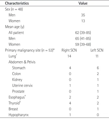
Characteristics of Supraclavicular Lymph Nodes The characteristics of patients with various malignancies are shown in Table 1. Fifty-three supraclavicular lymph nodes were found on CECT, US, and 18F-FDG PET/CT in 48 patients; five of whom had bilateral supraclavicular lymph nodes. Aspiration cytological examination confirmed the presence of supraclavic- ular lymph node metastasis in 43 (81%) of the 53 lymph nodes from the following cancer types: lung, 24; stomach, 10; thyroid, six; esophagus, four; colon, two; uterine cervix, one; breast, one;
kidney, one; hypopharynx, one; and prostate, one. Surgical ex- cision confirmed the presence of supraclavicular lymph node metastasis from lung cancer in one (2%) of the 53 lymph nodes, while the other nine (17%) lymph nodes proved to be benign. The finding was made at the aspiration cytological ex- amination in all benign lymph nodes.
Two experienced radiologists, who were blinded to any clini- cal information except that the patients had a primary malig- nancy prospectively read all of the CT scans. The supraclavic- ular nodal area was defined as the region that lies above the manubrium on the same image as the clavicle, lateral to the medial edge of the common carotid artery and medial to the clavicle and the lateral rib margin (20, 21). Metastatic supra- clavicular lymph nodes were defined as having a short-axis diameter ≥ 5 mm on CECT (1, 2, 6, 11). The long-axis diame- ter and its ratio with the short-axis diameter of the supracla- vicular lymph nodes were also recorded.
High-Resolution Thyroid Sonography and US-Guided Aspiration Biopsy
A sonographic examination was performed using a 5 to 12- MHz linear array transducer (iU22, Philips Medical Systems, Bothell, WA, USA) for the evaluation of the thyroid gland and neck in all patients. Before US-guided FNAB was performed, real-time sonography was performed by one of five radiolo- gists in the thyroid imaging department; this radiologist was unaware of any clinical information except that the patients had a primary malignancy. All US-guided FNABs in our se- ries were performed by one radiologist. Transverse and sagit- tal images were obtained from the submandibular gland to the acromioclavicular joint.
Interpretations of sonography were performed prospective- ly. The radiologist was unaware of the CT findings or of any clinical information except that the patients had a primary ma- lignancy. For imaging interpretation, several criteria were used for differentiating malignant from benign lymph nodes: long- axis diameter to short-axis diameter ratio ≤ 2; absence of a nodal hilum; and the presence of a nodal cortex abnormality, such as eccentric nodal thickening. The definition of ultrasonic malignancy criteria was based on previous literature (11, 22).
The sonography results, grouped into metastatic and benign, were compared with those of the final cytopathological reports for assessing the value of the test in diagnosing malignancy.
After the sonographic evaluation of the thyroid gland, US- guided FNABs were performed by one radiologist for re-eval- uating the cervical lymph nodes. The procedure was conduct- ed using a 23-gauge needle attached to a 10 mL disposable plastic syringe. Specimens obtained from the aspiration biop-
shown in Table 2. The sensitivities (p = 1.000) and specificities (p = 0.183) of 18F-FDG PET/CT and CECT did not differ sig- nificantly. In addition, the sensitivities (p = 0.337) and speci- ficities (p = 0.190) of 18F-FDG PET/CT and US did not differ significantly. The diagnostic accuracy was not significantly different between 18F-FDG PET/CT and CECT (p = 0.300) or
18F-FDG PET/CT and US (p = 1.200). The positive predictive values (p = 1.265) and negative predictive values (p = 0.090) were not significantly different between 18F-FDG PET/CT and CECT. Likewise, the positive predictive values (p = 2.500) and negative predictive values (p = 0.107) did not differ significant- ly between 18F-FDG PET/CT and US. However, 18F-FDG PET/
CT had a higher specificity and negative predictive value than CECT and had a higher specificity than US.
Correlation between Maximum SUV and Nodal Size The statistical assessment of variable measurements on 18F- FDG PET/CT and CECT according to the presence of metas- tasis in the supraclavicular lymph node is shown in Table 3.
No significant correlation was observed between long-axis di- ameter to short-axis diameter ratio on CECT and maximum SUV on 18F-FDG PET/CT (p = 0.198). However, significant dif- ferences were observed between the metastatic and benign groups of supraclavicular lymph node in terms of the maximum SUV of supraclavicular lymph nodes (p = 0.002), the nodal short-axis diameter on CECT (p = 0.001), and the nodal long- Diagnostic Efficacy of 18F-FDG PET/CT, CECT, and US
The accuracy, sensitivity, specificity, and positive and nega- tive predictive values of 18F-FDG PET/CT, CECT, and US in the detection of supraclavicular lymph node metastasis are Table 1. Characteristics of Patients with Malignancies
Characteristics Value
Sex (n = 48)
Men 35
Women 13
Mean age (y)
All patient 62 (39-85)
Men 65 (41-85)
Women 59 (39-68)
Primary malignancy site (n = 53)* Right SCN Left SCN
Lung† 14 11
Abdomen & Pelvis
Stomach 4 6
Colon 0 2
Kidney 0 1
Uterine cervix 1 1
Prostate 0 1
Esophagus‡ 2 2
Thyroid§ 4 2
Breast 0 1
Hypopharynx 1 0
Note.-Values in parentheses are ranges.
*Number of supraclavicular lymphnodes.
†Bilateral supraclavicular lymphnodes in two patients.
‡Bilateral supraclavicular lymphnodes in one patient.
§Bilateral supraclavicular lymphnodes in two patients.
SCN = supraclavicular lymphnode
Table 2. Diagnostic Efficacy of 18F-FDG PET/CT, CECT, and US
Finding Accuracy (%) Sensitivity (%) Specificity (%) PPV (%) NPV (%)
18F-FDG uptake on PET/CT 92 98 67 93 86
Short-axis diameter ≥ 5 mm on CECT 89 98 44 90 80
Malignant lymph node on US 91 98 56 91 83
Note.-CECT = contrast-enhanced CT, 18F-FDG PET/CT = 18F-fluorodeoxyglucose positron emission tomography, NPV = negative predictive value, PPV = positive predictive value, US = ultrasound
Table 3. Statistical Assessment of Measurements on 18F-FDG PET/CT and CECT According to the Presence of Metastatic SCN
Variable Measurement Metastatic SCN p value
Present Absent
Maximum SUV of SCN on 18F-FDG PET/CT Size on CECT (mm) 5.13 ± 2.26 1.87 ± 1.10 0.002*
Short axis diameter 1.25 ± 0.51 0.60 ± 0.21 0.001†
Long axis diameter 1.72 ± 0.60 0.95 ± 0.29 0.003*
Long-axis/short-axis 1.46 ± 0.39 1.65 ± 0.48 0.198*
Note.-*Mann-Whitney test.
†Student's t test.
CECT = contrast-enhanced CT, 18F-FDG PET/CT = 18F-fluorodeoxyglucose positron emission tomography, SCN = supraclavicular lymphnode, SUV = stan- dardized uptake value
One patient with left thyroid cancer had a false-positive find- ing of left supraclavicular lymph node metastasis. On US, the long-axis diameter to short-axis diameter ratio was 1.2 with no fatty hilum. The short-axis diameter was 10 mm on CECT, and FDG uptake in the supraclavicular area with a maximum SUV of 2.79 was observed on PET/CT. However, the result of aspiration cytological examination was found to be a benign reactive lymph node.
One patient with uterine cervical cancer had a false-posi- tive finding of right supraclavicular lymph node metastasis.
On US, the long-axis diameter to short-axis diameter ratio was 1.43 with no fatty hilum. The short-axis diameter was 7 mm on CECT, and FDG uptake in the supraclavicular area with a maximum SUV of 3.01 was observed on PET/CT. How- ever, the diagnosis of an aspiration cytological examination was a benign reactive lymph node. Two false-positive inter- pretations were made on CECT, and the mean short-axis di- axis diameter on CECT (p = 0.003).
False-Positive and False-Negative Interpretations on
18F-FDG PET/CT, CECT, and US
False-positive interpretations of metastatic lymph nodes were made on 18F-FDG PET/CT in three cases, on CECT in five cas- es, and on US in five cases. Three of these cases were interpret- ed as being false-positive on all modalities. One patient with stomach cancer had a false-positive finding for a right supracla- vicular lymph node metastasis. On US, the long-axis diameter to short-axis diameter ratio was 1.46 with no fatty hilum. The short-axis diameter was 7 mm on CECT, and a FDG uptake in the supraclavicular area with a maximum SUV of 3.89 was ob- served on PET/CT. However, lymphocytes and fibrous tissues were observed on aspiration cytological examination, and sur- gical excision of the lymph node confirmed the presence of tu- berculous caseating granuloma (Fig. 1).
D A
E B
F C
Fig. 1. A 49-year-old woman with stomach cancer and false-positive interpretation at US, contrast-enhanced CT, and 18F-FDG PET/CT.
A. US examination shows round supraclavicular lymph node without fatty hilum.
B. Contrast-enhanced CT scan shows right supraclavicular lymph node (arrow) with a short-axis diameter of 7 mm.
C-E. PET (C), CT (D), and PET/CT (E) show increased FDG uptake (arrow) in the right supraclavicular lymph node with a maximum SUV of 3.89.
F. Photomicrograph of lymph node biopsy specimen shows chronic granulomatous inflammation with caseation necrosis suggestive of tubercu- losis (H&E, × 40).
Note.-18F-FDG PET/CT = 18F-fluorodeoxyglucose positron emission tomography, SUV = standardized uptake value, US = ultrasound
cer showed FDG uptake of left supraclavicular lymph node on PET/CT with a maximum SUV of 2.31, but had a short-axis di- ameter of 4.2 mm on CECT and a long-axis diameter to short- axis diameter ratio of 2.42 with a fatty hilum on US.
DISCUSSION
Accurate staging is mandatory to ensure the selection of ap- propriate therapy in patients with malignancy. The supracla- vicular lymph nodes are an important component of lymphatic drainage and are involved in various malignancies. Therefore, the assessment of supraclavicular lymph nodes by using phys- ical palpation, US, CECT, or 18F-FDG PET/CT without un- necessary invasive procedures in patients with malignancy is important.
In several studies, examination of the supraclavicular lymph ameter of the supraclavicular lymph nodes was 7.5 mm (7
mm and 8 mm). Two false-positive interpretations were made on US, and the mean long-axis diameter to short-axis diame- ter ratio of the supraclavicular lymph nodes was 1.53 (1.95 and 1.1).
False-negative interpretations of metastatic supraclavicular lymph nodes were made on two patients. One patient with right thyroid cancer had a false-negative finding on 18F-FDG PET/
CT, while another patient with left breast cancer had a false-neg- ative interpretation on both CECT and US. The patient with right thyroid cancer had a false-negative finding on 18F-FDG PET/CT. A metastatic right supraclavicular lymph node with a long-axis diameter to short-axis diameter ratio of 1.94 with no fatty hilum on US and a short-axis diameter of 5.6 mm on CECT did not show significant FDG uptake (Fig. 2) in the same area on 18F-FDG PET/CT. The other patient with left breast can-
D A
E B
F C
Fig. 2. A 52-year-old woman with thyroid cancer and false negative interpretation at 18F-FDG PET/CT.
A. US examination of the right supraclavicular area reveals an ovoid lymph node without fatty hilum.
B. US examination of the left supraclavicular area shows a taller than wide lymph node (long-axis to short-axis diameter ratio < 2) without fatty hilum.
C. Contrast-enhanced CT scan shows right supraclavicular lymph node (black arrow) with a short-axis diameter of 5.6 mm and a left supraclavic- ular lymph node (white arrow) with a short-axis diameter of 7.6 mm.
D, E. CT (D) and PET/CT (E) show increased FDG uptake (white arrow) only in the left supraclavicular lymph node with maximum SUV of 4.57.
There is no FDG uptake in right supraclavicular area. Note the right thyroid show increased FDG uptake consistent with thyroid cancer.
F. Photomicrograph specimen from a sonographically-guided aspiration biopsy shows malignant cells suggestive of papillary cell carcinoma (H&E, × 200).
Note.-18F-FDG PET/CT = 18F-fluorodeoxyglucose positron emission tomography, US = ultrasound
nodes was defined as having a FDG uptake greater than that of the surrounding tissue, and 18F-FDG PET/CT had 92% ac- curacy, 98% sensitivity, 67% specificity, a 93% positive predic- tive value, and an 86% negative predictive value for the detec- tion of supraclavicular lymph node metastasis. Although the differences were not statistically different among imaging mo- dalities, 18F-FDG PET/CT had higher specificity than US, and had higher specificity and negative predictive value than CECT.
In one report (24), 18F-FDG PET/CT had higher sensitivity and negative predictive value than CECT in lung cancer with nonpalpable supraclavicular lymph nodes. In addition, no significant differences were observed in the report.
Although 18F-FDG PET/CT has the advantage of whole- body imaging for metastasis detection, one well-known limi- tation is the differentiation from the inflammatory process.
This can be problematic in countries with high endemic rates of tuberculosis, because reactive hyperplasia or inflammation due to granulomatous diseases such as tuberculosis shows in- creased FDG uptake. In our study, there was a false-positive interpretation of supraclavicular lymph node metastasis with a maximum SUV of 3.89 on 18F-FDG PET/CT and this case was confirmed by tuberculous caseating granuloma. Consider- ation of lymphatic drainage pathways and review of pre-con- trast CT imaging for high density lymph nodes may be help- ful in determining granulomatous lymphadenopathy.
Malignancies originating in the pelvis or abdomen are more likely to metastasize to the left supraclavicular lymph node (25). In the present case, all abdominal and pelvic malignan- cies metastasized to the left supraclavicular area. Right and left lower lung cancers had a tendency to metastasize to the right supraclavicular area. Moreover, we can predict the me- tastasis site by considering the location of the primary malig- nancy. Therefore if we consider the characteristics of lym- phatic drainage, we could reduce the number of false-positive cases in all imaging modalities.
Other possible causes of false-positive results are physiolog- ic muscle uptake and brown fat (26, 27). However, these pat- terns are typically bilateral, symmetric, fusiform, or elonagat- ed and are often confused with malignancy (28). In some cases, atherosclerotic plaques exhibit focal increased FDG uptake and manifest as malignancy (29, 30). However, these problems can be overcome with the use of 18F-FDG PET/CT.
nodes by palpation has been found to be unreliable (2-5). In various malignancies, such as head and neck cancer (5), mel- anoma (3), esophageal cancer (4), US and US-guided fine-nee- dle aspiration cytological analysis have proved to be superior to palpation for the detection and characterization of supra- clavicular lymph node metastasis. On US, a long-axis diame- ter to short-axis diameter ratio ≤ 2, absence of a nodal hilum, and nodal cortex abnormality are reliable criteria for differen- tiating malignant from benign lymph nodes (11, 22). In the application of these criteria as malignant lymph nodes, the following diagnostic characteristics were obtained: accuracy, 91%; sensitivity, 98%; specificity, 56%; positive predictive val- ue, 91%; and negative predictive value, 83%.
There are also reports of the successful use of CT for assess- ing supraclavicular lymph node metastasis in patients with lung cancer (2, 6) and esophageal cancer (23). These investigators obtained satisfactory sensitivity (82-85%) for supraclavicular lymph node metastasis on CT with the use of short-axis diame- ter criteria for the presence of metastases greater than or equal to 5 mm. In our study, CECT had 89% accuracy, 98% sensi- tivity, 44% specificity, a 90% positive predictive value, and an 80% negative predictive value.
In this study, several false-positive and false-negative inter- pretations on CECT were observed. The possible causes of these include the following: presence of normal-sized (short- axis diameter < 5 mm) metastatic lymph nodes; lymph node enlargement (short-axis diameter ≥ 5 mm) due to benign pro- cess; inaccurate measurements of short-axis diameter of su- praclavicular lymph nodes on axial CT scan; various interpre- tation with measurement error of borderline-size lymph nodes around 5 mm. These problems were partially solved in this study through the use of US and 18F-FDG PET/CT.
Several reports indicate that FDG PET is more accurate than CT for detecting or excluding nodal disease (12-16). These findings indicated that FDG PET was more sensitive for the detection of metastasis by increased metabolic activity. How- ever, its limitations include failure to depict anatomic land- marks and limited spatial resolution. In that regard, 18F-FDG PET/CT results in improved sensitivity, specificity, and accu- racy for the detection of malignant lymph nodes compared with FDG PET and CT (17).
In our study, the positive uptake of supraclavicular lymph
155-158
5. Baatenburg de Jong RJ, Rongen RJ, Laméris JS, Harthoorn M, Verwoerd CD, Knegt P. Metastatic neck disease. Palpa- tion vs ultrasound examination. Arch Otolaryngol Head Neck Surg 1989;115:689-690
6. Fultz PJ, Feins RH, Strang JG, Wandtke JC, Johnstone DW, Watson TJ, et al. Detection and diagnosis of nonpalpable supraclavicular lymph nodes in lung cancer at CT and US.
Radiology 2002;222:245-251
7. Monsó E, Montserrat JM, Abad J, Texidó A, Roig J, Morera J. Usefulness of supraclavicular ultrasonography in the staging of lung cancer. Lung 1992;170:243-244
8. Sugama Y, Kitamura S. Ultrasonographic evaluation of neck and supraclavicular lymph nodes metastasized from lung cancer. Intern Med 1992;31:160-164
9. Chang DB, Yang PC, Yu CJ, Kuo SH, Lee YC, Luh KT. Ultraso- nography and ultrasonographically guided fine-needle as- piration biopsy of impalpable cervical lymph nodes in pa- tients with non-small cell lung cancer. Cancer 1992;70:
1111-1114
10. Goldberg RP, Austin RM. Computed tomography of axil- lary and supraclavicular adenopathy. Clin Radiol 1985;36:
593-596
11. Vassallo P, Wernecke K, Roos N, Peters PE. Differentiation of benign from malignant superficial lymphadenopathy:
the role of high-resolution US. Radiology 1992;183:215- 220
12. Verhagen AF, Bootsma GP, Tjan-Heijnen VC, van der Wilt GJ, Cox AL, Brouwer MH, et al. FDG-PET in staging lung cancer: how does it change the algorithm? Lung Cancer 2004;44:175-181
13. Dwamena BA, Sonnad SS, Angobaldo JO, Wahl RL. Metas- tases from non-small cell lung cancer: mediastinal staging in the 1990s--meta-analytic comparison of PET and CT.
Radiology 1999;213:530-536
14. Marom EM, McAdams HP, Erasmus JJ, Goodman PC, Cul- hane DK, Coleman RE, et al. Staging non-small cell lung cancer with whole-body PET. Radiology 1999;212:803- 809
15. Wahl RL, Quint LE, Greenough RL, Meyer CR, White RI, Or- ringer MB. Staging of mediastinal non-small cell lung cancer with FDG PET, CT, and fusion images: preliminary In most instances, the FDG uptake of metastatic lesion is
proportional to that of primary lesion. Therefore, if the FDG uptake of the primary lesion is low, then the metastatic lesion might show low or no FDG uptake. In addition, FDG uptake is related to lesion size, cell density, and metabolic activity.
These are the main cause of false-negative results in 18F-FDG PET/CT.
Our study did have a limitation. Because patients who did not undergo cytological confirmation, US, CECT, or 18F-FDG PET/CT were excluded, the diagnostic efficacies of all imag- ing modalities may have been underestimated in the detec- tion of supraclavicular lymph node metastasis.
In conclusion, the presence of supraclavicular lymph node metastasis in various malignancies is important for accurate staging and appropriate therapy. 18F-FDG PET/CT is a more useful modality than the other abovementioned techniques for the detection and characterization of supraclavicular lymph node metastasis in various malignancies, because of its higher specificity and negative predictive value.
REFERENCES
1. Fultz PJ, Harrow AR, Elvey SP, Feins RH, Strang JG, Wandtke JC, et al. Sonographically guided biopsy of supraclavicular lymph nodes: a simple alternative to lung biopsy and other more invasive procedures. AJR Am J Roentgenol 2003;180:
1403-1409
2. van Overhagen H, Brakel K, Heijenbrok MW, van Kasteren JH, van de Moosdijk CN, Roldaan AC, et al. Metastases in supraclavicular lymph nodes in lung cancer: assessment with palpation, US, and CT. Radiology 2004;232:75-80 3. Blum A, Schlagenhauff B, Stroebel W, Breuninger H, Rass-
ner G, Garbe C. Ultrasound examination of regional lymph nodes significantly improves early detection of locoregional metastases during the follow-up of patients with cutane- ous melanoma: results of a prospective study of 1288 pa- tients. Cancer 2000;88:2534-2539
4. van Overhagen H, Laméris JS, Berger MY, van der Voorde F, Tilanus HW, Klooswijk AI, et al. Supraclavicular lymph node metastases in carcinoma of the esophagus and gas- troesophageal junction: assessment with CT, US, and US- guided fine-needle aspiration biopsy. Radiology 1991;179:
fine needle aspiration biopsy of supraclavicular lymph nodes in patients with esophageal carcinoma. Cancer 1991;67:
585-587
24. Sung YM, Lee KS, Kim BT, Kim S, Kwon OJ, Choi JY, et al.
Nonpalpable supraclavicular lymph nodes in lung cancer patients: preoperative characterization with 18F-FDG PET/
CT. AJR Am J Roentgenol 2008;190:246-252
25. Cervin JR, Silverman JF, Loggie BW, Geisinger KR. Virchow’s node revisited. Analysis with clinicopathologic correlation of 152 fine-needle aspiration biopsies of supraclavicular lymph nodes. Arch Pathol Lab Med 1995;119:727-730 26. Jackson RS, Schlarman TC, Hubble WL, Osman MM. Preva-
lence and patterns of physiologic muscle uptake detected with whole-body 18F-FDG PET. J Nucl Med Technol 2006;
34:29-33
27. Cohade C, Osman M, Pannu HK, Wahl RL. Uptake in supra- clavicular area fat (“USA-Fat”): description on 18F-FDG PET/CT. J Nucl Med 2003;44:170-176
28. Yeung HW, Grewal RK, Gonen M, Schöder H, Larson SM.
Patterns of (18)F-FDG uptake in adipose tissue and mus- cle: a potential source of false-positives for PET. J Nucl Med 2003;44:1789-1796
29. Dunphy MP, Freiman A, Larson SM, Strauss HW. Associa- tion of vascular 18F-FDG uptake with vascular calcifica- tion. J Nucl Med 2005;46:1278-1284
30. Ben-Haim S, Israel O. PET/CT for atherosclerotic plaque imaging. Q J Nucl Med Mol Imaging 2006;50:53-60 prospective evaluation. Radiology 1994;191:371-377
16. Steinert HC, Hauser M, Allemann F, Engel H, Berthold T, von Schulthess GK, et al. Non-small cell lung cancer: nodal staging with FDG PET versus CT with correlative lymph node mapping and sampling. Radiology 1997;202:441-446 17. Magnani P, Carretta A, Rizzo G, Fazio F, Vanzulli A, Lucig-
nani G, et al. FDG/PET and spiral CT image fusion for medis- tinal lymph node assessment of non-small cell lung cancer patients. J Cardiovasc Surg (Torino) 1999;40:741-748 18. Lardinois D, Weder W, Hany TF, Kamel EM, Korom S, Seif-
ert B, et al. Staging of non-small-cell lung cancer with in- tegrated positron-emission tomography and computed tomography. N Engl J Med 2003;348:2500-2507
19. Antoch G, Stattaus J, Nemat AT, Marnitz S, Beyer T, Kuehl H, et al. Non-small cell lung cancer: dual-modality PET/CT in preoperative staging. Radiology 2003;229:526-533 20. Som PM, Curtin HD, Mancuso AA. Imaging-based nodal
classification for evaluation of neck metastatic adenopa- thy. AJR Am J Roentgenol 2000;174:837-844
21. Madu CN, Quint DJ, Normolle DP, Marsh RB, Wang EY, Pierce LJ. Definition of the supraclavicular and infraclavic- ular nodes: implications for three-dimensional CT-based conformal radiation therapy. Radiology 2001;221:333-339 22. Ahuja AT, Ying M. Sonographic evaluation of cervical
lymph nodes. AJR Am J Roentgenol 2005;184:1691-1699 23. van Overhagen H, Laméris JS, Zonderland HM, Tilanus HW,
van Pel R, Schütte HE. Ultrasound and ultrasound-guided
다양한 원발암으로부터의 쇄골상림프절 전이:
18F-Fluorodeoxyglucose Positron Emission Tomography/CT, 조영증강 CT 그리고 초음파 간의 평가
류은비 · 오경승 · 정경순
목적: 전이성 쇄골상림프절의 진단에 있어서 18F-fluorodeoxyglucose positron emission/CT (이하 18F-FDG PET/CT), 조영증강 CT 그리고 초음파의 유용성을 비교하여 알아보고자 하였다.
대상과 방법: 다양한 원발암을 진단받은 환자 중에서 쇄골상림프절이 발견된 환자를 대상으로 연구를 시행하였다. 모든
환자는 18F-FDG PET/CT, 조영증강 CT, 그리고 초음파 검사를 시행하였고 최종적으로 48명의 환자에서 53개의 쇄골
상림프절을 평가하였다. 쇄골상림프절 발견은 18F-FDG PET/CT에서 주위 조직보다 FDG 섭취 증가가 있을 경우로 정
의하였고, 조영증강 CT에서는 림프절의 단축 직경이 5 mm보다 큰 경우로, 그리고 초음파에서는 림프절의 단축 직경에 대한 장축 직경의 비, 림프절 문, 림프절 겉질의 이상 유무로 정의하였다. 전이성 쇄골상림프절 발견에 있어 각각의 검사 법 종류에 대한 진단적 가치에 대해 비교하였다.
결과: 전이성 쇄골상림프절은 조직학적 검사를 통해 53개에서 44개(83%)가 진단되었다. 전이성 쇄골상림프절 발견에 있어서, 18F-FDG PET/CT, 조영증강 CT, 그리고 초음파의 진단 정확도는 각각 92%, 89%, 91%였다. 18F-FDG PET/
CT의 특이도(67%)와 음성 예측도(86%)가 조영증강 CT와 초음파의 것보다 높은 값을 나타냈다.
결론: 다양한 암환자에서 쇄골상림프절 전이 발견과 특성화에 있어서 18F-FDG PET/CT가 높은 특이도와 음성 예측도
를 나타내기 때문에 더욱 유용하다고 할 수 있다.
고신대학교 의과대학 복음병원 영상의학과학교실