ISSN 1225-7591(Print) / ISSN 2287-8173(Online)
The Synthesis and Photocatalytic activity of Carbon Nanotube-mixed TiO 2 Nanotubes
Chun Woong Park, Young Do Kim, Tohru Sekino
a, and Se Hoon Kim
b,*
Department of Materials Science and Engineering, Hanyang University, Seoul 04763, Republic of Korea
a
The Institute of Scientific and Industrial Research, Osaka University, Mihogaoka 8-1, Ibaraki, Osaka 567-0047, Japan
b
Materials Convergence & Design R&D Center, KATECH, 303 Poongsero, Poongsemyeon, Cheonan-si, Chungnam 31214, Republic of Korea
(Received August 11, 2017; Revised August 21, 2017; Accepted August 22, 2017)
···
Abstract The formation mechanism and photocatalytic properties of a multiwalled carbon nanotube (MWCNT)/TiO
2- based nanotube (TNTs) composite are investigated. The CNT/TNT composite is synthesized via a solution chemical route. It is confirmed that this 1-D nanotube composite has a core-shell nanotubular structure, where the TNT surrounds the CNT core. The photocatalytic activity investigated based on the methylene blue degradation test is superior to that of with pure TNT. The CNTs play two important roles in enhancing the photocatalytic activity. One is to act as a template to form the core-shell structure while titanate nanosheets are converted into nanotubes. The other is to act as an electron reservoir that facilitates charge separation and electron transfer from the TNT, thus decreasing the electron- hole recombination efficiency.
Keywords: CNT-TNT composite, Hydrothermal synthesis, Photocatalyst, TiO
2nanotube, Core-shell structure
···
1. Introduction
Titanium dioxide (TiO
2) has unique physicochemical properties and then is widely investigated and used in many applications such as photocatalyst, Li-battery anode, dye-sensitized solar cell, antimicrobial coating, gas sensor, hydrogen storage, and biocompatible surface due to their high photocatalytic activity, natural abundance, chemical stability and nontoxicity [1, 2].
Recently, TiO
2-based nanotubes have attracted much attention due to the unique combination of physico- chemical properties and low-dimensional nanostructures of TiO
2[3-5]. It has been prepared by replica- or tem- plate-assisted methods [5-9], templateless methods via a solution chemical synthesis [4, 6, 9-11], hydrothermal treatment [12, 13], and electrochemical anodic oxidation [14-16]. Among these, the solution chemical method, first reported by Kasuga et al. [4, 10], has the advantage of simple and low-cost fabrication of nanostructured crys- tallite materials. This synthesis easily allows to obtain
TNTs with diameter of around 10 nm and to hybridize with other materials by relatively low-temperature pro- cessing which is based on the refluxing TiO
2powder in the high concentration alkaline solution (10~20 M) at 100-150
oC. On the other hand, carbon nanotubes (CNTs) [17, 18] are also well known one-dimensional (1-D) nano- materials, and that not only can they facilitate the elec- tron-hole separation but also enhance the light absorption of the photocatalysts [12, 19], hence the synthesis of CNT-TiO
2nanoparticles are reported in some studies [13, 20]. In spite of unique characteristics of these two 1-D nanomaterials, as far as the authors know, there is no report on the synthesis and characterization of highly structure-controlled hybrids of TNT and CNT with core- shell morphology.
In this study, we demonstrate a simple route to fabri- cate the 1-D nanostructured core-shell composite of CNT and TNT for enhancing the photocatalytic properties.
This composite was synthesized via the solution chemi- cal synthesis route. The microstructural characteristics of
*Corresponding Author: Se Hoon Kim, TEL: +82-41-559-3377, FAX: +82-41-559-3288, E-mail: [email protected]
as-synthesized CNTs and TNTs composite, compared with pure TNTs, were investigated by several analytical techniques such as X-ray diffraction, scanning and trans- mission electron microscopy, thermo-gravimetric analy- sis, Raman spectroscopy, and the formation mechanism of as-prepared composite was discussed. The enhanced photocatalytic activity of CNT and TNT composite was also discussed by the results of adsorption and degrada- tion of methylene blue.
2. Materials and Methods
The commercial anatase-phase TiO
2(Wako pure chemi- cal, Japan, 99%) powder and MWCNTs (Sigma-Aldrich, USA, OD=10-20 nm, ID=5-10 nm, >95%) were used as starting materials. Next, MWCNT-TNT composite, hereaf- ter designated CNT-TNT, which had molar ratio of 5% of CNT to TNT was synthesized via a soft hydrothermal route. CNTs were dispersed in distilled (DI) water for 30 min by ultrasonic and magnetic stirring to achieve homo- geneous dispersions. TiO
2powder was added to the CNT dispersions, and then NaOH was added to reach 10 M NaOH solution, and the mixture was refluxed at control temperature of 135
oC for 24 h. The resultant product was washed with DI water and was treated 0.1 M HCl. The treated product was washed again with DI water until the solution conductivity reached below 10 μS/cm. Finally, the synthesized powder was dried in an oven at 70
oC for 48 h.
The phase analysis of synthesized samples was con- ducted by powder X-ray diffractometer (XRD, D8 Advance, Bruker AXS GmbH, Karlsruhe, Germany) using Cu K α radiation. Field emission scanning electron microscopy (FESEM) images were acquired using Nova NanoSEM 450 (FEI, Hillsboro, USA). Microstructure observation and com- position analysis were performed by a transmission elec- tron microscopy (TEM, JEM-2100F, JEOL, Japan) with an accelerating voltage of 200 kV equipped with an energy dispersive spectroscopy (EDS). The thermogravi- metric (TG, TG-DTA2000SA, MAC Science, Japan) analysis was performed under a heating rate of 1
oC/min up to 800
oC with Air flow of 1 l/min. RAMAN spectra of samples were acquired by NRS-2000A (JASCO, Japan).
The adsorption and degradation properties of TNT and CNT-TNT samples were analyzed using methylene blue (MB, Wako Pure Chemical Industries, Japan). 20 mg of
each sample was dispersed in 100 ml of 10 mg/l MB solu- tion. The adsorption was measured for 4 h in the dark, and then the degradation was evaluated in the UV light irradia- tion (mercury–xenon lamp with a center wavelength of 365 nm, UVF-204S, SAN-EI Electric, Japan) at room temperature. The MB solution was analyzed with UV-vis spectrophotometer (UV mini-1240, Shimadzu, Japan) by measuring the absorption band of 663 nm.
3. Results and Discussion
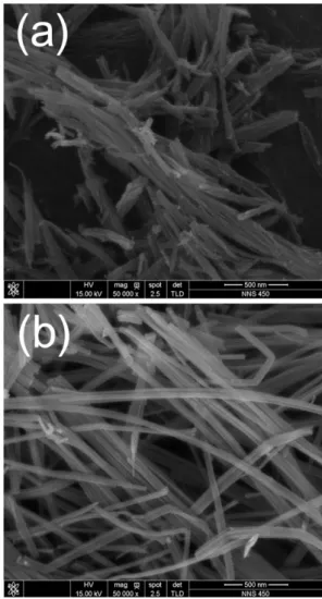
The solution chemical synthesis process successfully provided dark grayish powdery products. As shown in Fig. 1, FESEM observation was performed to confirm the morphologies of TNT and CNT-TNT samples. Fig.
1(a) shows a typical morphology of as-synthesized TNT sample. The nanotubes were bundled each other from
Fig. 1. FE-SEM images of (a) as-synthesized pure TNT and
(b) CNT-TNT composite.
several tens to several hundreds. Their outer diameter was approximately 10 nm and length was below 1 μm as is well-known. Contrastively, nanotubes of CNT-TNT composite were well-separated each other as presented in Fig. 1(b). They had outer diameter of 20~40 nm and maximum length of several micrometers. In case of the adding CNTs, the specific surface area was presumed to be slightly decreased to about quarter because the diame- ter of nanotubes was increased from 10 nm to more than 20 nm. It could be inferred that CNTs affected to the morphology of TNTs during the alkaline hydrothermal synthesis. However, CNT could not be detected easily through FESEM observation.
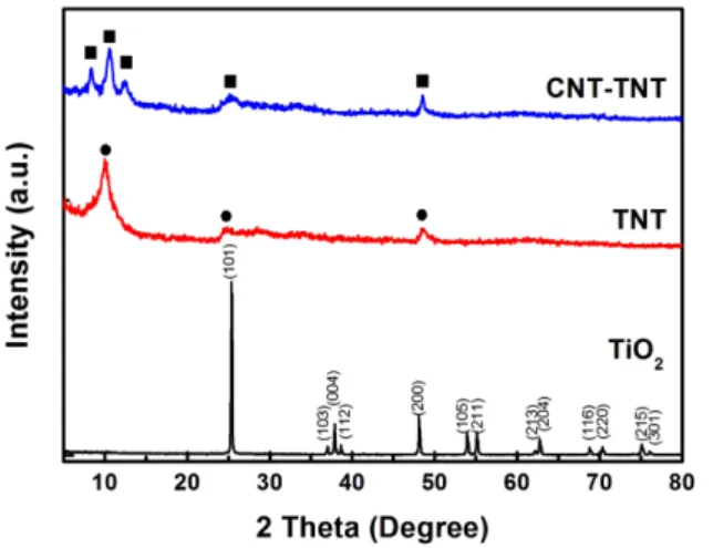
Fig. 2 shows the XRD patterns of the TiO
2powder as raw material and the TNTs synthesized with and without CNTs. Whereas the raw material TiO
2powder was shown as an anatase phase (JCPDS 21-1272), the as-syn- thesized TNT and CNT-TNT composite did not exhib- ited clear and sharp crystallite peaks. In fact, it was known that pure TNT synthesized by an alkaline hydro- thermal route was formed to H
2Ti
nO
2n+1or Na
xH
2-xTi-
n
O
2n+1(n=3, 4) [11]. In this study, observed diffraction peaks of the pure TNT at around 2 θ of 10
o, 24.5
oand 48.5
ocorresponding to the (200), (110), and (020) planes were in good agreement with the monoclinic H
2Ti
4O
9·H
2O (JCPDS 36-655) having four edge-sharing TiO
6octahe- dra in the unit cell. On the other hand, the XRD pattern of CNT-TNT sample (2 θ ~ 8.4
o, 10.6
o, 12.4
o, 25
oand 48.5
ocorresponding to (001), (200), (20 ), (110), and (020) planes, respectively) seemed to be monoclinic
H
2Ti
3O
7·nH
2O because two peaks of (001) and (20 ) planes were more similar to pattern of the H
2Ti
3O
7(JCPDS 47-561) having three edge-sharing TiO
6octahe- dra. The typical CNT peak that should exist around 25
ocould not be confirmed due to its small amount and peak overlap with TNT peak of (110) plane, and some peaks which were indexed to (001), (200), and (20 ) planes, were shifted to lower angles as compared with an anhy- drous H
2Ti
3O
7. It is well-known that the interlayer spac- ing of TNT is expanded with increasing the amount of adsorbed water because this adsorbed H
2O exists in the one-dimensional tunnel and skeletal crystal structure of nanotubes [21]. Thus, it was speculated that the peaks of (001), (200), and (20 ) planes were shifted due to adsorbed H
2O of sample.
To analyze more detail, TG analysis was conducted Fig. 3 shows the TG curves of TNT and CNT-TNT sam- ples. Some steps were observed at 30-150, 150-250, 250- 400
oC for TNT and CNT-TNT, and at 550-700
oC only for CNT-TNT. The first step up to 150
oC was desorption of H
2O, which was existed in interlayer space of nano- tube wall. The weight loss of CNT-TNT composite in this step was approximately 5.1% corresponding to 0.78H
2O in H
2Ti
3O
7·nH
2O. Pure TNT sample had the weight loss of 5.3% that was equivalent to one mole of H
2O in H
2Ti
4O
9·nH
2O. The weight losses of second and third steps from 150 to 450
oC meant that titanate (H
2Ti
-n
![Fig. 4 shows the RAMAN spectra of TNT and CNT-TNT samples. The RAMAN band at 190 cm -1 was attributed to the anatase TiO 2 mode [11] and bands at 275 cm -1 , 448 cm -1 , 670 cm -1 , 840 cm -1 were generally assigned to the TNTs [2, 22, 23]](https://thumb-ap.123doks.com/thumbv2/123dokinfo/4910447.536183/4.892.485.803.129.699/raman-spectra-samples-raman-attributed-anatase-generally-assigned.webp)
