선천성 고혈압 쥐에서 Imatinib Mesylate가 심근 섬유화에 미치는 영향
추 은 호, 임 상 현, 김 옥 란, 장 성 원, 박 찬 석, 김 희 열, 장 기 육, 윤 호 중, 정 욱 성, 승 기 배, 김 재 형
가톨릭대학교 의과대학 내과학교실
Imatinib Mesylate Attenuates Cardiac Fibrosis in Spontaneously Hypertensive Rats
Eun Ho Choo, MD, Sang-Hyun Ihm, MD, Ok-Ran Kim, MS, Sung-Won Jang, MD, Chan-Seok Park, MD, Hee-Yeol Kim, MD, Kiyuk Chang, MD, Ho-Joong Youn, MD,
Wook Sung Chung, MD, Ki-Bae Seung, MD, Jae-Hyeong Kim, MD
Division of Cardiology, Department of Internal Medicine, The Catholic University of Korea College of Medicine, Seoul, Korea
❙ABSTRACT❙
Background: Hypertensive myocardial fibrosis promotes abnormalities of cardiac function that may adversely affect the clinical outcome of hypertensive patients. Imatinib mesylate blocks receptor tyrosine kinase and is clinically used to treat leukemia.
Platelet-derived growth factor (PDGF) is a downstream target of receptor tyrosine kinases. Cardiac fibroblasts can be activated by PDGF. Thus we evaluated whether imatinib attenuate myocardial fibrosis and prevents diastolic dysfunction in spontaneously hypertensive rats (SHR). Methods: 8 weeks old male SHRs were subjected to treatment with 8 weeks of low dose imatinib (SHR-10; 10 mg/kg), high dose imatinib (SHR-30; 30 mg/kg) or saline (SHR-C; n = 6 in each group). At the age of 16 weeks, all rats underwent hemodynamic studies and Doppler echocardiography, and were sacrificed. Their hearts were extracted for histopathological, immunoblotting and quantitative reverse transcriptase-polymerase chain reaction analyses. Results: While imatinib did not affect blood pressure (BP), it markedly reduced perivascular and interstitial fibrosis in the hearts of SHR.
Echocardigram showed that high-dose imatinib significantly reduced left ventricular (LV) wall thickness (septal/posterior wall;
SHR-C vs. SHR-30: 18 ± 2/19 ± 2 mm vs. 15 ± 1/14 ± 1 mm; p < 0.05) and improved the parameters of LV diastolic function such as E/A ratio (SHR-C vs. SHR-30: 1.60 ± 0.10 vs. 1.86 ± 0.20; p < 0.05). Imatinib also significantly reduced mRNA expression of collagen III and PDGF β-receptor tyrosine phosphorylation in the hearts of SHR. Conclusions: These results suggest that imatinib, especially high dose, could attenuate myocardial fibrosis and prevent LV diastolic dysfunction in hypertensive rat model by decreased activity of PDGF. Imatinib may provide a potential therapeutic approach for hypertensive heart disease.
(J Korean Soc Hypertens 2011;17(2):48-56) Key words: Rats, Inbred SHR; Imatinib, Fibrosis; Echocardiography; Diastole
논문접수일: 2011. 6. 12, 심사완료일: 2011. 6. 22, 게재승인일: 2011. 6. 27 교신저자: 임상현
주소: 경기도 부천시 원미구 소사동 2번지 가톨릭대학교 부천성모병원 순환기내과 Tel: 032) 340-7027, Fax: 032) 340-2669
E-mail: [email protected]
본 연구는 2010년 대한고혈압학회 산학협동 연구비 수혜로 이루어짐.
서 론
고혈압 치료의 목적은 혈압 강하뿐만 아니라 향후 심혈 관 사건의 발생을 예방하는 데 있다. 심부전은 심혈관 사
건의 가장 흔한 원인 중 하나이며, 고혈압이 중요한 원인 질환 중 하나이다.1) 고혈압에 의한 심부전의 발생은 저항 의 증가에 의한 심비대와 관련되어 있을 뿐만 아니라 심근 의 섬유화와 이에 따른 강성(stiffness) 증가, 수축기능의 장애와 연관되어 있다.2) 섬유화는 심비대에서 심부전으로 이동하는데 있어 중요한 반응이다.3) 따라서 섬유화를 억 제함으로써 고혈압에 의한 심부전으로의 진행을 억제할 수 있을 것으로 생각되나, 현재까지 직접적인 치료적인 효 과가 있는 것으로 보이는 증거는 없다. 그 동안 앤지오텐 신 전환효소 억제제, 앤지오텐신 수용체 차단제, 알도스테 론 길항제 등에 초점이 맞춰져 왔으나 이들은 효과가 복잡 하며 여러 기전이 관여되어 있는 한계점이 있다.4) 섬유화의 발생은 전환성장인자(transforming growth factor, TGF) ß,5) 인터루킨(interleukin, IL)-1,6) 종양괴사인자(tumor necrosis factor, TNF) α,7) platelet derived growth factors (PDGFs)8)와 같은 사이토카인이나 성장인자들이 관여하는 것으로 보고 된 바 있다. PDGF는 섬유모세포(fibroblast)를 자극하여 주위 콜라겐 조직을 수축시키고 근섬유모세포(myofibro- blast)로 분화하도록 촉진한다.9,10) PDGF는 PDGF-AA, PDGF-AB, PDGF-BB, PDGF-CC, PDGF-DD와 같은 iso- form이 존재하며 PDGFRα와 PDGFRβ의 두 종류의 티로 신 키나아제(tyrosine kinase) 계열의 수용체에 결합하면서 세포 내의 작용이 나타난다.11)
Imatinib mesylate (Gleevec®, Novartis phameceuticals, Basel, Switzerland)은 선택적으로 티로신 키나아제 억제하 는 약물로 만성 골수성 백혈병 치료에 사용되고 있다.12) Imatinib은 bcr/abl의 티로신 키나아제 활성도를 억제함으 로써 만성 골수성 백혈병의 진행을 억제한다. 하지만, im- atinib은 c-kit, PDGFRα, PDGFRβ의 인산화 부위에 결합하 여 인산화를 억제하여 하위 단계의 연쇄반응을 차단하는 티로신 키나아제 억제제 역할도 한다.12)
선천성 고혈압 쥐(spontaneously hypertensive rat, SHR) 는 인간에서의 고혈압에 의한 심질환의 발생과 매우 유사한 경과를 보여 고혈압에 의한 병태 생리를 연구하는데 널리 사용되는 모델이다.3) SHR은 2–6주 정도 경과하면 고혈압 이 발생하고 그 후 고혈압에 의한 심장, 대동맥 및 혈관 등의 조직학적 변화가 오기 시작하여 심근섬유화, 심비대
및 심부전에 이르는 고혈압성 심질환이 발생한다.3,13,14) 그러므로 본 저자들은 SHR을 고혈압 쥐 모델로 시용하 여 티로신 키나아제 억제제인 imatinib가 심장의 PDGFR β의 티로신 키나아제 활성도를 억제함으로써 심장의 섬 유화를 억제하고 고혈압에 의한 심장의 이완기 장애를 완 화할 수 있는지를 알아보고자 하였다.
대상 및 방법
1. 실험동물
본 연구의 동물실험은 가톨릭대학교의 실험동물 윤리위 원회인 Institutional Animal Care and Use Committee로 부터 승인을 받아 진행하였으며, 고혈압 쥐 모델로 8주령 의 SHR과 정상대조군으로 Wistar Kyoto 쥐들(Wistar Kyoto rats, WKY)을 중앙실험동물로부터 구입하여 사용 하였다. 실험동물군은 정상대조군(WKY; n = 6), 고혈압대 조군(SHR-control; n = 6), 고혈압 치료군으로 저용량 im- atinib 투여군(SHR-10 mg: 하루 10 mg/kg; n = 6), 고용량 imatinib 투여군(SHR-30 mg: 하루 30 mg/kg; n = 6)으로 각 각 분류하였다. 모든 실험군은 생리 식염수를 음용하였고, Imatinib mesylate는 저용량과 고용량을 증류수에 용해시 켜 8주간 위관영양도관(gastric gavage tube)를 이용하여 경구 투여하였다. 약물을 투여하는 동안 모든 실험군의 실 험동물은 항온, 항습 조건에서 사육하였으며, 식이와 물을 자유롭게 허용하였다.
2. 체중, 혈압 및 심장초음파
모든 쥐는 약물 투약 직전 8주령과 8주간의 치료 후 16 주령에 체중을 쟀다. 그 후 실험이 끝나는 16주령에 12시 간 금식 후 안정 상태에서 체중을 잰 후 오전에 꼬리에서 tail-cuff plethysmography (BP-2000, Visitech system, Apex, NC, USA)를 이용하여 수축기 혈압을 측정하고 심 초음파를 시행하였다. Xylazine 10 mg/kg과 ketamine hydro- chloride 100 mg/kg을 근육 주사하여 전신마취 후 좌측 흉 부에 면도를 시행하여 선명한 영상을 얻었다. 15 MHz 고 주파 탐촉자(Acuson Sequoia 256, Acuson, Mountain views,
CA, USA)를 이용한 이면성 및 M형 심초음파 검사를 실 시하여 수축기 및 이완기말 좌심실 내경 및 내경 분획단축 (fractional shortening, FS)을 측정하였다. 그리고 좌심실 이완기능을 평가하기 위해 Pulsed wave 도플러를 이용하여 빠른 이완 초기 유입 혈류의 최고 속도(E velocity), 후기 유입 혈류의 최고 속도(A velocity), 감속시간(deceleration time)을 측정하였다. 모든 측정치는 3회 이상 반복 측정하 여 평균값을 사용하였다.
3. 실험동물 조직 적출 및 염색
16주령에 각 실험동물을 18.75 mg/kg의 tiletamine과 zo- lazepam (Virbec, Carros, France)을 혼합한 피하주사 마취 하에, 심장 조직을 적출하였다. 적출한 심장은 무게를 잰 후 좌심실 내에 생리식염수를 주입한 뒤 좌심실은 분리되 어, 심장 종축의 수직 방향으로 3등분하였다. 유두근육이 포함되어있는 중간부분을 10% 포르말린 용액에 24시간 고정 후에 paraffin 조직으로 5 μm 두께로 절제하였다. 콜 라겐 염색을 위해 콜라겐이 붉은색으로 염색되는 picrosir- ius red 염색을 시행하였다. 나머지 좌심실 근육조직은 냉동 시켜 실시간중합효소연쇄반응(real time polymerase chain reaction, RT-PCR) 및 Western blot 분석에 사용하였다.
4. 염색된 심근 조직의 정량적 측정
염색된 조직은 혈관 주위 부분과 간질 부분을 현미경 (Olympus AX70 TR62A02, Olympus, Tokyo, Japan)으로 관찰한 뒤 영상을 Olympus DP70 디지털 카메라(Olympus) 을 사용하여 촬영하였다. 모든 슬라이드는 100배와 200배 배율에서 관찰하였고, Image pro plus ver. 4.5 (Media Cybernetics, Silver Spring, MD, USA) 프로그램을 사용하 여 측정하였다. 콜라겐 부피 분할(collagen volume fraction,
%)는 콜라겐 특수 염색인 picrosirius red 염색을 한 전체 심근조직에서 양성 염색 부위의 백분율로 정의하였다.
5. 섬유화 정도를 측정하기 위한 RT-PCR
각 심근조직으로부터 전체 RNA를 TRIZOL 용액(Invit- rogen, Carlsbad, CA, USA)를 사용하여 추출하였다. 전체
RNA는 Onestep RT kit (Invitrogen)을 사용하여 역전사 (reverse transcription) 하였다. Collagen type III, glycer- aldehyde 3-phosphate dehydrogenase (GAPDH)의 발현 정 도를 SYBR green dye를 사용하여 RT-PCR을 통하여 확인 하였다. 각각의 primer는 다음과 같다. collagen type III (sense, 5’-GCC AGTCCCATGACTGTCCCGCGG-3’ and antisense, 5’-AGTGCAGCCATCCTCTAGAACTGTG-3’), rat GAPDH (sense, 5’-AATGCATCCTGCACCACCAA-3’ and antisense, 5’-GTAGCCATATTCATTGTCATA-3’).
Collagen type III의 발현양은 GAPDH의 발현 양으로 반 정량화(semi-quantify) 하였다.
6. Western blotting
심근 조직을 RIPA buffer (0.1% SDS, 1% deoxycholate, 1% Triton X-100, 150 mM NaCl, 5 mM EDTA, 10 mM Tris pH7.4, 10% protease inhibitor cocktails [Sigma–
Aldrich])에 4℃에서 30분간 용해 한 뒤 4℃에서 3분간 13,000 rpm으로 원심 분리하였다. 상층 액을 채취하여 Bio-Rad Protein assay reagent (Bio-Rad, Regents Park, NSW, Australia) 을 사용하여 단백농도를 측정하였다. 각 샘플로부터 같은 양의 단백을 8% sodium dodecylsulfatepolyacrylamide gel에 서 분리하여 Immobilon-PVDF membranes (Millipore, Billerica, MA, USA)으로 transfer 한 뒤 5% skim milk (Difco, Sparks, MD, USA)에서 실온에서 1시간 blocking 하였다.
PDGFRβ의 tyrosine phosphorylation 정도는 immobilized phosphotyrosine antibody (cellsignaling)를 사용하여 측정하 였다. Membrane을 phosphotyrosine antibody에 4℃에서 반 응 시킨뒤 Tris-buffered saline with 0.1% Tween-20 (TBST) 으로 세척한 후 HRP-conjugated anti-rabbit IgG (Santa Cruz Biotechnology, Santa Cruz, CA, USA)로 다시 1시간 동안 반응하여 상용화된 chemiluminesence detection system을 이용하여 blot을 관찰하였다. 동량의 단백에 대하여 PDGFR β (cellsignaling)의 western blotting도 시행하여 대조군으로 사용하였다.
7. 통계처리
모든 측정치는 평균± 표준편차로 표시하였으며, 각 군
WKY SHR SHR-10 mg SHR-30 mg
Pre-BWt (g) 206 ± 10 184 ± 11* 187 ± 6* 183 ± 13*
Post-BWt (g) 390 ± 12 346 ± 8* 334 ± 24* 339 ± 16*
Systolic BP (mm Hg) 114 ± 8 166 ± 14* 163 ± 8* 170 ± 13*
Heart weight (g) 1.2 ± 0.1 1.5 ± 0.1* 1.3 ± 0.1 1.4 ± 0.1*
Tibia length (mm) 5.2 ± 0.4 4.8 ± 0.2 4.8 ± 0.3 4.8 ± 0.2
Heart/Tibia ratio (g/mm) 0.23 ± 0.01 0.31 ± 0.03* 0.28 ± 0.02* 0.30 ± 0.02*
Data are expressed as mean ± standard deviation.
WKY, Wista Kyoto rat; SHR, spontaneous hypertensive rat; SHR-10 mg, SHR treated with 10 mg/kg of imatinib; SHR-30 mg, SHR treated with 30 mg/kg of imatinib; Pre-BWt, body weight before treatment; Post-BWt. body weight after treatment; BP, blood pressure.
*p < 0.05 vs. WKY.
†p < 0.05 vs. SHR.
Table 1. General characteristics
WKY SHR SHR-10 mg SHR-30 mg
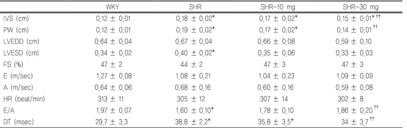
IVS (cm) 0.12 ± 0.01 0.18 ± 0.02* 0.17 ± 0.02* 0.15 ± 0.01*††
PW (cm) 0.12 ± 0.01 0.19 ± 0.02* 0.17 ± 0.02* 0.14 ± 0.01††
LVEDD (cm) 0.64 ± 0.04 0.67 ± 0.04 0.66 ± 0.08 0.59 ± 0.10
LVESD (cm) 0.34 ± 0.02 0.40 ± 0.02* 0.35 ± 0.06 0.33 ± 0.03
FS (%) 47 ± 2 44 ± 2 47 ± 3 47 ± 3
E (m/sec) 1.27 ± 0.08 1.08 ± 0.21 1.04 ± 0.23 1.09 ± 0.09
A (m/sec) 0.64 ± 0.06 0.68 ± 0.16 0.60 ± 0.16 0.59 ± 0.08
HR (beat/min) 313 ± 11 305 ± 12 307 ± 14 302 ± 8
E/A 1.97 ± 0.07 1.60 ± 0.10* 1.78 ± 0.10 1.86 ± 0.20††
DT (msec) 29.7 ± 3.3 38.8 ± 2.2* 35.8 ± 3.5* 34 ± 3.7††
Data are expressed as mean ± standard deviation.
WKY, Wista Kyoto rat; SHR, spontaneous hypertensive rat; SHR-10 mg, SHR treated with 10 mg/kg of imatinib; SHR-30 mg, SHR treated with 30 mg/kg of imatinib; IVS, interventricular septal thickness; PW, posterior wall thickness; LVEDD, left ventricular end diastolic dimension; LVESD, left ventricular end systolic dimension; FS, fractional shortening of LV diameter; E, peak velocity of early transmitral inflow; A, peak velocity of late transmitral inflow; HR, heart rat; DT, deceleration time.
*p < 0.05 vs. WKY.
†p < 0.05 vs. SHR.
Table 2. Echocardiographic parameters
간의 비교는 student t-test와 ANOVA를 이용한 통계적 분 석을 시행하였다. 모든 통계 값에서 0.05 미만인 경우 통 계적 의의가 있는 것으로 하였다.
결 과
1. 치료 전 후 실험 대상의 특징
치료 전 후 WKY에 비해 SHR의 몸무게가 현저히 적었 다(Table 1). SHR에서 imatinib 치료는 몸무게 변화에 영 향을 주지 않았다. 또한 WKY에 비하여 SHR의 혈압이 높았으나, imatinib의 치료가 혈압 변화를 일으키지 않았
다. 심장의 무게도 SHR에서 높았지만, imatinib 치료 후 에 심장의 무게와 심장 무게 대 경골(tibia) 길이 비율에 변화는 관찰되지 않았다.
2. 심초음파를 통한 심실 벽 두께 측정 및 심기능 평가
치료에 따른 심비대의 변화 및 심기능을 평가하기 위해 심장 초음파를 시행하였다. SHR은 WKY에 비하여 좌심 실 벽의 두께가 증가되어 있으며, 이완기능의 저하가 관 찰되었다(Table 2). 8주간 imatinib으로 치료한 SHR은 특 히 고용량군에서 SHR-control군에 비해 좌심실 벽 두께의 감소 및 E/A 비율의 증가, 감속시간의 감소가 관찰되었으
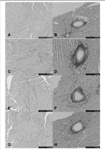
Fig. 1. Light micrographs of the myocardium (×100, picrosirius red stain) reveal that the fibrosis of perivascular area and inter- stitial area is increased in SHR compared to WKY. The fibrosis of both areas was decreased after imatinib treatment in SHR (A, B). interstitial and perivascular area of WKY. (C, D). interstitial and perivascular area of SHR-control. (E, F). interstitial and perivascular area of SHR 10-mg. (G, H). interstitial and peri- vascular area of SHR 30-mg. WKY, Wistar Kyoto rats; SHR-con- trol, spontaneously hypertensive rats treated with normal saline;
SHR-10 mg, spontaneously hypertensive rats treated with 10 mg/kg of imatinib; SHR-30 mg, spontaneously hypertensive rats treated with 30 mg/kg of imatinib.
Fig. 2. Collagen volume fraction of Left ventricle shows that the fibrosis of perivascular area and interstitial area is increased in SHR compared to WKY. Treatment with 30 mg/kg of imatinib in SHR significantly reduced the fibrosis of perivascular area and interstitial area. (A) Interstitial area. (B) Perivascular area.
WKY, Wistar Kyoto rats; SHR-control, spontaneously hyper- tensive rats treated with normal saline; SHR-10 mg, sponta- neously hypertensive rats treated with 10 mg/kg of imatinib;
SHR-30 mg, spontaneously hypertensive rats treated with 30 mg/kg of imatinib. *p < 0.05 vs. WKY. †p < 0.05 vs. SHR.
며 저용량군에서는 차이가 관찰되었으나 통계적 의의는 없었다. 반면 수축기 기능을 대변하는 내경 분획단축(FS) 에는 WKY군과 SHR은 모든 군에서 차이가 없었다.
3. 심근의 조직학적 섬유화 정도 측정
Picrosirius red 염색을 하여 광학 현미경으로 심근의 혈 관 주위와 간질을 관찰하였다(Fig. 1).
WKY에 비해 SHR에서 혈관 주위 및 간질에서 섬유화가 증가되어 있었다. imatinib을 투여 한 SHR에서는 혈관 주위
및 간질에서 심근 섬유화의 정도가 감소되어 있었다. 콜라 겐 부피 분율을 측정한 결과, 혈관 주위에서는 SHR-control 에 비하여 30 mg/kg으로 imatinib을 투여한 SHR-30 mg군 에서 현저한 감소 소견을 보였고(SHR-control vs. SHR-30 mg, 14.56 ± 1.73 vs. 6.27 ± 1.88; p = 0.004), 간질에서도 대 조군인 SHR-control군에 비하여 imatinib 30 mg/kg 투약군 이 현저한 섬유화의 감소 소견을 보였다(SHR-control vs.
SHR-30 mg, 1.26 ± 0.19 vs. 0.77 ± 0.07; p = 0.01). 한편 저용 량 10 mg/kg을 투약한 SHR-10 mg군은 대조군인 SHR-con- trol에 비해 섬유화 정도가 감소하기는 하였으나 통계적 의 의는 없었다(Fig. 2).
4. 콜라겐 생산 측정
심장의 섬유모세포에 의한 콜라겐 생산에 따른 섬유화
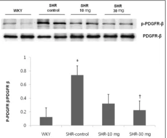
Fig. 4. The effect of imatinib treatment on tyrosine phosphor- ylation level of platelet derived growth factor (PDGF)β. Tyrosine phosphorylation level of PDFGRβ was increased in SHR com- pared to WKY. Treatment with 30 mg/kg of imatinib decreased the tyrosine phosphorylation level of PDFGRβ. WKY, Wistar Kyoto rats; SHR-control, spontaneously hypertensive rats treated with normal saline; SHR-10 mg, spontaneously hyper- tensive rats treated with 10 mg/kg of imatinib; SHR-30 mg, spontaneously hypertensive rats treated with 30 mg/kg of imatinib.*p < 0.05 vs. WKY. †p < 0.05 vs. SHR.
Fig. 3. Myocardial expression of mRNA of collagen type III was increased in SHR compared to WKY. Expression of collagen type III was significantly decreased after treatment with 30 mg/kg of imatinib. Normalization relative GAPDH was performed. (A) Electrophoresis. (B) Expression of collagen type III. GAPDH, glyceride 3-phosphate dohydroqenase; WKY, Wistar Kyoto rats; SHR-control, spontaneously hypertensive rats treated with normal saline; SHR-10 mg, spontaneously hypertensive rats treated with 10 mg/kg of imatinib; SHR-30 mg, spontaneously hypertensive rats treated with 30 mg/kg of imatinib. *p < 0.05 vs. WKY. †p < 0.05 vs. SHR.
정도를 측정하기 위해 각 실험 쥐의 심근에서 collagen type III의 발현을 RT-PCR을 통하여 측정하였다. WKY에 비해 SHR에서 collagen type III의 발현이 증가되어 있었다.
Imatinib을 투여 한 SHR에서는 고용량의 SHR 30 mg군이 대조군에 비하여 collagen type III의 발현이 감소되었다 (Fig. 3).
5. PDGFR의 활성화 정도 측정
PDGFRβ의 인산화 정도를 측정하기 위해, PDGFRβ의 tyrosine residue의 인산화된 단백과 PDGFRβ에 대해 Western blotting을 시행하였다. SHR에서 WKY에 비해 PDGFRβ의 인산화가 증가되어 있었으며, SHR의 PDGFRβ 인산화는 30 mg/kg의 imatinib을 투여하였을 때 현저하게 감소됨을 관찰할 수 있다(Fig. 4).
고 찰
본 연구를 통해 백혈병 치료제로 사용되는 tyrosine kin- ase 억제제인 imatinib mesylate (Gleevec)가 고혈압 실험 쥐 모델인 SHR에서 심근의 섬유화를 감소시켰고 이로 인 해 좌심실 비대 및 좌심실 이완기 기능 등의 심장 기능의 악화를 개선하였다는 것을 알 수 있었다. Imatinib투여함 으로써 심장 초음파상에서 SHR군의 좌심실 비대 및 좌심 실 이완기능부전의 호전 소견을 관찰 할 수 있었으며, 이 는 10 mg/kg 보다 30 mg/kg를 투여하였을 때 명확히 관찰 할 수 있었다. 이는 심근 조직소견에서 혈관 주위 및 간질 의 섬유화 정도의 개선과 일치하는 소견으로, imatinib 치 료가 실제 좌심실의 콜라겐 생산을 억제하였음을 관찰할 수 있었다. 이러한 섬유화 감소는 imatinib의 티로신 키나 아제의 억제로 인한 PDGFRβ의 인산화가 감소와 연관되 어 있었다. 이는 고혈압에 의한 심근의 섬유화와 이로 인 한 좌심실 비대와 심부전으로의 진행을 imatinib이 억제 할 수 있음을 제시한다.
Imatinib의 섬유화 억제 효과는 이미 타 연구에서도 입증 된 바 있다. Bleomycin15,16)와 방사선17)에 의한 폐의 섬유화 가 억제되었다. 정상 섬유모세포 및 공피증(scleroderma)에 서 섬유모세포 증식을 imatinib이 억제하여, 피부의 섬유화
를 감소시켰다.18,19) 신부전 쥐 모델에서 신장의 섬유화도 억제한다는 보고도 있다.20) 심장에 대한 imatinib의 효과에 대한 연구는 만성 심근염 쥐 모델에서 심근의 섬유화를 감소시켰다는 연구21)와 안지오텐신 II가 항진된 TGR (mRen2) 27 쥐 모델에서 심장기능 악화와 신장의 미세혈 관손상을 완화시켰다는 연구22)가 있었다.
그러나 본 연구는 imatinib의 고혈압성 심질환에 미치는 영향을 인간에서 고혈압에 의한 심혈관 합병증의 발생과 유사한 실험쥐 모델인 SHR (선천성 고혈압 쥐)을 이용하 여 imatinib의 섬유화 억제 효과 및 좌심실 비대의 퇴축, 나아가서 좌심실 기능 개선 효과를 확인 한 유일한 연구 라 할 수 있다.
고혈압이 지속됨에 따라 섬유모세포가 근섬유모세포로 전환되며, 이로 인해 extracellular matrix에 섬유화가 진행 된다.23) PDGF는 근섬유모세포로의 전환을 유도하며, im- atinib는 PDGF 수용체를 선택적으로 차단함으로써 섬유 화 억제효과가 나타난다. 또한 imatinib가 TGFβ1의 down- stream target 인 c-abl을 억제하는 효과가 있어 이에 의한 섬유화 억제효과도 생각할 수 있다.24) 뿐만 아니라 PDGF 활성도 증가 시 TGFβ1의 활성도도 증가하는 보고도 있어 PDGFR의 억제에 의한 TGFβ1 활성도 감소로 생각할 수 도 있다.25)
고혈압이 지속됨으로써 심장의 섬유화뿐만 아니라 좌심 실 비대가 동반되게 된다. 좌심실 비대가 발생되는 기전이 과거부터 연구되어 왔으며 병태생리 중에 티로신 키나아제 의 공통적인 downstream target인 PI3K-phosphatase and tensin homologue 이 관여되어 있음이 밝혀졌다.26) 또한 PDGF가 extracellular signal-regulated kinase 1/2를 자극 하여 심근세포의 성장을 촉진한다는 동물실험 결과도 있
다.27,28) Epidermal growth factor receptor 을 통해서 세포
내에서 티로신 키나아제 또는 serine/threonine kinase가 항 진될 때 병적인 심근 비후가 발생되는 것이 보고된 바도
있다.29,30) 따라서 imatinib을 비롯한 티로신 키나아제 억제
제가 좌심실 비대 치료에 대한 새로운 목표가 될 수 있을 것으로 보인다.
그러나 최근 imatinib가 만성 골수성 백혈병에 대한 치 료제로 사용되면서 울혈성 심부전 및 무증상 좌심실 기능
장애를 유발하는 심독성에 대한 의심이 되어 문제점이 제 시되기도 하였다.31,32) 이는 imatinib에 의해 미토콘드리아 의 장애가 나타나고 그로 인한 에너지 생산이 감소되어 세포괴사로 이어지는 것으로 생각되었다.31,32) 그러나 5년 이상 장기간 실제 만성 골수성 백혈병 환자에게 사용한 결과 imatinib의 심독성은 위약에 비해 증가는 소견을 보 이지 않아서 이러한 심독성이 실제 환자의 치료에 문제가 되지는 않았다.33,34) imatinib가 만성 골수성 백혈병 이외 에 위장관 기질 종양(gastrointestinal stroma tumor)에 사 용되면서 연구가 이루어졌으나 심독성에 대한 명확한 증 거는 나타난 바 없다.35,36)
본 연구에서 한계점으로 가장 중요한 심장초음파를 사 용하여 심장의 이완기능 평가함에 있어 가장 민감한 tissue Doppler를 사용하지 못하였다는 점이 있다. 그러나 일반 적으로 널리 사용되는 이완기능 지표인 E/A ratio와 감속 시간을 사용하였고 측정할 때 심 박동수가 실험군 간에 차이가 없도록 하여 그 오차를 최소화 하여 사용하였으므 로 그 오차가 크지 않을 것으로 생각된다. 또한 imatinib 투여 전 후의 심장 무게나 심장 무게 대 경골 비율의 변화 는 통계적으로 의미가 없었으나 심장 초음파에서는 심근 벽 두께가 의미 있게 줄어들었다. 이는 심장무게 변화 보 다는 심근 벽 두께 측정이 심비대의 변화를 입증하는데 더욱 민감하기 때문으로 생각되며 실험쥐 심장의 무게를 측정 할 때 심장 무게가 매우 적기 때문에 보다 민감하고 정확한 저울을 사용하지 않아 미묘한 차이를 측정하지 못 했을 가능성이 많으며 그러지 않더라도 실험 쥐의 숫자가 증가한다면 심장무게에서도 통계적으로 의미 있는 결과를 보일 것으로 생각된다.
따라서 본 연구는 imatinib가 PDGF 수용체의 인산화를 억제함으로써 심근의 섬유화를 억제하여 그로 인하여 좌 심실 비대를 완화하고, 나아가서 좌심실 이완기능을 개선 시킴으로써 고혈압에 의한 중요한 합병증 중 하나인 심부 전으로의 진행을 억제할 수 있음을 제시하였다.
요 약
연구배경: 고혈압성 좌심실 섬유화는 고혈압 환자의 예
후와 관련된 다양한 심질환을 일으키는 원인이 된다. Imatinib mesylate는 tyrosine kinase 수용체 차단제로써 임상적으로 백혈병 치료에 사용되고 있다. Platelet-derived growth fac- tor (PDGF)는 티로신 키나아제 수용체의 하위 단계의 반응 물질로서 심근의 섬유화는 이런 PDGF에 의해 활성화 될 수 있다. 따라서 우리는 고혈압 실험 쥐 모델인 선천성 고혈압 쥐에서 티로신 키나아제 수용체 차단제인 imatinib가 PDGF 수용체의 활성화를 감소시켜 심근의 섬유화를 억제하고 그 로 인해 심장의 이완기 장애를 완화할 수 있는지를 알아보 고자 하였다.
방법: 8주령의 선천성 고혈압 쥐를 6마리씩 8주 동안 저 용량 imatinib (하루 10 mg/kg), 고용량 imatinib (하루 30 mg/kg), 생리식염수로 치료하였다. 8주간의 치료 후 16주 령이 된 모든 쥐를 혈압측정을 하고 심장초음파를 시행하 였다. 검사가 끝난 후 희생하여 심장을 적출 한 후 조직 염색, western blotting과 reverse transcriptase-polymerase chain reaction을 시행하였다.
결과: Imatinib은 선천성 고혈압 쥐의 혈압에는 영향을 주지 않았다. Imatinib 치료 후 특히 고용량(30 mg/kg) 투 약군에서 혈관 주위 및 간질에서 콜라겐이 현저히 감소하 였다. 심장초음파에서 역시 고용량 imatinib을 투여하였을 때 좌심실 비대와 이완기 장애의 악화를 억제하였다.
Imatinib은 collagen의 발현을 억제하였고 PDGF receptor β의 tyrosine phosphorylation을 감소시켰다.
결론: 이 실험을 통해 imatinib 치료 특히 고용량 사용시 고혈압 실험 쥐 모델인 선천성 고혈압 쥐에서의 심장의 섬유화와 이완기 장애를 완화시켰으며, 이는 PDGF의 활 성도 감소와 관련이 있을 것으로 생각된다. 그러므로 im- atinib는 고혈압성 심질환의 새로운 예방적 약물이 될 수 있을 것으로 기대된다.
References
1. Ruy KH, Han SW. Clinical characterisrics and prognositic factors of patients with congestive heart failure in Korea (epidemiology of congestive heart failure in Korea). J Korean Soc Hypertens. 2002;8:38-47.
2. Kim TS, Youn HJ. From hypertension to heart failure. J Korean Soc Hypertens 2009;15:1-11.
3. Conrad CH, Brooks WW, Hayes JA, Sen S, Robinson KG, Bing OH. Myocardial fibrosis and stiffness with hyper- trophy and heart failure in the spontaneously hypertensive rat. Circulation. 1995;91:161-70.
4. Creemers EE, Pinto YM. Molecular mechanisms that con- trol interstitial fibrosis in the pressure-overloaded heart.
Cardiovasc Res. 2011;89:265-72.
5. Rosenkranz S, Flesch M, Amann K, Haeuseler C, Kilter H, Seeland U, et al. Alterations of beta-adrenergic signaling and cardiac hypertrophy in transgenic mice overexpressing TGF-beta(1). Am J Physiol Heart Circ Physiol. 2002;283:
H1253-62.
6. Isoda K, Kamezawa Y, Tada N, Sato M, Ohsuzu F. Myo- cardial hypertrophy in transgenic mice overexpressing hu- man interleukin 1alpha. J Card Fail. 2001;7:355-64.
7. Bryant D, Becker L, Richardson J, Shelton J, Franco F, Peshock R, et al. Cardiac failure in transgenic mice with myocardial expression of tumor necrosis factor-alpha.
Circulation. 1998;97:1375-81.
8. Ponten A, Li X, Thoren P, Aase K, Sjoblom T, Ostman A, et al. Transgenic overexpression of platelet-derived growth factor-C in the mouse heart induces cardiac fibrosis, hyper- trophy, and dilated cardiomyopathy. Am J Pathol. 2003;
163:673-82.
9. Heldin CH, Westermark B. Mechanism of action and in vivo role of platelet-derived growth factor. Physiol Rev.
1999;79:1283-316.
10. Uutela M, Wirzenius M, Paavonen K, Rajantie I, He Y, Karpanen T, et al. PDGF-D induces macrophage recruit- ment, increased interstitial pressure, and blood vessel ma- turation during angiogenesis. Blood. 2004;104:3198-204.
11. Fredriksson L, Li H, Eriksson U. The PDGF family: four gene products form five dimeric isoforms. Cytokine Growth Factor Rev. 2004;15:197-204.
12. Savage DG, Antman KH. Imatinib mesylate--a new oral targeted therapy. N Engl J Med. 2002;346:683-93.
13. Bing OH, Brooks WW, Robinson KG, Slawsky MT, Hayes JA, Litwin SE, et al. The spontaneously hypertensive rat as a model of the transition from compensated left ventricular hypertrophy to failure. J Mol Cell Cardiol. 1995;27:383-96.
14. Cingolani OH, Yang XP, Liu YH, Villanueva M, Rhaleb NE, Carretero OA. Reduction of cardiac fibrosis decreases systolic performance without affecting diastolic function in hypertensive rats. Hypertension. 2004;43:1067-73.
15. Daniels CE, Wilkes MC, Edens M, Kottom TJ, Murphy SJ, Limper AH, et al. Imatinib mesylate inhibits the profibro- genic activity of TGF-beta and prevents bleomycin-medi- ated lung fibrosis. J Clin Invest. 2004;114:1308-16.
16. Aono Y, Nishioka Y, Inayama M, Ugai M, Kishi J, Uehara H, et al. Imatinib as a novel antifibrotic agent in bleomy- cin-induced pulmonary fibrosis in mice. Am J Respir Crit Care Med. 2005;171:1279-85.
17. Abdollahi A, Li M, Ping G, Plathow C, Domhan S, Kiessling F, et al. Inhibition of platelet-derived growth fac- tor signaling attenuates pulmonary fibrosis. J Exp Med.
2005;201:925-35.
18. Distler JH, Jungel A, Huber LC, Schulze-Horsel U, Zwerina J, Gay RE, et al. Imatinib mesylate reduces pro- duction of extracellular matrix and prevents development of experimental dermal fibrosis. Arthritis Rheum. 2007;
56:311-22.
19. Soria A, Cario-Andre M, Lepreux S, Rezvani HR, Pasquet JM, Pain C, et al. The effect of imatinib (Glivec) on scle- roderma and normal dermal fibroblasts: a preclinical study.
Dermatology. 2008;216:109-17.
20. Wang S, Wilkes MC, Leof EB, Hirschberg R. Imatinib me- sylate blocks a non-Smad TGF-beta pathway and reduces renal fibrogenesis in vivo. FASEB J. 2005;19:1-11.
21. Leipner C, Grun K, Muller A, Buchdunger E, Borsi L, Kosmehl H, et al. Imatinib mesylate attenuates fibrosis in coxsackievirus b3-induced chronic myocarditis. Cardiovasc Res. 2008;79:118-26.
22. Schellings MW, Baumann M, van Leeuwen RE, Duisters RF, Janssen SH, Schroen B, et al. Imatinib attenuates end-organ damage in hypertensive homozygous TGR (mRen2)27 rats. Hypertension. 2006;47:467-74.
23. Berk BC, Fujiwara K, Lehoux S. ECM remodeling in hy- pertensive heart disease. J Clin Invest. 2007;117:568-75.
24. Bhattacharyya S, Ishida W, Wu M, Wilkes M, Mori Y, Hinchcliff M, et al. A non-Smad mechanism of fibroblast activation by transforming growth factor-beta via c-Abl and Egr-1: selective modulation by imatinib mesylate.
Oncogene. 2009;28:1285-97.
25. Tuuminen R, Nykanen AI, Krebs R, Soronen J, Pajusola K, Keranen MA, et al. PDGF-A, -C, and -D but not PDGF-B increase TGF-beta1 and chronic rejection in rat cardiac allografts. Arterioscler Thromb Vasc Biol. 2009;29:691-8.
26. Oudit GY, Penninger JM. Cardiac regulation by phosphoi- nositide 3-kinases and PTEN. Cardiovasc Res. 2009;82:
250-60.
27. Shimizu T, Kinugawa K, Yao A, Sugishita Y, Sugishita K, Harada K, et al. Platelet-derived growth factor induces cel- lular growth in cultured chick ventricular myocytes.
Cardiovasc Res. 1999;41:641-53.
28. Liu J, Wu LL, Li L, Zhang L, Song ZE. Growth-promoting effect of platelet-derived growth factor on rat cardiac myocytes. Regul Pept. 2005;127:11-8.
29. Kagiyama S, Qian K, Kagiyama T, Phillips MI. Antisense to epidermal growth factor receptor prevents the develop- ment of left ventricular hypertrophy. Hypertension. 2003;
41:824-9.
30. Smith NJ, Chan HW, Osborne JE, Thomas WG, Hannan RD. Hijacking epidermal growth factor receptors by angio- tensin II: new possibilities for understanding and treating cardiac hypertrophy. Cell Mol Life Sci. 2004;61:2695-703.
31. Kerkela R, Grazette L, Yacobi R, Iliescu C, Patten R, Beahm C, et al. Cardiotoxicity of the cancer therapeutic agent imatinib mesylate. Nat Med. 2006;12:908-16.
32. Force T, Krause DS, Van Etten RA. Molecular mechanisms of cardiotoxicity of tyrosine kinase inhibition. Nat Rev Cancer. 2007;7:332-44.
33. Druker BJ, Guilhot F, O'Brien SG, Gathmann I, Kantarjian H, Gattermann N, et al. Five-year follow-up of patients re- ceiving imatinib for chronic myeloid leukemia. N Engl J Med. 2006;355:2408-17.
34. Hochhaus A, Druker B, Sawyers C, Guilhot F, Schiffer CA, Cortes J, et al. Favorable long-term follow-up results over 6 years for response, survival, and safety with imatinib me- sylate therapy in chronic-phase chronic myeloid leukemia after failure of interferon-alpha treatment. Blood. 2008;
111:1039-43.
35. Perik PJ, Rikhof B, de Jong FA, Verweij J, Gietema JA, van der Graaf WT. Results of plasma N-terminal pro B-type na- triuretic peptide and cardiac troponin monitoring in GIST patients do not support the existence of imatinib-induced cardiotoxicity. Ann Oncol. 2008;19:359-61.
36. Ribeiro AL, Marcolino MS, Bittencourt HN, Barbosa MM, Nunes Mdo C, Xavier VF, et al. An evaluation of the car- diotoxicity of imatinib mesylate. Leuk Res. 2008;32:1809- 14.