Patency of Hemashield grafts versus ringed Gore-Tex grafts in middle hepatic vein reconstruction for living
donor liver transplantation
Sang Hoon Kim, Shin Hwang, Minjae Kim, Tae-Yong Ha, Gi-Won Song, Dong-Hwan Jung, Chul-Soo Ahn, Deok-Bog Moon, Ki-Hun Kim, Gil-Chun Park, and Sung-Gyu Lee
Division of Hepatobiliary Surgery and Liver Transplantation, Department of Surgery, Asan Medical Center, University of Ulsan College of Medicine, Seoul, Korea
Backgrounds/Aims: Owing to the short supply of homologous vein allografts, we previously used ringed Gore-Tex vas- cular grafts for middle hepatic vein (MHV) reconstruction in living donor liver transplantation. When ringed Gore-Tex grafts became unavailable, we used Hemashield vascular grafts. This study aimed to compare the patency and compli- cation rates of Hemashield and ringed Gore-Tex grafts. Methods: This retrospective two-arm study compared the study group that received Hemashield grafts (n=157) and the propensity score-matched control group that received ringed Gore-Tex grafts (n=157). Results: In the Hemashield and Gore-Tex groups, the recipient age was 54.7±9.4 and 53.3±6.3 years; Model for End-stage Liver Disease scores were 15.9±9.2 and 16.9±8.3; and graft-recipient weight ratios were 1.07±0.24 and 1.10±0.23, respectively. In the Hemashield group, V5 reconstruction was performed using single (n=113, 72.0%), double (n=39, 24.8%), and triple (n=3, 1.9%) anastomoses. The proportion of double and triple anasto- moses for V5 and V8 was higher in the Hemashield group than in the Gore-Tex group. Two (1.3%) patients required MHV conduit stenting owing to early thrombosis of the Hemashield graft. There was no difference in conduit occlu- sion-free patient survival rates between groups (p=0.91). The incidence of accidental conduit migration in the Hemashield and Gore-Tex groups was 0 (0%) and 2 (1.3%), respectively. Conclusions: Hemashield grafts used in MHV reconstruction demonstrated acceptably high short- and mid-term patency rates, no incidences of conduit migra- tion, easy handling, and good flexibility for length adjustment. Therefore, we suggest that the Hemashield graft is the preferentially suitable prosthetic material for MHV reconstruction. (Ann Hepatobiliary Pancreat Surg 2021;25:46-53) Key Words: Prosthetic graft; Hepatic venous congestion; Patency; Thrombosis; Graft migration
Received: January 11, 2021; Revised: February 1, 2021; Accepted: February 2, 2021 Corresponding author: Shin Hwang
Department of Surgery, Asan Medical Center, University of Ulsan College of Medicine, 88 Olympic-ro 43-gil, Songpa-gu, Seoul 05505, Korea Tel: +82-2-3010-3930, Fax: +82-2-3010-6701, E-mail: [email protected]
Copyright Ⓒ 2021 by The Korean Association of Hepato-Biliary-Pancreatic Surgery
This is an Open Access article distributed under the terms of the Creative Commons Attribution Non-Commercial License (http://creativecommons.org/
licenses/by-nc/4.0) which permits unrestricted non-commercial use, distribution, and reproduction in any medium, provided the original work is properly cited.
Annals of Hepato-Biliary-Pancreatic Surgery ∙ pISSN: 2508-5778ㆍeISSN: 2508-5859
INTRODUCTION
Middle hepatic vein (MHV) reconstruction with vas- cular graft interposition has been accepted as a standard procedure for living donor liver transplantation (LDLT) using modified right lobe grafts. Various vascular conduit materials have been used for this procedure.1-4 Sizable vein allografts recovered from deceased organ or tissue donors are ideal materials for MHV reconstruction, but their supply does not meet the demand, especially in countries where LDLT is frequently performed.
Prosthetic vascular grafts are alternatives to sizable vein allografts, with comparable patency.5-7 There are many types
of commercially available prosthetic vascular grafts; thus, it is important to determine the most suitable graft for MHV reconstruction. We have previously reported long- term patency rates of MHV reconstruction using ringed ex- panded polytetrafluoroethylene (ePTFE; Gore-Tex vascular graft, Gore Medical; Newark, DE, USA; hereafter referred to as Gore-Tex) grafts.5,6 Since ringed Gore-Tex grafts be- came unavailable in Korea due to discontinuation of the product, we have used collagen-impregnated woven dou- ble-velour polyester (Hemashield Platinum, Maquet; Rastatt, Germany; hereafter referred to as Hemashield) grafts.6,7
We have reported the short-term patency rates of MHV reconstruction using Hemashield vascular grafts in a pre-
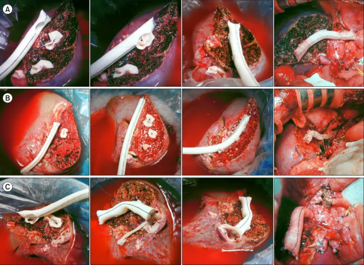
Fig. 1. Intraoperative photographs showing techniques for middle hepatic vein reconstruction using a Hemashield vascular graft.
(A) Vascular patches are attached at the V5 and V8 openings, and a 12-mm Hemashield graft is anastomosed on the back table. The conduit is anastomosed to the recipient’s inferior vena cava (IVC) in a side-to-end fashion. (B) A 10-mm Hemashield graft is attached to the liver graft using a single or double V5 anastomosis and a single V8 anastomosis, and this is anastomosed to the recipient’s IVC in an end-to-end fashion. (C) A 12-mm Hemashield graft is attached to the liver graft with a single V5 anastomosis and a single V8 anastomosis, and this is conjoined with the liver graft’s right hepatic vein orifice. A saphenous vein patch is attached around the conjoined orifice. The conjoined right and middle hepatic vein orifice is anastomosed to the recipient’s IVC.
vious study;6 however, the sample size was too small and follow-up period was too short to fully assess the suit- ability of Hemashield grafts for MHV reconstruction.
Therefore, in this study, we aimed to compare the short- and mid-term patency rates and complications of Hema- shield and ringed Gore-Tex grafts in MHV reconstruction.
MATERIALS AND METHODS
Study design
This was a retrospective two-arm observational study of the outcomes of Hemashield and ringed Gore-Tex graft interposition in MHV reconstruction. The primary purpose of this study was to determine the short- and mid-term
patency rates of Hemashield grafts, and the secondary purpose was to assess the incidence of Hemashield graft- associated complications.
To assess the outcomes of Hemashield graft inter- position, we selected all patients who underwent MHV re- construction using Hemashield grafts during a 26-month study period from November 2017 to December 2019 (Hemashield group; n=157). As a control group, we se- lected an equal number of patients who underwent MHV reconstruction using ringed Gore-Tex grafts during a 48 month period from January 2011 to December 2014 using 1:1 propensity score matching (Gore-Tex group; n=157).
The patency of ringed Gore-Tex grafts was used as refer- ence data for prosthetic vascular grafts; thus, we selected
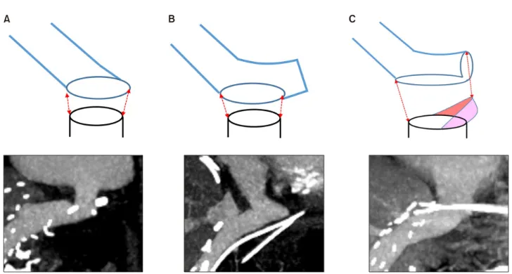
Fig. 2. Schematic illustration of reconstruction techniques for anastomosis of a Hemashield graft conduit to the recipient’s left-middle hepatic vein trunk stump. There are three types of anastomosis: (A) end-to-end anastomosis, (B) side-to-end anasto- mosis, and (C) oblique cutting of the conduit end and patch venoplasty. The colored area represents a vein patch.
patients who survived for at least 2 years without hep- atocellular carcinoma recurrence. Patients in both groups were followed up until the end of August 2020.
The study protocol was approved by the Institutional Review Board of our institution (IRB No. 2019-1347), which waived the requirement for informed consent owing to the retrospective nature of the study. This study was performed in accordance with the ethical guidelines of the 2013 World Medical Association Declaration of Helsinki.
Selection of prosthetic vascular grafts for MHV reconstruction
After establishing the techniques for MHV reconstruction in 1997, we have attempted to reconstruct most of the MHV branches with a diameter of ≥5 mm, including the segment V vein (V5) and the segment VIII vein (V8). The indications for MHV reconstruction have been described elsewhere.1-6 Previously, when large-sized vessel allog- rafts were not available at our institutional tissue bank, we used 10-mm or 12-mm ringed Gore-Tex grafts. Subse- quently, during the study period, ringed Gore-Tex grafts were no longer available; hence, we used 10-mm or 12-mm Hemashield grafts. The indications for the use of Hemashield grafts were the same as those for the use of
ringed Gore-Tex grafts.
Surgical techniques for MHV reconstruction We used the same surgical techniques for MHV re- construction regardless of the graft type. A prosthetic vas- cular graft with an internal diameter of 10-12 mm was selected. After making a small cut to enlarge the orifice of the V5 or V8 stump, a bridging allograft patch was attached for end-to-side anastomosis of the MHV branch (Fig. 1).5,6
If the orifice diameter of V5 or V8 was >8 mm and the stump cuff of the V5/V8 orifice was thick, a Hema- shield graft was directly anastomosed to the V5/V8 orifice without placing a bridging allograft patch.
We used three different methods to anastomose the Hemashield graft to the inferior vena cava (IVC) opening — end-to-end anastomosis, side-to-end anastomosis, and ob- lique cutting of the conduit end and patch venoplasty (Fig.
2). The anastomosis method was selected according to the relative alignment of the Hemashield graft and the IVC and the surgeon’s preference.
Table 1. Clinical profiles of patients who underwent middle hepatic vein reconstruction using hemashield or Gore-Tex vascular grafts
Hemashield group Gore-Tex group p-value
Patients (n) 157 157
Age (years) 54.7±9.4 53.3±6.3 0.12
Sex (n) 0.9
Male 114 (72.6%) 115 (73.2%)
Female 43 (27.4%) 42 (26.8%)
MELD score 15.9±9.2 16.9±8.3 0.47
Primary disease (n) 0.002*
HBV infection 74 (47.1%) 102 (65.0%)
HCV infection 9 (5.7%) 7 (4.5%)
Alcoholic liver disease 51 (32.5%) 25 (15.9%)
Cryptogenic cirrhosis 10 (6.4%) 12 (7.6%)
Acute liver failure 5 (3.2%) 5 (3.2%)
Autoimmune hepatitis 3 (1.9%) 1 (0.6%)
Primary sclerosing cholangitis 3 (1.9%) 1 (0.6%)
Wilson’s disease 2 (1.3%) 2 (1.3%)
Polycystic liver disease 0 2 (1.3%)
Concurrent hepatocellular carcinoma (n) 80 (50.9%) 77 (49.1%) 0.74
ABO-incompatible transplantation (n) 28 (17.8%) 26 (16.6%) 0.77
Graft-recipient weight ratio 1.07±0.24 1.10±0.23 0.26
*Represents comparison between HBV infection and other primary diseases
MELD, model for end-stage liver disease; HBV, hepatitis B virus; HCV, hepatitis C virus Evaluation of MHV-interposed prosthetic graft
patency and indications for interventional stenting
After the transplant, dynamic computed tomography (CT) was performed weekly during hospitalization and ev- ery 3-6 months thereafter at the outpatient clinic for the first 3 years. Thereafter, follow-up CT was performed an- nually for 5 years and biannually after 5 years. CT scan was performed more frequently in patients diagnosed with hepatocellular carcinoma.
We defined occlusion of the MHV conduit as non-visu- alization of blood flow in the prosthetic graft conduit be- tween V8 (or V5 in cases where only V5 was recon- structed) and the IVC on liver CT. In cases where V5 was occluded but V8 remained patent, we considered the MHV conduit to be patent. In cases where it was not pos- sible to perform CT owing to impaired renal function, Doppler ultrasonography was performed.
Interventional stenting of the thrombosed MHV conduit was indicated if significant MHV conduit occlusion-asso- ciated perfusion abnormality occurred in the graft liver.8,9 Interventional conduit stenting was regarded as conduit graft occlusion, regardless of the post-stenting patency.
Statistical analysis
The parameters for propensity score matching were re- cipient age and sex, Model for End-stage Liver Disease score, graft-recipient weight ratio, and number of V5/V8 anastomoses. All numerical data are presented as mean values with standard deviations. Continuous variables were compared using Student’s t-test, and frequency vari- ables were compared using the chi-square test or Fisher’s exact test. Patency rates were determined using the Kaplan- Meier method and were compared using the log-rank test.
A p-value of <0.05 was considered statistically sig- nificant. Statistical analyses were performed using SPSS version 22 (IBM; New York, NY, USA).
RESULTS
Patient profiles
The clinical profiles of all 314 patients who underwent LDLT using a modified right lobe graft along with MHV reconstruction using either Hemashield or ringed Gore- Tex vascular grafts are summarized in Table 1. In the Hemashield and Gore-Tex groups, the mean recipient age was 54.7±9.4 and 53.3±6.3 years; the frequency of hep-
Table 2. Configurations of middle hepatic vein reconstruction performed using Hemashield or ringed Gore-Tex vascular grafts
Hemashield group (n=157)
Gore-Tex group (n=157)
V5 reconstruction (n)
No reconstruction 2 (1.3%) 5 (3.2%) Single anastomosis 113 (72.0%) 128 (81.5%) Double anastomoses 39 (24.8%) 24 (15.3%) Triple anastomoses 3 (1.9%) 0
V8 reconstruction (n)
No reconstruction 11 (7.0%) 37 (23.6%) Single anastomosis 124 (79.0%) 119 (75.8%) Double anastomoses 20 (12.7%) 1 (0.6%) Triple anastomoses 2 (1.3%) 0 V5, liver segment V vein; V8, liver segment VIII vein
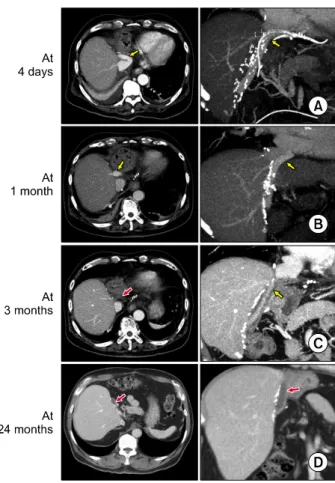
Fig. 3. Serial computed tomography (CT) images showing progressive occlusion of the lumen within the interposed Hemashield graft. Liver CT scans were performed at: (A) 4 days, (B) 1 month, (C) 3 months, and (D) 24 months after transplantation. Middle hepatic vein outflow was nearly com- pletely occluded around 3 months, but no hepatic venous con- gestion occurs, due to the development of intrahepatic venous collaterals. The thrombosed Hemashield graft conduit is visi- ble (D). Yellow arrows indicate luminal blood flow within the interposed Hemashield graft. Red arrows indicate the thrombus-filled Hemashield graft conduit.
atitis B virus infection was 74 (47.1%) and 102 (65.0%);
the Model for End-stage Liver Disease score was 15.9±9.2 and 16.9±8.3; the frequency of ABO-incompatible LDLT was 28 (17.8%) and 26 (16.6%); and the graft-recipient weight ratio was 1.07±0.24 and 1.10±0.23, respectively.
Except primary liver diseases, all the investigated parame- ters were similar between groups.
Configurations of MHV reconstruction using prosthetic vascular grafts
All the available types of vessel fragment, including cryopreserved iliac arteries and veins and autologous sa- phenous and portal veins, were used for vessel patches that were attached to the V5/V8 orifices. Patch unification of two or three small V5/V8 orifices was preferentially performed because it enabled the performance of a single anastomosis to the interposed prosthetic graft.
In the Hemashield graft group, V5 reconstruction was performed with a single anastomosis in 113 patients (72.0%, including unification venoplasty), double anasto- moses in 39 patients (24.8%), and triple anastomoses in 3 patients (1.9%). V8 reconstruction was performed with a single anastomosis in 124 patients (79.0%, including unification venoplasty), double anastomoses in 20 patients (12.7%), and triple anastomoses in 2 patients (1.3%).
These configurations of V5 and V8 reconstruction are summarized in Table 2. The internal diameter of the Hemashield graft was 10 mm in 102 cases (65.0%) and 12 mm in 55 cases (35.0%).
In the ringed Gore-Tex group, V5 reconstruction was performed with a single anastomosis in 128 patients (81.5%, including unification venoplasty) and double anastomoses in 24 patients (15.3%). V8 reconstruction was performed with a single anastomosis in 119 patients (75.8%, including unification venoplasty) and double anastomoses in 1 patient (0.6%) (Table 2). The internal diameter of the Gore-Tex vascular graft was 10 mm in 149 cases (94.9%) and 12 mm in 8 cases (5.1%).
The proportion of double and triple anastomose for V5 or V8 was higher in the Hemashield group than in the ringed Gore-Tex group (p=0.009 for V5 and p<0.001 for V8) because it was less technically challenging to make multiple anastomoses with the Hemashield grafts than
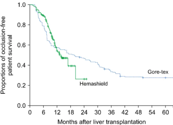
Fig. 4. Comparison of occlusion-free patient survival curves for different prosthetic vascular graft materials.
with the ringed Gore-Tex grafts.
Sequences of prosthetic graft occlusion Serial follow-up CT scans showed that luminal occlu- sion occurred within the prosthetic grafts around the V5 anastomosis. V5 outflow was gradually reduced, which re- sulted in concentric thickening of the luminal thrombus.
Consequently, the prosthetic graft with an original internal diameter of 10 mm or 12 mm was transformed into a nar- row conduit with a small inner diameter. Thereafter, the lumen of the graft conduit between the V5 and V8 orifices was occluded. V8 outflow was maintained for a longer period than V5 outflow. The progression of MHV conduit occlusion that occurred after using a Hemashield graft is depicted in Fig. 3. The sequence of progressive occlusion of the interposed ringed Gore-Tex vascular graft has been reported previously.5,6
Patency of prosthetic graft conduits
During posttransplant follow-up, 3 patients in the Hemashield group died (in-hospital mortality) within 3 months. The Hemashield conduits in these patients were patent until patient death, according to their final fol- low-up imaging studies.
In the Hemashield graft group, 2 (1.3%) patients re- quired MHV conduit stenting owing to early thrombosis of the conduit. The suspected site of stenosis in these pa- tients was the anastomosis site between the conduit and IVC. These patients underwent early stenting at posttrans- plant days 1 and 3, and the patency of the MHV conduit was restored. The conduit occlusion-free patient survival rates in the Hemashield group were 94.9% at 3 months, 88.9% at 6 months, 55.3% at 12 months, and 26.1% at 24 months (Fig. 4).
In the ringed Gore-Tex graft group, 4 patients (2.5%) required MHV stenting. These patients underwent early stenting within 3 weeks of liver transplant (LT), and the MHV flow was successfully restored. The conduit occlu- sion-free patient survival rates in the ringed Gore-Tex group were 93.0% at 3 months, 79.0% at 6 months, 57.3%
at 12 months, and 47.1% at 24 months, which were sim- ilar to those in the Hemashield group (Fig. 4, p=0.91).
Prosthetic vascular graft-associated complications
In the Hemashield graft group, no accidental migration of the conduit graft into adjacent visceral structures oc- curred over a mean follow-up period of 14.5±5.9 months.
In contrast, in the ringed Gore-Tex graft group, there were 2 cases of accidental migration of the graft into the gastric wall at 6 months and 3 years after LT,6,10 resulting in an incidence of 1.3% over a mean follow-up period of 108.7±13.4 months. These 2 patients underwent exploratory laparotomy to remove the migrated conduit. The incidence of accidental conduit migration was not significantly dif- ferent between two groups (p=0.50).
DISCUSSION
MHV reconstruction in LDLT using a modified right lobe graft results in improved recipient survival outcomes and contributes to a reduction in donor complications.
Since MHV reconstruction was established as a routine procedure for most adult LDLT operations in our in- stitution, the demand for sizable vein allografts has increased. In Korea, the number of deceased organ and tissue donors is very limited; thus, the supply of vein al- lografts is much smaller than the demand from LDLT.
Prosthetic vascular grafts have no such supply limitations and, thus, offer very good availability. The long-term pa- tency rates of the ringed Gore-Tex grafts were acceptably high;5,11 therefore, we previously used them whenever siz- able vascular allografts were not available. After ringed Gore-Tex grafts became unavailable, they were substituted
with Hemashield grafts in our institution.6,7
The physical features of Hemashield grafts are similar to those of ringed Gore-Tex vascular grafts. The circular pleats (ConcentricrimpTM, Maquet) of the Hemashield grafts prevent wall collapse caused by extrinsic com- pression. The wall is a thin, flexible structure of woven double-velour polyester; thus, it is easy to handle and su- ture the Hemashield grafts. Its bellows-like circular pleats offer flexible shortening or lengthening longitudinally to allow adjustment of the conduit length during anasto- mosis, and the morphological changes induced by graft re- generation are readily accommodated. The woven struc- ture of these bovine collagen-impregnated grafts also pre- vents needle-hole bleeding; thus, the usual Prolene sutures can be used. Its longitudinal colored lines (GuidelineTM stripe, Maquet) are useful for adjusting the alignment dur- ing anastomosis. The bovine collagen-impregnated and woven double-velour structure facilitates reduction of ear- ly luminal thrombus formation and tissue reaction at the anastomosis sites. The commercially available Hemashield Platinum vascular graft has a diameter of 10 or 12 mm and a length of 25 cm, which are optimal dimensions for an MHV conduit.
We have previously tested other prosthetic vascular grafts.6 We found that ePTFE grafts with outer spi- ral-wound rings (ImpraTM, Bard; New Providence, NJ, USA) provide flexibility in longitudinal length control, but products with a diameter >10 mm are not currently in commercial production. A thin-walled ePTFE graft with an inner carbon lining (Impra CarbofloTM, Bard) appears suitable for MHV reconstruction, but the commercially available product is only 10 cm in length. Owing to these critical inadequacies, the abovementioned prosthetic vas- cular grafts are not viable alternatives to ringed Gore-Tex grafts.
This study revealed that the short- and mid-term pa- tency rates of Hemashield grafts are comparable to those of ringed Gore-Tex grafts. This is the most important point in this study because it indicates that ringed Gore- Tex grafts can be reliably replaced with Hemashield grafts. This is the first report to present the mid-term pa- tency rates of Hemashield grafts in MHV reconstruction.
The primary reasons for the high patency rates of Hema- shield grafts include the large internal conduit diameter, circular pleats resistant to extrinsic compression, bridging
allograft patch that reduces tissue reactions, and low-throm- bogenic bovine collagen-impregnated lumen that induces slow construction of an endothelial cell-lined internal tun- nel within the luminal thrombus.5,6,11
There were some cases of accidental migration of a Gore-Tex graft into the hollow viscera.5,6,11-13 A ringed Gore-Tex graft is converted to a rock-hard foreign body if the lumen is filled with thrombosis. Its accidental mi- gration into the stomach or duodenum can induce life- threatening complications; thus, the migrated conduit should be removed through re-exploration. We have pre- viously reported that the incidence of ringed Gore-Tex con- duit migration was 1.6% at 5 years,6 while in Taiwanese study, it was 1.5%.12 However, we have not encountered any cases of accidental migration of Hemashield grafts yet.
As our observation period for Hemashield grafts was much shorter than that for ringed Gore-Tex grafts, long- term observation is necessary to assess the real risk of conduit-associated complications for Headshield grafts be- cause all occluded prosthetic vascular grafts remain as foreign bodies. In our current analysis of morphological changes after thrombotic occlusion, thrombosed Hemashield grafts partially collapsed, forming a flattened foreign body, but the shape of ringed Gore-Tex grafts was unchanged. We conjecture that such differences in mor- phology may alter the incidence of accidental graft migra- tion into the adjacent hollow viscera. The findings of this study suggest that Hemashield grafts are more suitable than ringed Gore-Tex grafts for MHV reconstruction con- sidering their patency rates and incidence of con- duit-associated complications.
This study has some limitations. This study was a sin- gle-center study, which could potentially introduce se- lection bias. The follow-up period was relatively short;
thus, the incidence of conduit-associated complications was not completely evaluated.
In conclusion, Hemashield grafts used in MHV re- construction demonstrated acceptably high short- and mid-term patency rates, no incidences of conduit migra- tion, easy handling, and good flexibility for length adjustment. Therefore, we suggest that the Hemashield graft is the preferentially suitable prosthetic material for MHV reconstruction.
CONFLICT OF INTEREST
No potential financial conflicts or other conflicts of in- terest exist for any of the authors of this article.
ORCID
Sang Hoon Kim: https://orcid.org/0000-0002-8025-1816 Shin Hwang: https://orcid.org/0000-0002-9045-2531 Minjae Kim: https://orcid.org/0000-0001-6743-0636 Tae-Yong Ha: https://orcid.org/0000-0001-9932-0212 Gi-Won Song: https://orcid.org/0000-0002-4235-0434 Dong-Hwan Jung: https://orcid.org/0000-0001-5984-023X Chul-Soo Ahn: https://orcid.org/0000-0002-3844-3646 Deok-Bog Moon: https://orcid.org/0000-0002-8209-3540 Ki-Hun Kim: https://orcid.org/0000-0002-4016-0995 Gil-Chun Park: https://orcid.org/0000-0003-1631-3258 Sung-Gyu Lee: https://orcid.org/0000-0001-9161-3491
AUTHOR CONTRIBUTIONS
Conceptualization: SH. Data curation: CSA, DBM, KHK, SGL. Methodology: SHK, MK, TYH, GWS, DHJ, GCP. Visualization: SH. Writing - original draft: SH, SHK.
Writing - review & editing: SH.
REFERENCES
1. Hwang S, Lee SG, Lee YJ, Sung KB, Park KM, Kim KH, et al. Lessons learned from 1,000 living donor liver transplantations in a single center: how to make living donations safe. Liver Transpl 2006;12:920-927.
2. Hwang S, Lee SG, Ahn CS, Park KM, Kim KH, Moon DB, et al. Cryopreserved iliac artery is indispensable interposition graft
material for middle hepatic vein reconstruction of right liver grafts. Liver Transpl 2005;11:644-649.
3. Sugawara Y, Makuuchi M, Akamatsu N, Kishi Y, Niiya T, Kaneko J, et al. Refinement of venous reconstruction using cry- opreserved veins in right liver grafts. Liver Transpl 2004;10:
541-547.
4. Hwang S, Lee SG, Song GW, Lee HJ, Park JI, Ryu JH. Use of endarterectomized atherosclerotic artery allograft for hepatic vein reconstruction of living donor right lobe graft. Liver Transpl 2007;13:306-308.
5. Hwang S, Jung DH, Ha TY, Ahn CS, Moon DB, Kim KH, et al.
Usability of ringed polytetrafluoroethylene grafts for middle hep- atic vein reconstruction during living donor liver transplantation.
Liver Transpl 2012;18:955-965.
6. Park GC, Hwang S, Ha TY, Song GW, Jung DH, Ahn CS, et al. Hemashield vascular graft is a preferable prosthetic graft for middle hepatic vein reconstruction in living donor liver transplantation. Ann Transplant 2019;24:639-646.
7. Jeong IJ, Hwang S, Ha TY, Song GW, Jung DH, Park GC, et al. Technical refinement of prosthetic vascular graft anastomosis to recipient inferior vena cava for secure middle hepatic vein re- construction in living donor liver transplantation. Ann Hepatobiliary Pancreat Surg 2020;24:144-149.
8. Ko GY, Sung KB, Yoon HK, Kim JH, Song HY, Seo TS, et al. Endovascular treatment of hepatic venous outflow obstruction after living-donor liver transplantation. J Vasc Interv Radiol 2002;13:591-599.
9. Ko GY, Sung KB, Yoon HK, Kim KR, Kim JH, Gwon DI, et al. Early posttransplant hepatic venous outflow obstruction:
long-term efficacy of primary stent placement. Liver Transpl 2008;14:1505-1511.
10. Ha TY, Hwang S, Jung DH, Ahn CS, Kim KH, Moon DB, et al. Complications analysis of polytetrafluoroethylene grafts used for middle hepatic vein reconstruction in living-donor liver transplantation. Transplant Proc 2014;46:845-849.
11. Papanicolaou G, Beach KW, Zierler RE, Detmer PR, Strandness DE Jr. Hemodynamics of stenotic infrainguinal vein grafts: theo- retic considerations. Ann Vasc Surg 1995;9:163-171.
12. Hsu SC, Thorat A, Yang HR, Poon KS, Li PC, Yeh CC, et al.
Assessing the safety of expanded polytetrafluoroethylene syn- thetic grafts in living donor liver transplantation: graft migration into hollow viscous organs- diagnosis and treatment options.
Med Sci Monit 2017;23:3284-3292.
13. Sultan AM, Shehta A, Salah T, Elshoubary M, Wahab MA.
Spontaneous migration of thrombosed synthetic vascular graft to the duodenum after living-donor liver transplantation: a case- report. Int J Surg Case Rep 2018;45:42-44.