Solitary Osseous Metastasis of Rectal Carcinoma Masquerading as Osteogenic Sarcoma on Post- Chemotherapy Imaging: A Case Report
Amar Udare, MBBS
1, Nilesh Sable, MD
1, Rajiv Kumar, MD
2, Meenakshi Thakur, MD
1, Shashikant Juvekar, MD
1Departments of 1Radiodiagnosis and 2Pathology, Tata Memorial Hospital, Mumbai 400012, India
Solitary metastases from colorectal carcinoma in the absence of hepatic or pulmonary metastases are rare. These can have a diverse imaging appearance, particularly after chemotherapy. It is important identify patients with solitary skeletal metastases, as they have a better prognosis than those with multiple skeletal or visceral metastases. We describe an unusual case of a solitary metastasis to the femur in a case of colon carcinoma that went undiagnosed and later presented with imaging features of osteogenic sarcoma.
Index terms: Colorectal neoplasms; Skeletal metastases; Osteosarcoma; Radiography; Computed tomography; MRI
Received September 1, 2013; accepted after revision October 28, 2014.
Corresponding author: Amar Udare, MBBS, Department of Radiodiagnosis, Tata Memorial Hospital, Dr E. Borges Marg, Parel, Mumbai 400012, India.
• Tel: (9122) 24177000 Ext: 4157 • Fax: (9122) 24146937
• E-mail: amarudare@gmail.com
This is an Open Access article distributed under the terms of the Creative Commons Attribution Non-Commercial License (http://creativecommons.org/licenses/by-nc/3.0) which permits unrestricted non-commercial use, distribution, and reproduction in any medium, provided the original work is properly cited.
Korean J Radiol 2015;16(1):175-179 http://dx.doi.org/10.3348/kjr.2015.16.1.175 pISSN 1229-6929 · eISSN 2005-8330
Case Report
| Musculoskeletal ImagingINTRODUCTION
Colorectal cancer is one of the leading causes of cancer- related death worldwide (1). Skeletal metastases in patients with colon carcinoma are rare, and the occurrence of isolated an solitary skeletal metastasis in the absence of visceral metastases is even rarer (2). These metastases can have bizarre radiological appearances due to post-treatment changes. We herein describe a case in which the post- chemotherapy imaging features of a metastatic lesion from a patient with colorectal carcinoma mimicked the radiologic appearance of osteosarcoma.
This case report has been approved by the Institutional Review Board.
CASE REPORT
A 26-year-old woman presented to the outpatient department with a chief complaint of pain in the left lower limb of 1 month duration. The patient had a known case of rectal carcinoma for which she had undergone a diagnostic laparoscopy and transverse colostomy and was on neoadjuvant chemotherapy thereafter.
A clinical examination revealed painful swelling of a unilateral lower limb. A radiograph of the femur was obtained which showed ill-defined lytic area in the proximal meta-diaphyseal region of the left femur with spiculated periosteal reaction (Fig. 1A). The patient underwent a computed tomography (CT) study of the pelvis and the proximal lower limbs, which showed a lytic lesion with a circumferential spiculated sunburst type of periosteal reaction and an ill-defined associated vascular soft tissue component in the proximal third of the meta-diaphysis of the left femur (Fig. 1B). A provisional diagnosis of osteogenic sarcoma was made based on the radiograph and CT scan findings.
A magnetic resonance imaging (MRI) examination of the thigh was performed to evaluate the local disease. MRI revealed altered marrow signal intensity in the proximal
A
B
C
Fig. 1. Solitary osseous rectal carcinoma metastasis in 26-year-old woman.
A. Plain radiograph of left femur (anteroposterior and lateral views) shows ill-defined lytic area in proximal meta-diaphyseal region of left femur with spiculated periosteal reaction (white arrow). B. Post-contrast axial computed tomography (CT) scan shows ill-defined lytic lesion with circumferential spiculated sunburst type of periosteal reaction and ill-defined soft tissue component in proximal third of diaphysis of left femur.
Lesion shows intense neovasculartiy on reformatted volume-rendered coronal images. C. Magnetic resonance image of left thigh. Hypointensity is seen in radial distribution of proximal aspect of femur on axial gradient echo sequences. Peripheral enhancement was noted in soft tissue component of femoral lesion on post-contrast gadolinium T1-weighted images.
Solitary Osseous Metastasis of Rectal Carcinoma Masquerading as Osteosarcoma on Post-Chemotherapy Imaging
meta-diaphysis of the left femur with a circumferential soft tissue mass. Hypointensity was seen in the radial distribution on gradient echo sequences, corroborating the “sunburst” calcification noted on the radiograph and CT scan (Fig. 1C). The lesion showed significant peripheral enhancement in the soft tissue component (Fig. 1C). These findings supported a diagnosis of a second primary lesion in the form of osteogenic sarcoma.
A CT-guided biopsy was performed to confirm the diagnosis and revealed an extremely tiny focus malignant tumor composed of scattered large polygonal cells with a moderate amount of vacuolated cytoplasm, hyperchromatic nuclei, and occasional prominent nucleoli. The differential diagnoses considered were high grade carcinoma and lymphoma (anaplastic large cell lymphoma or plasmablastic lymphoma) but this was unlikely to be osteogenic sarcoma.
A special stain for mucicarmine was positive. The tumor cells were immunopositive for epithelial markers, including cytokeratin (CK) and epithelial membrane antigen but negative for lymphoma and plasma cell markers, i.e., leukocyte common antigen, CD30, and CD138. The tumor cells were also immunopositive for CK20 and Cdx2, but negative for CK7, confirming metastatic poorly differentiated adenocarcinoma primarily of colonic origin (Fig. 1D).
Based on the histology, we reviewed the positron emission tomography (PET)-CT staging scan and prior radiographs taken at the time of presentation 6 months previously. The radiographs showed an ill-defined lytic lesion in the proximal femur that appeared to be an ill- defined lytic area on the PET-CT images and was mildly
18F-fluorodeoxyglucose (FDG)-avid, with a standardized uptake value (SUV) of 3.2 (Fig. 1E). Thus, the lesion was present prior to chemotherapy. These findings were inadvertently not communicated to the treating physician.
The patient underwent a re-staging PET-CT scan, which showed no other skeletal or visceral metastasis, and the lesion in the proximal meta-diaphyseal region of the femur had a decreased SUV value compared to that of the previous staging PET-CT, and it now measured 1.2. Thus, the radiological appearance represented healed metastases from the primary rectal carcinoma with calcification, which mimicked the classic pattern of a spiculated sunburst periosteal reaction with a lytic sclerotic appearance of an osteogenic sarcoma.
DISCUSSION
Colorectal cancer is the third most commonly diagnosed cancer in males and the second most common in females (3).
It is the most common malignancy in the gastrointestinal tract and is the fourth leading cause of cancer-associated death in the world (1). Colorectal cancer is a disease of middle age and elderly; younger patients often have an advanced stage at presentation and present with more poorly differentiated lesions leading to a worse prognosis (1).
Skeletal metastases in patients with colorectal carcinoma are relatively uncommon with an incidence of 4.7–10.9%
(2). A large retrospective study found an incidence of approximately 10% (4). Incidence is particularly higher in rectal carcinomas compared to that of other parts of the colon and in carcinomas with a non-mucinous histopathology (4). The most common sites for metastases include the vertebral column, pelvis, and long bones (4).
Metastases to the bone are primarily via the paravertebral venous plexus of Batson (5) after direct invasion of the veins by the lesion, which further explains the higher incidence of skeletal metastases from recto-sigmoid carcinoma (6). The prognosis of patients with skeletal metastases from colorectal carcinoma is poor (6, 7).
Isolated solitary skeletal metastases from primary colonic carcinoma are even rarer. A study conducted by Roth et al. (7) using whole-body 18F-FDG PET showed that no patient had osseous metastasis at the time of diagnosis, and no patient had isolated skeletal metastases without other organ development. A few earlier studies reported a 1–2% incidence of solitary osseous metastases, although none of them utilized PET-CT scans for the evaluation (8, 9). Osseous metastases from colon carcinoma are predominantly osteolytic in nature, and osteoblastic or mixed osteoblastic-osteolytic lesions are rare (4). The lytic lesions are associated with reactive changes within the bone, periosteum, and adjacent soft tissue and often simulate a primary bone lesion (10). Notably, patients with isolated skeletal metastases have a better prognosis than those with multiple skeletal or visceral metastases (3).
The lesion in our case was predominantly osteolytic on CT images and radiographs obtained prior to chemotherapy.
The same lesion now appeared osteoblastic with significant soft tissue and a sunburst type of periosteal reaction mimicking a primary bone lesion, such as osteosarcoma, rather than metastases. However in our setting where the
patient had a known primary; even though the imaging appearance as suggestive of osteogenic sarcoma, the first differential considered was metastasis, as primary bone tumors are relatively rare and should be considered only after metastasis has been ruled out. Hence, a biopsy was performed, which confirmed the presence of metastases.
The characteristic pseudo-sarcomatous appearance of the lesion was attributed to post-treatment changes.
The purpose of our case report is to highlight the unusual radiological appearance of these osseous metastases after
chemotherapy. We emphasize that the occurrence of a second osseous primary malignant lesion in an already diagnosed case of colorectal carcinoma is far less likely than a solitary metastasis, although the incidence of the latter is very low.
In conclusion, it is imperative for radiologists to analyze imaging findings in light of available clinical data to arrive at the correct diagnosis. Skeletal metastases can have varied imaging appearances, particularly after chemotherapy; thus, it is essential to diagnose such lesions D
E
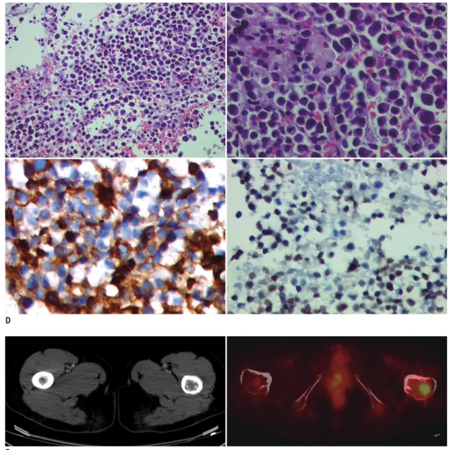
Fig. 1. Solitary osseous rectal carcinoma metastasis in 26-year-old woman.
D. Histopathological examination shows malignant tumor composed of dispersed population of large polygonal cells with moderate amount of vacuolated cytoplasm (H&E; x 100 [upper right image], x 200 [upper left image]). Tumor cells were immunopositive cytokeratin 20 (lower right image) and Cdx2 (lower left image) confirming metastatic adenocarcinoma of colonic origin. E. Positron emission tomography (PET)-CT scan was taken for staging prior to chemotherapy. Axial CT images confirm ill-defined lytic lesion seen in proximal left femur on plain radiograph, which is
18F-fluorodeoxyglucose-avid, as seen on fused axial PET-CT images.
Solitary Osseous Metastasis of Rectal Carcinoma Masquerading as Osteosarcoma on Post-Chemotherapy Imaging
at the earliest for accurate staging and prompt management to ensure a better prognosis.
REFERENCES
1. Chan KK, Dassanayake B, Deen R, Wickramarachchi RE, Kumarage SK, Samita S, et al. Young patients with colorectal cancer have poor survival in the first twenty months after operation and predictable survival in the medium and long- term: analysis of survival and prognostic markers. World J Surg Oncol 2010;8:82
2. Chalkidou AS, Boutis AL, Padelis P. Management of a Solitary Bone Metastasis to the Tibia from Colorectal Cancer. Case Rep Gastroenterol 2009;3:354-359
3. Jemal A, Bray F, Center MM, Ferlay J, Ward E, Forman D.
Global cancer statistics. CA Cancer J Clin 2011;61:69-90 4. Santini D, Tampellini M, Vincenzi B, Ibrahim T, Ortega C,
Virzi V, et al. Natural history of bone metastasis in colorectal cancer: final results of a large Italian bone metastases study.
Ann Oncol 2012;23:2072-2077
5. Batson OV. The function of the vertebral veins and their role in the spread of metastases. Ann Surg 1940;112:138-149 6. Nozue M, Oshiro Y, Kurata M, Seino K, Koike N, Kawamoto T,
et al. Treatment and prognosis in colorectal cancer patients with bone metastasis. Oncol Rep 2002;9:109-112
7. Roth ES, Fetzer DT, Barron BJ, Joseph UA, Gayed IW, Wan DQ.
Does colon cancer ever metastasize to bone first? a temporal analysis of colorectal cancer progression. BMC Cancer 2009;9:274
8. Kanthan R, Loewy J, Kanthan SC. Skeletal metastases in colorectal carcinomas: a Saskatchewan profile. Dis Colon Rectum 1999;42:1592-1597
9. Besbeas S, Stearns MW Jr. Osseous metastases from carcinomas of the colon and rectum. Dis Colon Rectum 1978;21:266-268
10. Oh YK, Park HC, Kim YS. Atypical bone metastasis and radiation changes in a colon cancer: a case report and a review of the literature. Jpn J Clin Oncol 2001;31:168-171