Comparison of Optical Coherence Tomographic Assessment between First- and Second-Generation Drug-Eluting Stents
Byeong-Keuk Kim,
1Jung-Sun Kim,
1Junbeom Park,
1Young-Guk Ko,
1Donghoon Choi,
1Yangsoo Jang,
1,2and Myeong-Ki Hong
1,21Division of Cardiology, Severance Cardiovascular Hospital, Yonsei University College of Medicine, Seoul;
2Severance Biomedical Science Institute, Yonsei University College of Medicine, Seoul, Korea.
Received: July 11, 2011 Revised: August 31, 2011 Accepted: September 8, 2011
Corresponding author: Dr. Myeong-Ki Hong, Division of Cardiology, Severance Cardiovascular Hospital, Yonsei University College of Medicine, 50 Yonsei-ro, Seodaemun-gu, Seoul 120-752, Korea.
Tel: 82-2-2228-8458, Fax: 82-2-393-2041 E-mail: [email protected]
∙ The authors have no financial conflicts of interest.
© Copyright:
Yonsei University College of Medicine 2012 This is an Open Access article distributed under the terms of the Creative Commons Attribution Non- Commercial License (http://creativecommons.org/
licenses/by-nc/3.0) which permits unrestricted non- commercial use, distribution, and reproduction in any medium, provided the original work is properly cited.
Purpose: There is a lack of sufficient data in comparison of optical coherence to- mographic (OCT) findings between first- and second-generation drug-eluting stents (DES). Compared to first-generation (i.e., sirolimus- or paclitaxel-eluting stents), second-generation DESs (i.e., everolimus- or biolinx-based zotarolimus- eluting stents) might have more favorable neointimal coverage. Materials and Methods: Follow-up OCT findings of 103 patients (119 lesions) treated with sec- ond-generation DESs were compared with those of 139 patients (149 lesions) treated with first-generation DESs. The percentage of uncovered or malapposed struts, calculated as the ratio of uncovered or malapposed struts to total struts in all OCT cross-sections, respectively, was compared between the two groups. Results:
Both DES groups showed similar suppression of neointimal hyperplasia (NIH) on OCT (mean NIH cross-sectional area; second- vs. first-generation=1.1±0.5 versus 1.2±1.0 mm2, respectively, p=0.547). However, the percentage of uncovered struts of second-generation DESs was significantly smaller than that of first-generation DESs (3.8±4.8% vs.7.5±11.1%, respectively, p<0.001). The percentage of malap- posed struts was also significantly smaller in second-generation DESs than in first- generation DESs (0.4±1.6% vs.1.4±3.7%, respectively, p=0.005). In addition, in- tra-stent thrombi were less frequently detected in second-generations DESs than in first-generation DESs (8% vs. 20%, respectively, p=0.004). Conclusion: This fol- low-up OCT study showed that second-generation DESs characteristically had greater neointimal coverage than first-generation DESs.
Key Words: Optical coherence tomography, stent
INTRODUCTION
With the introduction of first-generation drug-eluting stents (DES), a significant reduction in restenosis rates and an improvement in short-term clinical outcomes were reported.1,2 However, the use of first-generation DESs, e.g., sirolimus- and paclitaxel-eluting stents, has been shown to be strongly related with the occurrence of late or very late stent thrombosis, raising safety concerns.3,4 Several attempts to develop newer DESs have set out to prevent the occurrence of stent thrombosis by
tion, all patients received dual antiplatelet therapy with aspi- rin and clopidogrel until the follow-up OCT was conducted.
This study was approved by the Institutional Review Board of our institute, and written informed consent was obtained from each patient.
OCT imaging and analysis
Detailed explanations regarding the OCT system and meth- ods for imaging have been described in our previous stud- ies.12,13 OCT examination was performed using a conven- tional OCT system (Model M2 Cardiology Imaging System, LightLab Imaging, Westford, MA, USA) with a motorized pull-back system at 1.0 mm/s. The occlusion catheter was positioned proximal to the stent, and a 0.014-inch wire-type imaging catheter (ImageWire, LightLab Imaging) was po- sitioned distal to the stent. During image acquisition, the occlusion balloon (Helios, Avantec Vascular, Sunnyvale, CA, USA) was inflated to 0.4-0.6 atm, and lactated Ringer’s solution was infused at a rate of 1.0 mL/s. The imaging wire was pulled from distal to proximal, and continuous images were acquired and stored digitally for subsequent analy- sis.12,13 OCT analysis was performed by an independent in- vestigator blinded to patient and procedural information.
Cross-sectional OCT images were analyzed at 1-mm inter- vals (every 15 frames). Stent and luminal cross-sectional ar- eas (CSAs) were measured at 1-mm intervals, and NIH CSA was calculated as the stent CSA minus the luminal CSA. Per- cent NIH CSA was calculated as NIH CSA×100/stent CSA.
Mean values are reported in this study. The thickness of NIH, defined as the distance between the endoluminal sur- face of neointima and the strut, was measured inside the struts at a line as perpendicular as possible to the neointima and strut.10 An uncovered strut was defined as having a NIH thickness of 0 μm.10,14 A malapposed strut was defined as a strut that had detached from the vessel wall (CypherTM,
≥160 μm; TaxusTM, ≥130 μm; Endeavor Resolute®, ≥110 μm; XienceTM, ≥100 μm).15,16 The percentage of uncovered or malapposed struts was investigated for evaluation of the healing responses of DESs as shown on OCT. The percent- age of malapposed or uncovered struts in each stented le- sion was calculated as the (number of malapposed or un- covered struts/total number of struts in all cross-sections of the lesion)×100, respectively. Cross sections with major side branches (diameter ≥2 mm) were excluded from this analy- sis. The neointimal coverage of stent struts in each cross- section was evaluated, and then the percentage of uncov- ered struts was analyzed and compared between the two modifying the eluted drugs, drug carrying systems, and stent
design. Of these, second-generation DESs, e.g., everolim- us-eluting stents (EES) and Biolinx-based zotarolimus-elut- ing stents (Bx-ZES), have been reported to suppress neo- intima hyperplasia (NIH) effectively and simultaneously demonstrate favorable long-term outcomes.5-9 Optical co- herence tomography (OCT) has enabled researchers to evaluate neointimal coverage of DESs in detail, even at the strut level.10,11 However, no sufficient OCT data has been reported comparing neointimal coverage between first- and second-generation DESs. Therefore, using OCT, we sought to compare healing responses, including neointimal cover- age, between the two groups.
MATERIALS AND METHODS
Study population
We used data submitted to the Yonsei OCT registry, evaluat- ing neointimal coverage in patients who underwent coro- nary stent implantation for de novo lesions.12,13 General ex- clusion criteria for the follow-up OCT study were as follows:
1) untreated significant left main coronary artery disease, 2) apparent congestive heart failure, 3) renal insufficiency (baseline creatinine ≥2.0 mg/dL), and 4) lesions unsuitable for OCT imaging (vessel size ≥3.5 mm or lesions within 10 mm of the ostium of a major epicardial artery). Between September 2007 and October 2010, a total of 242 patients with 268 lesions were selected from the OCT registry data- base. Inclusion criteria of the current study comprised le- sions treated with EES, Bx-ZES, sirolimus- or paclitaxel- eluting stents, as well as those followed with a follow-up OCT examination at 12±4 months after stent implantation.
Exclusion criteria were 1) bifurcation treated with 2-stent techniques, 2) angiographic evidence of restenosis, 3) le- sions with repeated revascularization, 4) bare-metal stent implantation, and 5) poor OCT image quality. Second-gen- eration DESs were deployed in 103 patients for 119 lesions including EES (Xience VTM, Abbott Vascular, Santa Clara, CA, USA) and Bx-ZES (Endeavor Resolute®, Medtronic, Santa Rosa, CA, USA), while first-generation DESs were implanted in 139 patients for 149 lesions including sirolim- us-eluting stents (CypherTM, Cordis, Miami, FL, USA) and paclitaxel-eluting stents (TaxusTM, Boston scientific, Natick, MA, USA). DES implantation was performed using current, conventional techniques, and the choice of DES was made according to the operators’ discretion. After DES implanta-
of p<0.05 was considered statistically significant.
RESULTS
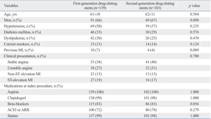
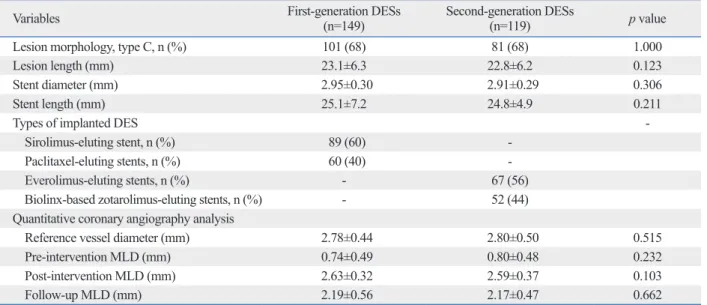
Baseline clinical characteristics are shown in Table 1. There were no significant differences in baseline clinical charac- teristics between the two groups. Baseline angiographic and procedural characteristics are listed in Table 2. There were also no significant differences in baseline angiograph- ic and procedural characteristics between the two groups.
Table 3 summarizes the OCT findings of both groups.
There were no significant differences in mean lumen and stent CSA, as well as time to follow-up OCT (days) be- tween the two groups. Although both groups showed a nearly similar suppression of NIH, the percentage of un- covered struts of second-generation DESs was significantly smaller than that of first-generation DESs (3.8±4.8% vs.
7.5±11.1%, respectively, p<0.001). The percentage of malapposed struts was also significantly smaller in second- generation DESs than in first-generation DESs (0.4±1.6%
vs. 1.4±3.7%, respectively, p=0.005). In addition, intra- stent thrombi were less frequently detected in second-gen- erations DESs than in first-generation DESs (8% vs. 20%, respectively, p=0.004).
groups. Intra-stent thrombi were defined as a signal-rich, low-backscattering protrusions or high-backscattering pro- trusions inside the lumen of the artery with signal-free shadowing on OCT images (dimension ≥250 μm).13 Angiographic analysis
Quantitative coronary angiography analysis was performed using an offline quantitative coronary angiography system (CASS system II, Pie Medical Imaging, Nuenen, the Neth- erlands) before and after stent implantation and at the fol- low-up angiogram. The minimal luminal diameter of the coronary lesions treated with DESs and a reference diame- ter were measured in the view that was the most severe and not foreshortened.
Statistical analysis
All statistical analyses were performed using the Statistical Analysis System software (SAS; 9.1.3., SAS Institute, Cary, NC, USA). Categorical data were presented as a number (%) and compared with Chi-square statistics or Fisher’s exact test.
Continuous data were presented as mean±standard deviation and compared with Student’s t-test. Comparisons among the four different DESs were performed by analysis of variance with post-hoc analysis by the Bonferroni method. If the distri- butions were skewed, a non-parametric test was used. A value Table 1. Baseline Clinical Characteristics
Variables First-generation drug-eluting
stents (n=139) Second-generation drug-eluting
stents (n=103) p value
Age, yrs 61±10 62±11 0.584
Men, n (%) 91 (66) 69 (67) 0.890
Hypertension, n (%) 69 (50) 59 (57) 0.235
Diabetes mellitus, n (%) 46 (33) 30 (29) 0.576
Dyslipidemia, n (%) 42 (30) 26 (25) 0.470
Current smokers, n (%) 15 (11) 14 (14) 0.124
Previous MI, n (%) 10 (7) 4 (4) 0.089
Clinical presentation, n (%) 0.780
Stable angina 53 (38) 41 (40)
Unstable angina 38 (27) 32 (31)
Non-ST elevation MI 21 (15) 13 (13)
ST-elevation MI 27 (19) 16 (17)
Medications at index procedure, n (%)
Aspirin 139 (100) 102 (100) 1.000
Clopidogrel 138 (99) 101 (98) 1.000
Beta-blockers 115 (83) 86 (83) 0.856
ACEI or ARB 100 (72) 80 (78) 0.279
Statins 137 (99) 101 (98) 1.000
MI, myocardial infarction; ACEI, angiotensin converting enzyme inhibitors; ARB, angiotensin receptor blockers; SD, standard deviation.
Values are expressed as mean±SD for quantitative variables or as n (%) for qualitative variables.
growth was not different from that of first-generation DESs.
As concerns regarding the safety issues of first-genera- tion DESs have increased, newly developing DESs have fo- cused more on safety with a similar efficacy to that of first- generation DESs through the long-term follow-up studies.3,4 Among many newly developed DESs, EES and Bx-ZES have been shown to have both excellent efficacy and safe- ty.5-9 Clinical Evaluation of the Xience V Everolimus Elut- ing Coronary Stent System in the Treatment of Patients
DISCUSSION
This follow-up OCT study demonstrated that second-genera- tion DESs lead to a lower percentage of uncovered and malapposed struts, as well as a lower incidence of intra-stent thrombi, compared with first-generation DESs. In spite of su- perior healing responses of second-generation DESs, the effi- cacy of second-generation DESs in the suppression of NIH Table 2. Baseline Angiographic and Procedural Characteristics
Variables First-generation DESs
(n=149) Second-generation DESs
(n=119) p value
Lesion morphology, type C, n (%) 101 (68) 81 (68) 1.000
Lesion length (mm) 23.1±6.3 22.8±6.2 0.123
Stent diameter (mm) 2.95±0.30 2.91±0.29 0.306
Stent length (mm) 25.1±7.2 24.8±4.9 0.211
Types of implanted DES -
Sirolimus-eluting stent, n (%) 89 (60) -
Paclitaxel-eluting stents, n (%) 60 (40) -
Everolimus-eluting stents, n (%) - 67 (56)
Biolinx-based zotarolimus-eluting stents, n (%) - 52 (44)
Quantitative coronary angiography analysis
Reference vessel diameter (mm) 2.78±0.44 2.80±0.50 0.515
Pre-intervention MLD (mm) 0.74±0.49 0.80±0.48 0.232
Post-intervention MLD (mm) 2.63±0.32 2.59±0.37 0.103
Follow-up MLD (mm) 2.19±0.56 2.17±0.47 0.662
DES, drug-eluting stents; MLD, minimal lumen diameter; SD, standard deviation.
Values are expressed as mean±SD for quantitative variables or as n (%) for qualitative variables.
Table 3. Follow-Up Optical Coherence Tomography Measurements
Variables First-generation DESs (n=149) Second-generation DESs (n=119) p value
Number of cross sections 3709 2665
Total number of analyzable struts 32972 23750
Time to follow-up OCT (days) 296±60 290±43 0.273
Mean stent CSA (mm2) 7.1±1.7 7.0±1.6 0.925
Mean lumen CSA (mm2) 5.9±1.6 5.9±1.7 0.923
Mean NIH CSA (mm2) 1.2±1.0 1.1±0.5 0.547
Mean percent NIH CSA (%) 16.9±12.4 17.1±8.3 0.884
Mean NIH thickness (µm) 148±111 142±63 0.666
Percentage of uncovered struts, % 7.5±11.1 3.8±4.8 <0.001
Percentage of malapposed struts, % 1.4±3.7 0.4±1.6 0.005
Presence of intra-stent thrombi, n (%) 30 (20) 9 (8) 0.004
Comparisons among the DES Sirolimus-eluting
stents (n=89) Paclitaxel-eluting
stents (n=60) Everolimus-eluting stents (n=67)
Biolinx-based zotarolimus-eluting
stents (n=52) p value Percentage of uncovered struts, % 10.2±12.9 3.5±6.0* 3.6±4.1* 3.9±5.6* <0.001
Percentage of malapposed struts, % 1.7±4.1 0.8±3.1 0.3±0.6† 0.6±2.3 0.014
Presence of intra-stent thrombi, n (%) 25 (28) 5 (8)* 8 (12)† 1 (2)* <0.001
CSA, cross-sectional area; DES, drug-eluting stent; NIH, neointimal hyperplasia; OCT, optical coherence tomography; SD, standard deviation.
Values are expressed as mean±SD for quantitative variables or as n (%) for qualitative variables.
*p<0.01 and †p<0.05 when compared to sirolimu-eluting stents.
was significantly lower than that of first-generation DESs.
Stent malapposition has been also regarded as an important predictor of DES thrombosis in intravascular ultrasound studies.20,21 DES type has been suggested as one of the most determining factors of stent malapposition.21,22 A difference in the incidence of stent malapposition dependent upon the type of DES might be associated with differences in long- term outcomes. Namely, a lower rate of malapposition in second-generation DESs may translate to more favorable clinical outcomes after DES implantation thereof.
Study limitations
This study has several limitations. First, selection bias might affect the results because this was a non-randomized regis- try study. Second, because this study was not a controlled comparative one, direct comparisons among four different DESs could not be performed. However, there were no sig- nificant differences in the baseline clinical and angiograph- ic parameters between the two groups. Third, because the study population of the current study was free of major ad- verse events after DES implantation until follow-up OCT, the lesions of this study might not represent those seen in real world practice. Fourth, in this study, the first-generation DES group, which consisted of sirolimus-eluting stents and paclitaxel-eluting stents, was compared with second-gener- ation DESs. However, because sirolimus-eluting stents and paclitaxel-eluting stents showed different outcomes in some pathologic and imaging studies,12,13,23,24 careful attention must be given when interpreting the results. Finally, no clin- ical follow-up data was provided due to the short duration of clinical follow-up after OCT evaluation.
In conclusion, this follow-up OCT study showed that second-generation DESs characteristically had greater neo- intimal coverage than first-generation DESs. For more defi- nite conclusions, long term clinical and serial OCT follow- up with a larger population will be needed in the future.
ACKNOWLEDGEMENTS
This study was partly supported by grants from the Korea Healthcare Technology R&D Project, Ministry for Health, Welfare & Family Affairs, Republic of Korea (No.
A085012 and A102064), the Korea Health 21 R&D Proj- ect, Ministry of Health & Welfare, Republic of Korea (No.
A085136), and the Cardiovascular Research Center, in Seoul, Korea.
with de novo Native Coronary Artery Lesions (SPIRIT) III randomized study showed that EES implantation resulted in a statistically significant reduction of angiographic late loss at 8-month follow-up and showed significantly im- proved event-free survival, compared with implantation of paclitaxel-eluting stents at 2-year follow-up.5,6 In addition, compared with patients treated with paclitaxel-eluting stents, those treated with EES tended to have fewer epi- sodes of late stent thrombosis at 1 and 2 years (0.2% versus 1.0%; p=0.10) thereafter.6 Recently, Bx-ZES, which com- prises a low-profile, thin-strut platform, and a Biolinx tri- polymer, also showed favorable short-term angiographic outcomes with a comparable low late loss.7
Many potential mechanisms or factors are expected to be related with the favorable outcomes demonstrated in second- generation DESs.5-8,17 Of these, the degree of reendothelial- ization, regarded as the most powerful predictor of stent thrombosis, might be strongly related with better outcomes in DESs.11,18,19 Although some studies have evaluated neointi- mal coverage for various types of DES, they were conducted as an autopsy or animal study.17,18 A new imaging tool, OCT, has enabled researchers to evaluate the reendothelialization of DES in live patients with a superior resolution capacity.10,11 Therefore, using OCT, healing responses, including neointi- mal coverage of stent struts, was evaluated between second- generation and first-generation DESs in this study.
In the current OCT study, the rate of uncovered struts as shown on OCT was significantly different between the two groups; second-generation DESs showed a higher % of un- covered struts, meaning more complete neointimal cover- age. However, the amount of NIH was similar between the first- and second-generation DESs. The superior nature of second-generation DESs, showing better endothelialization and healing responses compared with first-generation DESs, while maintaining similar efficacy represented by the sup- pression of NIH on OCT or a low late loss on follow-up an- giogram, might be caused by the unique components com- prising DESs, including the use of novel drugs, superior biocompatibility and morphology of polymers, reduced polymer layers, and thin-strut design.17 As a result, newer DESs are both safe and have equal efficacy to first-genera- tion DESs, and this study, in comparison of the OCT find- ings thereof, suggests that second-generation DESs are close to the ideal DES.
The degree of stent malapposition, evaluated by OCT, was also significantly different between the two groups; the percentage of malapposed struts of second-generation DESs
treated with drug-eluting stent: the usefulness of optical coherence tomography. Yonsei Med J 2010;51:332-8.
13. Kim JS, Hong MK, Fan C, Kim TH, Shim JM, Park SM, et al. In- tracoronary thrombus formation after drug-eluting stents implan- tation: optical coherence tomographic study. Am Heart J 2010;
159:278-83.
14. Barlis P, Dimopoulos K, Tanigawa J, Dzielicka E, Ferrante G, Del Furia F, et al. Quantitative analysis of intracoronary optical coher- ence tomography measurements of stent strut apposition and tis- sue coverage. Int J Cardiol 2010;141:151-6.
15. Tanigawa J, Barlis P, Di Mario C. Intravascular optical coherence tomography: optimisation of image acquisition and quantitative as- sessment of stent strut apposition. EuroIntervention 2007;3:128-36.
16. Tanigawa J, Barlis P, Dimopoulos K, Dalby M, Moore P, Di Ma- rio C. The influence of strut thickness and cell design on immedi- ate apposition of drug-eluting stents assessed by optical coherence tomography. Int J Cardiol 2009;134:180-8.
17. Joner M, Nakazawa G, Finn AV, Quee SC, Coleman L, Acampado E, et al. Endothelial cell recovery between comparator polymer- based drug-eluting stents. J Am Coll Cardiol 2008;52:333-42.
18. Finn AV, Joner M, Nakazawa G, Kolodgie F, Newell J, John MC, et al. Pathological correlates of late drug-eluting stent thrombosis:
strut coverage as a marker of endothelialization. Circulation 2007;115:2435-41.
19. Cook S, Wenaweser P, Togni M, Billinger M, Morger C, Seiler C, et al. Incomplete stent apposition and very late stent thrombosis af- ter drug-eluting stent implantation. Circulation 2007;115:2426-34.
20. Hassan AK, Bergheanu SC, Stijnen T, van der Hoeven BL, Snoep JD, Plevier JW, et al. Late stent malapposition risk is higher after drug-eluting stent compared with bare-metal stent implantation and associates with late stent thrombosis. Eur Heart J 2010;31:
1172-80.
21. Hong MK, Mintz GS, Lee CW, Park DW, Park KM, Lee BK, et al. Late stent malapposition after drug-eluting stent implantation:
an intravascular ultrasound analysis with long-term follow-up.
Circulation 2006;113:414-9.
22. Kim JS, Jang IK, Kim JS, Kim TH, Takano M, Kume T, et al. Op- tical coherence tomography evaluation of zotarolimus-eluting stents at 9-month follow-up: comparison with sirolimus-eluting stents. Heart 2009;95:1907-12.
23. Kim BK, Ko YG, Oh S, Kim JS, Kang WC, Jeon DW, et al. Com- parisons of the effects of stent eccentricity on the neointimal hy- perplasia between sirolimus-eluting stent versus paclitaxel-eluting stent. Yonsei Med J 2010;51:823-31.
24. Kim JS, Kim JS, Kim TH, Fan C, Lee JM, Kim W, et al. Compar- ison of neointimal coverage of sirolimus-eluting stents and pacli- taxel-eluting stents using optical coherence tomography at 9 months after implantation. Circ J 2010;74:320-6.
REFERENCES
1. Moses JW, Leon MB, Popma JJ, Fitzgerald PJ, Holmes DR, O’Shaughnessy C, et al. Sirolimus-eluting stents versus standard stents in patients with stenosis in a native coronary artery. N Engl J Med 2003;349:1315-23.
2. Stone GW, Ellis SG, Cox DA, Hermiller J, O’Shaughnessy C, Mann JT, et al. A polymer-based, paclitaxel-eluting stent in patients with coronary artery disease. N Engl J Med 2004;350:221-31.
3. Stone GW, Moses JW, Ellis SG, Schofer J, Dawkins KD, Morice MC, et al. Safety and efficacy of sirolimus- and paclitaxel-eluting coronary stents. N Engl J Med 2007;356:998-1008.
4. Daemen J, Wenaweser P, Tsuchida K, Abrecht L, Vaina S, Morger C, et al. Early and late coronary stent thrombosis of sirolimus- eluting and paclitaxel-eluting stents in routine clinical practice:
data from a large two-institutional cohort study. Lancet 2007;369:
667-78.
5. Stone GW, Midei M, Newman W, Sanz M, Hermiller JB, Wil- liams J, et al. Comparison of an everolimus-eluting stent and a pa- clitaxel-eluting stent in patients with coronary artery disease: a randomized trial. JAMA 2008;299:1903-13.
6. Stone GW, Midei M, Newman W, Sanz M, Hermiller JB, Williams J, et al. Randomized comparison of everolimus-eluting and pacli- taxel-eluting stents: two-year clinical follow-up from the Clinical Evaluation of the Xience V Everolimus Eluting Coronary Stent System in the Treatment of Patients with de novo Native Coronary Artery Lesions (SPIRIT) III trial. Circulation 2009;119:680-6.
7. Meredith IT, Worthley S, Whitbourn R, Walters DL, McClean D, Horrigan M, et al. Clinical and angiographic results with the next- generation resolute stent system: a prospective, multicenter, first- in-human trial. JACC Cardiovasc Interv 2009;2:977-85.
8. Mukherjee D, Moliterno DJ. Second-generation drug-eluting stents and the continuous need for rapidly available real-world data. JACC Cardiovasc Interv 2009;2:1236-9.
9. Meredith IT, Worthley SG, Whitbourn R, Walters D, McClean D, Ormiston J, et al. Long-term clinical outcomes with the next-gener- ation Resolute Stent System: a report of the two-year follow-up from the RESOLUTE clinical trial. EuroIntervention 2010;5:692-7.
10. Takano M, Inami S, Jang IK, Yamamoto M, Murakami D, Seimi- ya K, et al. Evaluation by optical coherence tomography of neo- intimal coverage of sirolimus-eluting stent three months after im- plantation. Am J Cardiol 2007;99:1033-8.
11. Guagliumi G, Sirbu V. Optical coherence tomography: high reso- lution intravascular imaging to evaluate vascular healing after cor- onary stenting. Catheter Cardiovasc Interv 2008;72:237-47.
12. Kim U, Kim JS, Kim JS, Lee JM, Son JW, Kim J, et al. The initial extent of malapposition in ST-elevation myocardial infarction