A ccording to a previous pathological study using direc- tional coronary atherectomy specimens, the components of neointimal tissue in drug-eluting stent (DES) in-stent reste- nosis were similar to those of bare-metal stent in-stent reste- nosis at 4 to 36 months after implantation, and the components were mainly composed of smooth muscle cells.1 Recently, neoatherosclerosis, the atherosclerotic change within neo- intima, has been introduced as an important variable in both ex- and in-vivo studies.
2–7 Neoatherosclerosis is observed more frequently and occurs significantly earlier in DES- treated lesions when compared with bare-metal stent-treated lesions.
2–7 Furthermore, compared with patients without
neoatherosclerosis, those with neoatherosclerosis have more severe coronary artery disease, such as acute coronary syn- drome or stent thrombosis.
5,8Accordingly, neoatherosclerosis has been regarded as a primary mechanism for late stent fail- ure after DES implantation.
9From recent registry studies with large study populations, the second-generation DES showed similar efficacy, but lower incidence of stent thrombosis com- pared with the first-generation DES.
10,11However, the devel- opment of neoatherosclerosis in second-generation DESs has not been sufficiently evaluated. Although the previous stud- ies suggested several predictors for neoatherosclerosis, it did not compare first- with second-generation DESs or included Background—Despite the enhanced properties of the second-generation drug-eluting stent (DES), its association with neoatherosclerosis has not been sufficiently evaluated. Therefore, we sought to evaluate and compare neoatherosclerosis in second-generation DESs to first-generation DESs.
Methods and Results—A total of 212 DES-treated patients with >50% percent neointimal cross-sectional area stenosis were retrospectively enrolled from the Korean multicenter optical coherence tomography (OCT) registry. Within this population, 111 patients had a second-generation DES (40 zotarolimus, 36 everolimus, and 35 biolimus) and 101 patients had a first- generation (65 sirolimus and 36 paclitaxel) DES. Neoatherosclerosis on OCT was defined as neointima formation with the presence of lipids or calcification. OCT-determined neoatherosclerosis was identified in 27.4% (58/212) of all DES- treated lesions. The frequency of neoatherosclerosis increased with the stent age. Stent age was shorter in the second- generation DES group (12.4 months versus 55.4 months, P<0.001), and neoatherosclerosis was less frequently observed in that group (10.8% versus 45.5%, P<0.001). However, after adjusting for cardiovascular risk factors, chronic kidney disease (odds ratio, 4.113; 95% confidence interval, 1.086–15.575; P=0.037), >70 mg/dL of low-density cholesterol at follow-up OCT (odds ratio, 2.532; 95% confidence interval, 1.054–6.084; P=0.038), and stent age (odds ratio, 1.710;
95% confidence interval, 1.403–2.084; P<0.001) were all independent predictors for neoatherosclerosis, whereas the type of DES (first- versus second-generation) was not. Patients with neoatherosclerosis showed a higher rate of acute coronary syndrome at follow-up OCT (19.0% versus 3.9%, respectively, P=0.001).
Conclusions—The second-generation DES is not more protective against neoatherosclerosis compared with the first- generation DES. (Circ Cardiovasc Interv. 2015;8:e001878. DOI: 10.1161/CIRCINTERVENTIONS.114.001878.)
Key Words: atherosclerosis ◼ drug-eluting stent ◼ optical coherence tomography
© 2015 American Heart Association, Inc.
Received August 5, 2014; accepted November 19, 2014.
From the Department of Cardiology, International St. Mary’s Hospital, Incheon, Korea (S.-Y.L.); Department of Cardiology, Keimyung University College of Medicine, Daegu, Korea (S.-H.H.); Department of Cardiology, Ulsan University College of Medicine, Ulsan, Korea (S.-G.L.); Department of Cardiology, Chung-Ang University Medical Center, Seoul, Korea (S.-W.K.); Division of Cardiology, Severance Cardiovascular Hospital, Yonsei University Health System, Seoul, Korea (D.-H.S., J.-S.K., B.-K.K., Y.-G.K., D.C., Y.J., M.-K.H.); and Cardiovascular Institute (D.-H.S., J.-S.K., B.-K.K., Y.-G.K., D.C., Y.J., M.-K.H.) and Severance Biomedical Science Institute (Y.J., M.-K.H.), Yonsei University College of Medicine, Seoul, Korea.
*Drs S.-Y. Lee and Hur contributed equally to this work.
Correspondence to Myeong-Ki Hong, MD, PhD, Division of Cardiology, Severance Cardiovascular Hospital, Yonsei University College of Medicine, 250 Seongsanno, Seodaemun-gu, Seoul 120–752, Korea. E-mail [email protected]
Optical Coherence Tomographic Observation of In-Stent Neoatherosclerosis in Lesions With More Than 50%
Neointimal Area Stenosis After Second-Generation Drug-Eluting Stent Implantation
Seung-Yul Lee, MD*; Seung-Ho Hur, MD*; Sang-Gon Lee, MD; Sang-Wook Kim, MD;
Dong-Ho Shin, MD, MPH; Jung-Sun Kim, MD; Byeong-Keuk Kim, MD;
Young-Guk Ko, MD; Donghoon Choi, MD; Yangsoo Jang, MD; Myeong-Ki Hong, MD
lesions with a small burden of neointima that were not related to clinical events.
2,4–7Therefore, we sought to investigate the neoatherosclerosis in DES-treated lesions with larger burden of neointima and compare first- and second-generation DES- treated lesions using optical coherence tomography (OCT).
Methods Study Design and Patients
The Korean multicenter OCT registry consisted of 5 hospitals in South Korea, and it investigated a variety of vascular reactions of the coro- nary artery after stent implantation. All patients who received postint- ervention or follow-up OCT evaluation after stent implantation were eligible for this registry. Between January 2008 and March 2014, a total of 2219 patients were included in this OCT registry. Among 2219 patients, 1983 patients were excluded for following reasons: (1) 1624 patients with <50% neointimal cross-sectional area (CSA) stenosis; (2) 152 patients with postintervention OCT alone; (3) 50 patients treated with bare-metal stents; (4) 83 patients for poor quality of OCT image;
and (5) 24 patients for insufficient demographic data. Consequently, 236 patients who underwent follow-up OCT and had DES-treated le- sions with >50% neointimal CSA stenosis at the tightest segment were
initially identified. Among these patients, 24 patients were excluded for the following reasons: 13 patients had poor quality of OCT images;
9 underwent OCT evaluation after angioplasty; and 2 had insufficient demographic data. Consequently, 212 patients were finally enrolled in this study. The reasons for follow-up angiography were the following:
(1) evidence of myocardial ischemia or symptoms of coronary artery disease (n=179), or (2) planned follow-up angiography for other stent- ed lesions (n=33). At the time of OCT follow-up, 17 patients presented with acute coronary syndrome, 162 presented with stable, and the rest of the patients were asymptomatic. There were 22 patients with stent thrombosis in the Korean multicenter OCT registry; 15 patients with
<50% neointimal CSA stenosis and 7 patients with >50% neointimal CSA stenosis. Seven of 22 patients with stent thrombosis were includ- ed in the group of 17 patients with acute coronary syndrome in this study. The general inclusion and exclusion criteria for OCT examina- tion were previously reported.12 The study protocol was approved by the Institutional Review Board of each institute, and written informed consent was obtained from all enrolled patients.
The selection of DES at the time of coronary intervention was at the discretion of the physician. The 212 DES examined in this study were composed of 65 sirolimus-eluting stents (Cypher, Cordis, Miami Lakes, FL), 36 paclitaxel-eluting stents (Taxus, Boston Scientific, Natick, MA), 40 zotarolimus-eluting stents (Endeavor sprint or Resolute, Medtronic, Santa Rosa, CA), 36 everolimus-eluting stents (Xience, Abbott Vascular, Abott Park, IL), and 35 biolimus-eluting stents (Nobori, Terumo Corporation, Tokyo, Japan or BioMatrix, Biosensors Inc, Singapore). First-generation DESs are defined as sirolimus- or paclitaxel-eluting stents; second-generation DESs are defined as zotarolimus-, everolimus-, or biolimus-eluting stents.
DES implantation was performed using conventional techniques.
A minimum dose of 100 mg aspirin and a loading dose of 300 mg clopidogrel were administered ≥12 hours before DES implantation.
Unfractionated heparin was administered as an initial bolus of 100 IU/kg, with additional boluses administered during the procedure to achieve an activated clotting time of 250 to 300 seconds during stent implantation. Dual antiplatelet therapy (aspirin and clopidogrel) was recommended to all patients for ≥12 months after DES implantation.
Stent thrombosis was defined according to the recommendations of the Academic Research Consortium.13 Target-lesion revascularization was defined as a repeat percutaneous intervention or bypass surgery of the target lesions with the following findings: ischemic symptoms or positive stress test and angiographic minimal lumen diameter ste- nosis ≥50% assessed by quantitative coronary angiographic analysis or an angiographic diameter stenosis ≥70% assessed by quantitative coronary angiographic analysis without either ischemic symptoms or a positive stress test.
Patient history and prescribed medications were investigated through medical record and interview. Laboratory evaluations in- cluded total cholesterol, triglycerides, and high-density lipoprotein and low-density lipoprotein (LDL) cholesterol. Estimated glomeru- lar filtration rate was calculated using the Chronic Kidney Disease Epidemiology Collaboration equation.14 Chronic kidney disease was indicated if a patient had <60 mL/min/1.73 m2 of estimated
Figure 1. Representative images of neoath- erosclerosis. A, Lipid-laden neointima.
B, Neointima with calcification.
WHAT IS KNOWN
• Neoatherosclerosis is observed more frequently and occurs significantly earlier in first-generation drug- eluting stent–treated lesions when compared with bare-metal stent-treated lesions.
• The incidence of neoatherosclerosis increases with stent age.
• The possible predictors for in-stent neoatherosclero- sis were usage of first-generation drug-eluting stent, longer stent age, chronic kidney disease, and use of angiotensin-converting enzyme inhibitors/angioten- sin receptor blocker.
WHAT THE STUDY ADDS
• The second-generation DES is not more protective against neoatherosclerosis compared with the first- generation DES.
• More than 70 mg/dL of low-density cholesterol
at follow-up was an independent predictor for
neoatherosclerosis.
glomerular filtration rate, which is consistent with the National Kidney Foundation classification stages 3 to 5.15
Quantitative Angiographic Analysis
Quantitative coronary angiography analysis was performed using an offline computerized quantitative coronary angiographic system (CASS system, Pie Medical Imaging, Maastricht, Netherlands) in an independent core laboratory (Cardiovascular Research Center, Seoul, Korea). The minimal lumen diameter and reference diameters of treated coronary lesions were measured in the view with the narrow- est lumen and the least amount of foreshortening.
OCT Procedure and Analysis
OCT was performed with either the Model M2 imaging system or the C7-XR imaging systems (LightLab Imaging, Inc., St. Jude Medical, St. Paul, MN). In the former system (Model M2), the occlusion cath- eter was positioned proximal to the stent and a 0.014 inch wire-type imaging catheter was positioned distal to the stent. During image acquisition, the occlusion balloon (Helios, Avantec Vascular Corp., CA) was inflated to 0.4 to 0.6 atm and lactated Ringer’s solution was infused at 0.5 to 1.0 mL/s. The imaging wire was pulled from dis- tal to proximal with a motorized pull-back system at 1.0 mm/s. The
frequency-domain OCT system (Model C7-XR) has been developed to generate frames at much higher rates and, thus, allow faster pull- back speeds. OCT images were generated at 100 frames/s, whereas the catheter was pulled back at 20 mm/s. A contrast medium was continuously flushed through a guiding catheter at a rate of 4 to 5 mL/s for 3 to 4 seconds. With both systems continuous images were acquired and digitally stored for subsequent analysis.
All OCT images were analyzed using certified offline software (QIvus, Medis medical imaging system, the Netherlands) at a core laboratory (Cardiovascular Research Center, Seoul, Korea) by 2 analysts who were blinded to patient and procedural information.
Neointimal CSA was defined as stent CSA−luminal CSA, and the percent neointimal CSA stenosis was defined as ([neointimal CSA/
stent CSA]×100). OCT images were measured at 1-mm intervals.
We separately identified consecutive cross-sections with >50% of percent neointimal CSA stenosis. These cross-sections included the frame with minimal lumen area. Using Simpson’s rule, neointimal volume was calculated as ∑(stent CSA−lumen CSA), and the per- centage of neointimal volume was calculated as ∑(stent CSA−lumen CSA)×100/∑ stent CSA.16 The volume index (mm3/mm) was calcu- lated as the volume divided by the measured length.16 Lipid-laden neointima was defined as a diffusely bordered, signal-poor region with overlying signal-rich bands corresponding to fibrous caps.16 Of
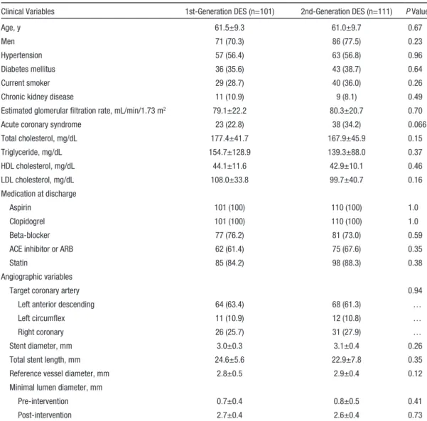
Table 1. Baseline Characteristics
Clinical Variables 1st-Generation DES (n=101) 2nd-Generation DES (n=111) P Value
Age, y 61.5±9.3 61.0±9.7 0.67
Men 71 (70.3) 86 (77.5) 0.23
Hypertension 57 (56.4) 63 (56.8) 0.96
Diabetes mellitus 36 (35.6) 43 (38.7) 0.64
Current smoker 29 (28.7) 40 (36.0) 0.26
Chronic kidney disease 11 (10.9) 9 (8.1) 0.49
Estimated glomerular filtration rate, mL/min/1.73 m2 79.1±22.2 80.3±20.7 0.70
Acute coronary syndrome 23 (22.8) 38 (34.2) 0.066
Total cholesterol, mg/dL 177.4±41.7 167.9±45.9 0.15
Triglyceride, mg/dL 154.7±128.9 139.3±88.0 0.37
HDL cholesterol, mg/dL 44.1±11.6 42.9±10.1 0.46
LDL cholesterol, mg/dL 108.0±33.8 99.7±40.7 0.16
Medication at discharge
Aspirin 101 (100) 110 (100) 1.0
Clopidogrel 101 (100) 110 (100) 1.0
Beta-blocker 77 (76.2) 81 (73.0) 0.59
ACE inhibitor or ARB 62 (61.4) 75 (67.6) 0.35
Statin 85 (84.2) 98 (88.3) 0.38
Angiographic variables
Target coronary artery 0.94
Left anterior descending 64 (63.4) 68 (61.3) …
Left circumflex 11 (10.9) 12 (10.8) …
Right coronary 26 (25.7) 31 (27.9) …
Stent diameter, mm 3.0±0.3 3.1±0.4 0.26
Total stent length, mm 24.6±5.6 22.9±7.8 0.35
Reference vessel diameter, mm 2.8±0.5 2.9±0.4 0.12
Minimal lumen diameter, mm
Pre-intervention 0.7±0.4 0.8±0.5 0.41
Post-intervention 2.7±0.4 2.6±0.4 0.73
Data are presented as mean±SD or n (%). ACE indicates angiotensin converting enzyme; ARB, angiotensin receptor blocker; DES,
lipid-laden neointima, thin-cap fibroatheroma was defined as a fi- brous cap thickness ≤65 µm at the thinnest part and an angle of the lipid ≥180°.16 Neointimal rupture was a break in the fibrous cap that connected the lumen with the underlying lipid pool.5 Calcification inside the neointima was defined as a well-delineated, signal-poor region with sharp borders.16 The criteria for the diagnosis of neoath- erosclerosis were lesions with lipid-laden neointima, neointima with calcification, a thin-cap fibroatheroma-like neointima, or neointimal rupture.4,5 Intraluminal material was defined as visible material inside the lumen.16 The inter- and intraobserver agreement for the evaluation of neointimal tissue in this core laboratory was previously reported.17 Representative images of neoatherosclerosis are shown in Figure 1.
Statistical Analysis
Statistical analysis was performed using SPSS (version 18.0.0, SPSS Inc, Chicago, IL). Data are expressed as number (%), mean±standard
deviation, or median (interquartile range). Comparisons of categori- cal data were made using χ2 test or the Fisher exact test. Continuous variables were analyzed with Student’s t test or Mann–Whitney U test. Multivariable logistic regression model was applied to determine an independent predictor for neoatherosclerosis. Variables with a P value <0.1 resulting from univariate analysis or traditional cardiovas- cular risk factors were entered into the multivariable analysis. A P value <0.05 was considered statistically significant.
Results
There were no statistically significant differences in base- line clinical and angiographic characteristics in patients who received a first- or second-generation DES (Table 1). Table 2 shows clinical, angiographic, and OCT characteristics between the 2 groups at the time of follow-up OCT. Compared with
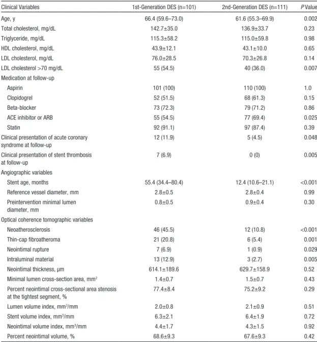
Table 2. Follow-Up Characteristics
Clinical Variables 1st-Generation DES (n=101) 2nd-Generation DES (n=111) P Value
Age, y 66.4 (59.6–73.0) 61.6 (55.3–69.9) 0.002
Total cholesterol, mg/dL 142.7±35.0 136.9±33.7 0.23
Triglyceride, mg/dL 115.3±58.2 115.0±59.8 0.98
HDL cholesterol, mg/dL 43.9±12.1 43.1±10.0 0.65
LDL cholesterol, mg/dL 76.0±28.5 70.3±26.8 0.14
LDL cholesterol >70 mg/dL 55 (54.5) 40 (36.0) 0.007
Medication at follow-up
Aspirin 101 (100) 110 (100) 1.0
Clopidogrel 52 (51.5) 68 (61.3) 0.15
Beta-blocker 73 (72.3) 79 (71.2) 0.86
ACE inhibitor or ARB 55 (54.5) 77 (69.4) 0.025
Statin 92 (91.1) 97 (87.4) 0.39
Clinical presentation of acute coronary syndrome at follow-up
12 (11.9) 5 (4.5) 0.048
Clinical presentation of stent thrombosis at follow-up
7 (6.9) 0 (0) 0.005
Angiographic variables
Stent age, months 55.4 (34.4–80.4) 12.4 (10.6–21.1) <0.001
Reference vessel diameter, mm 2.8±0.5 2.8±0.4 0.99
Preintervention minimal lumen diameter, mm
0.8±0.5 0.9±0.4 0.30
Optical coherence tomographic variables
Neoatherosclerosis 46 (45.5) 12 (10.8) <0.001
Thin-cap fibroatheroma 21 (20.8) 6 (5.4) 0.001
Neointimal rupture 7 (6.9) 1 (0.9) 0.029
Intraluminal material 13 (12.9) 3 (2.7) 0.005
Neointimal thickness, µm 614.1±189.6 629.7±158.9 0.52
Minimal lumen cross-section area, mm2 1.4±0.7 1.5±0.7 0.43
Percent neointimal cross-sectional area stenosis at the tightest segment, %
77.4±8.4 75.2±9.2 0.29
Lumen volume index, mm3/mm 2.0±0.8 2.1±0.9 0.51
Stent volume index, mm3/mm 6.3±2.1 6.4±1.9 0.72
Neointimal volume index, mm3/mm 4.4±1.7 4.3±1.5 0.92
Percent neointimal volume, % 68.6±9.3 67.6±9.3 0.42
Data are presented as mean±SD, median (interquartile range) or n (%). ACE indicates angiotensin converting enzyme; ARB, angiotensin receptor blocker; DES, drug-eluting stent; HDL, high-density lipoprotein; and LDL, low-density lipoprotein.
first-generation DES-treated patients, those treated with a second-generation DES showed a lower frequency of acute myocardial infarction (11.9% versus 4.5%, respectively;
P=0.048) and stent thrombosis (6.9% versus 0%, respectively;
P=0.005). LDL cholesterol levels >70 mg/dL were less fre-
quently observed in second-generation DES-treated patients (36.0% versus 55.4%; P=0.007). Angiotensin-converting enzyme inhibitor/angiotensin receptor blockers were more fre- quently used in patients treated with second-generation DES (69.4% versus 54.5% in first-generation DES; P=0.025). OCT- derived neoatherosclerosis was identified in 27.4% (58/212) of all DES-treated lesions. Neoatherosclerosis was less common in patients treated with second-generation DES (10.8% versus 45.5% in first-generation DES; P<0.001). However, the stent age of the second-generation DES was significantly shorter [12.4 (10.6–21.1) months versus 55.4 (34.4–80.4) months;
P<0.001] and may in part contribute to this observed differ-
ence. The frequency of neoatherosclerosis among the zotaro- limus-, everolimus-, and biolimus-eluting second-generation DESs was similar (10.0% versus 13.9% versus 8.6%, respec- tively; P=0.80). The frequency of neoatherosclerosis increased with the follow-up period (Figure 2). Neoatherosclerosis was found in 1.6% (1/64) of the lesions under 1 year. On the con- trary, neoatherosclerosis was observed in 73.9% (17/23) of the lesions over 7 years. Among the 189 lesions treated by repeat target-lesion revascularization, 8 were treated with a plain bal- loon, 140 were treated with a drug-eluting balloon, and 41 were treated with a DES.
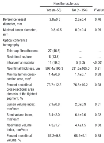
Table 3 shows the baseline and follow-up clinical, angio- graphic, and OCT characteristics of patients with and with- out neoatherosclerosis. At the index procedure, patients with neoatherosclerosis had a higher incidence of both chronic kidney disease and use of first-generation DES and a lower rate of statin treatment. At the follow-up OCT, patients with neoatherosclerosis presented with a higher rate of acute coro- nary syndrome, stent thrombosis, and an LDL cholesterol level >70 mg/dL. These patients also had a lower rate of statin
treatment. The duration of stent age was significantly longer in patients with neoatherosclerosis (66.1 [45.1–87.8] months versus 12.9 [10.5–33.8] months, P<0.001).
Predictors for Neoatherosclerosis
Table 4 lists the predictors for neoatherosclerosis after DES implantation. In univariate logistic regression analy- sis, chronic kidney disease, LDL cholesterol >70 mg/dL at follow-up, usage of first-generation DES, and stent age were significantly associated with neoatherosclerosis. In multivari- able analysis, chronic kidney disease (odds ratio [OR], 4.113;
95% confidence interval [CI], 1.086–15.575; P=0.037), LDL cholesterol >70 mg/dL at follow-up (OR, 2.532; 95% CI, 1.054–6.084; P=0.038), and stent age (OR, 1.710; 95% CI, 1.403–2.084; P<0.001) were independent factors for neoath- erosclerosis. However, compared with first-generation DESs, second-generation DESs were not associated with neoath- erosclerosis after adjusting for cardiovascular risk factors.
Notably, no individual subtype of second-generation DES was associated with neoatherosclerosis (OR, 0.384; 95% CI, 0.092–1.600; P=0.19 in zotarolimus-; OR, 0.844; 95% CI, 0.221–3.221; P=0.80 in everolimus-; and OR, 0.475; 95% CI, 0.097–2.341; P=0.36 in biolimus-eluting stent).
Discussion
This study shows that neoatherosclerosis was observed in about a quarter of all DES-treated lesions with >50% percent neointimal CSA stenosis. The incidence of neoatherosclero- sis increases with stent age. The clinical presentation of neo- atherosclerosis was significantly associated with the onset of acute coronary syndrome or stent thrombosis at follow-up.
Although neoatherosclerosis was less frequently detected in second-generation DES in univariate analysis, multivariable analysis revealed that chronic kidney disease, an LDL choles- terol level of >70 mg/dL at follow-up, and stent age were each an independent predictor for neoatherosclerosis. DES type
Figure 2. The prevalence of neoatherosclerosis between first- and second-generation drug-eluting stent (DES) is shown according to stent age.
(first- versus second-generation) was not a significant predic- tor for neoatherosclerosis.
DES and Neoatherosclerosis
Previous studies have shown that neoatherosclerosis was detected earlier in DES-treated lesions compared with bare- metal stent-treated lesions.
2–7In the present era where the use of the DES is so common, this observation may raise the con- cern that atherosclerotic changes inside DES-neointima may facilitate the sudden onset of an adverse cardiac event (ie, stent thrombosis) during the extended follow-up period. Compared with the first-generation DES, the second-generation DES has significantly improved, in many regards. They are coated with new antiproliferative drugs, constructed with a biodegrad- able, and designed in a thin stent strut. These properties were associated with better strut coverage of second-generation DES
18and anticipated the enhanced property regarding in- stent neoatherosclerosis. However, most of published OCT or autopsy studies did not compare the in-stent neoatheroscle- rosis between first- and second-generation DES because of smaller number of second-generation DES-treated lesions.
2–7A recent autopsy study revealed the superiority of cobalt- chromium everolimus-eluting stent over first-generation DES in terms of vascular inflammation, fibrin deposition, and stent thrombosis.
18Nevertheless, the frequency of neoatheroscle- rosis was similar between 2 devices.
18This is consistent with
Table 3. Baseline and Follow-Up Characteristics According tothe Presence of Neoatherosclerosis
Neoatherosclerosis
P Value Yes (n=58) No (n=154)
Baseline characteristics
Age, y 61.7±10.1 61.1±9.3 0.70
Male 43 (74.1) 114 (74.0) 0.99
Hypertension 35 (60.3) 85 (55.2) 0.50
Diabetes mellitus 20 (34.5) 59 (38.3) 0.61 Current smoker 15 (25.9) 49 (31.8) 0.40 Chronic kidney disease 11 (19.0) 9 (5.8) 0.004 Estimated glomerular
filtration rate, mL/min/1.73 m2
72.7±27.4 80.5±22.5 0.036
Acute coronary syndrome 20 (34.5) 41 (26.6) 0.26 Total cholesterol, mg/dL 175.3±38.5 171.2±46.1 0.59 Triglyceride, mg/dL 165.7±158.1 139.5±84.2 0.17 HDL cholesterol, mg/dL 44.3±12.0 43.1±10.4 0.53 LDL cholesterol, mg/dL 111.1±36.8 100.8±37.8 0.12 Medication at discharge
Beta-blocker 45 (77.6) 113 (73.4) 0.53
ACE inhibitor or ARB 35 (60.3) 102 (66.2) 0.42
Statin 46 (79.3) 137 (89.0) 0.068
Target coronary artery 0.70
Left anterior descending 35 (60.3) 97 (63.0) …
Left circumflex 8 (13.8) 15 (9.7) …
Right coronary 15 (25.9) 42 (27.3) …
Stent type <0.001
First-generation DES 46 (79.3) 55 (35.7) … Second-generation DES 12 (20.7) 99 (64.3) … Reference vessel diameter,
mm
2.9±0.4 2.9±0.4 0.50
Minimal lumen diameter, mm
Preintervention 0.8±0.4 0.7±0.5 0.63
Postintervention 2.7±0.4 2.6±0.4 0.17
Follow-up characteristics
Age, y 69.5 (60.7–74.4) 62.5 (56.0–70.9) 0.003 Clinical presentation of
acute coronary syndrome at follow-up
11 (19.0) 6 (3.9) 0.001
Clinical presentation of stent thrombosis at follow-up
7 (12.1) 0 (0) <0.001
Total cholesterol, mg/dL 147.0±36.5 136.9±33.2 0.064 Triglyceride, mg/dL 123.2±57.9 111.2±59.2 0.29 HDL cholesterol, mg/dL 43.2±12.4 43.6±10.5 0.82 LDL cholesterol, mg/dL 79.6±29.7 70.5±26.6 0.038 LDL cholesterol >70 mg/dL 38 (65.5) 57 (37.0) <0.001 Medication at follow-up
Beta-blocker 41 (70.7) 111 (72.1) 0.84
ACE inhibitor or ARB 32 (55.2) 100 (64.9) 0.19
Statin 48 (82.8) 141 (91.6) 0.066
Stent age, months 66.1 (45.1–87.8) 12.9 (10.5–33.8) <0.001 (Continued)
Reference vessel diameter, mm
2.8±0.5 2.8±0.4 0.76
Minimal lumen diameter, mm
0.8±0.5 0.9±0.4 0.29
Optical coherence tomography
Thin-cap fibroatheroma 27 (46.6) … …
Neointimal rupture 8 (13.8) … …
Intraluminal material 11 (19.0) 5 (3.2) <0.001 Neointimal thickness, µm 597.4±195.3 631.5±165.0 0.21 Minimal lumen cross-
section area, mm2
1.4±0.6 1.4±0.7 0.88
Percent neointimal cross-sectional area stenosis at the tightest segment, %
73.7±12.3 76.8±10.2 0.28
Lumen volume index, mm3/mm
2.1±0.8 2.0±0.9 0.61
Stent volume index, mm3/mm
6.4±2.0 6.4±2.0 0.92
Neointimal volume index, mm3/mm
4.3±1.7 4.4±1.5 0.88
Percent neointimal volume, %
67.2±9.8 68.4±9.1 0.38
Data are presented as mean±SD, median (interquartile range) or n (%). ACE indicates angiotensin converting enzyme; ARB, angiotensin receptor blocker; DES, drug-eluting stent; HDL, high-density lipoprotein; and LDL, low-density lipoprotein.
Table 3. Continued
Neoatherosclerosis
P Value Yes (n=58) No (n=154)
the results in the current study. The current study investigated the largest number of second-generation DES-treated lesions compared with the previous studies
2–7and also directly com- pared second- with first-generation DES. A preclinical study reported that cobalt-chromium everolimus-eluting stent shows greater expression of platelet endothelial cell adhesion mole- cule-1 versus first-generation DES.
19However, compared with bare-metal stent, endothelial maturation without impairment of endothelial barrier function is still insufficient in cobalt- chromium everolimus-eluting stent, as well as first-genera- tion DES.
18,19Although the second-generation DES failed to improve in-stent neoatherosclerosis compared with the first- generation DES, a large registry data showed the superiority of second-generation DES regarding stent thrombosis.
10,11This phenomenon may be partly explained by the finding that the incompetent endothelization can cause both late stent throm- bosis and in-stent neoatherosclerosis.
20,21Although improved strut coverage of second-generation DES may reduce the occurrence of late stent thrombosis by poor strut coverage, it seems still insufficient for second-generation DES to inhibit the development of neoatherosclerosis.
Predictors for Neoatherosclerosis
Previous studies suggested possible predictors for in-stent neo- atherosclerosis.
2,4–7According to pathological study by Naka- zawa et al, younger age, longer implant durations, usage of first-generation DES (compared with bare-metal stent), and underlying unstable plaques were independent determinants for neoatherosclerosis.
2Using OCT, Yonetsu et al suggested that
≥48 months of stent age, all subtypes of DES (compared with bare-metal stent), current smoking, chronic kidney disease, and use of angiotensin-converting enzyme inhibitors/angiotensin receptor blocker were risk factors.
4Ali and colleagues reported that baseline serum creatinine and high-density lipoprotein cholesterol tended to be associated with neoatherosclerosis in
factors, LDL cholesterol >70 mg/dL at follow-up and chronic kidney disease, are related to neoatherosclerosis.
The association between renal dysfunction and in-stent neo- atherosclerosis was founded by previous studies.
4,6The present study supports these data and additionally shows this association is relevant in a relatively large burden of neointima as well. As possible explanations, previous reports have suggested that oxi- dative stress and inflammation might mediate the observed high frequency of cardiovascular disease in patients with chronic kidney disease.
22,23These systemic responses may also lead to the atherosclerotic changes observed inside neointima over the course of an extended follow-up period. Neoatherosclerosis may be associated with incomplete or delayed endothelization of the DES.
19,21,24Because the endothelium generally acts as a barrier against the excessive uptake of circulating lipids,
25pro- longed exposure to even a modest level of LDL cholesterol may potentially lead to the accumulation of lipids inside the neo- intima with incompetent endothelium.
26Higher levels of LDL cholesterol have been regarded as an important risk factor for the development of atherosclerosis in de novo coronary lesions.
This is the first study to report that there may be a significant relationship between neoatherosclerosis and higher levels of LDL cholesterol at follow-up even in DES-treated lesions.
Study Limitations
Because this is a retrospective registry study, only patients with OCT evaluation at follow-up were included in the present study.
The use of particular type of DES was at the discretion of the physician. The indications for follow-up OCT were different from myocardial ischemia to planned follow-up angiography.
Thus, selection bias may have affected the results. Unmeasured confounders (ie, missing of cardiovascular events or death in the interim before an OCT follow-up) can exit and may influence the results of present study. Although the detection of lipid-rich plaque using OCT has been validated by histopathology studies,
Table 4. Predictors of NeoatherosclerosisUnivariate Analysis Multivariable Analysis
Odds Ratio 95% CI P Value Odds Ratio 95% CI P Value
Baseline characteristics
Age, y 1.006 0.975–1.039 0.70 1.017 0.969–1.067 0.50
Male 1.006 0.505–2.004 0.99 1.077 0.393–2.947 0.89
Hypertension 1.235 0.668–2.284 0.50 1.088 0.446–2.658 0.85
Diabetes mellitus 1.180 0.628–2.219 0.61 1.004 0.426–2.365 0.99
Chronic kidney disease 3.771 1.472–9.656 0.006 4.113 1.086–15.575 0.037
Usage of second- generation DES
0.145 0.071–0.296 <0.001 0.538 0.196–1.481 0.23
Follow-up characteristics
LDL cholesterol >70 mg/dL 3.233 1.718–6.087 <0.001 2.532 1.054–6.084 0.038
Medication at follow-up
ACE inhibitor or ARB 0.665 0.360–1.228 0.19 1.581 0.635–3.937 0.33
Statin 0.443 0.182–1.075 0.072 0.502 0.141–1.784 0.29
Stent age, y 1.769 1.506–2.078 <0.001 1.710 1.403–2.084 <0.001
ACE indicates angiotensin converting enzyme; ARB, angiotensin receptor blocker; CI, confidence interval; DES, drug-eluting stent; and LDL, low-density lipoprotein.
consensus among publications as to the OCT criteria for neoath- erosclerosis, and this might affect the incidence of this phenome- non. The time interval from DES implantation to follow-up, OCT was different between first- and second-generation DES; differ- ent stent age (longer time interval in first-generation DES versus shorter time interval in second-generation DES) may affect the statistical analysis in multivariable logistic regression analysis even after control for stent age. Because stent type is unavoidably confounded by stent age, there might be no statistical significance of stent type in multivariable logistic regression analysis. In addi- tion, the statistical analysis may have limited ability to control for all necessary confounders and to detect an OR as low as 0.05 because small size of study populations. Therefore, further stud- ies with larger number of DES-treated patients and comparable stent age are required. The study patients in our prior publication
5were included in the analysis of this study.
Conclusions
Neoatherosclerosis accounts for about a quarter of DES-treated lesions. Using multivariable logistic regression analysis, the presence of chronic kidney disease, LDL cholesterol of >70 mg/dL at follow-up, and stent age are independently associ- ated with neoatherosclerosis. The use of first- or second-gen- eration DES held no predictive value of neoatherosclerosis.
These findings suggest that the continuous protection against cardiovascular risks may suppress neoatherosclerotic changes and subsequently avoid the clinical deterioration related to neoatherosclerosis during the extended follow-up period after DES implantation.
Sources of Funding
This study was supported by a grant from the Korea Healthcare Technology R&D Project, Ministry for Health, Welfare and Family Affairs, Republic of Korea (Nos. A085012 and A102064), and a grant from the Korea Health 21 R&D Project, Ministry of Health and Welfare, Republic of Korea (No. A085136), and the Cardiovascular Research Center, Seoul, Korea.
Disclosures
None.
References
1. Chieffo A, Foglieni C, Nodari RL, Briguori C, Sangiorgi G, Latib A, Montorfano M, Airoldi F, Michev I, Carlino M, Colombo A, Maseri A.
Histopathology of clinical coronary restenosis in drug-eluting versus bare metal stents. Am J Cardiol. 2009;104:1660–1667. doi: 10.1016/j.
amjcard.2009.07.041.
2. Nakazawa G, Otsuka F, Nakano M, Vorpahl M, Yazdani SK, Ladich E, Kolodgie FD, Finn AV, Virmani R. The pathology of neoatherosclerosis in human coronary implants bare-metal and drug-eluting stents. J Am Coll Cardiol. 2011;57:1314–1322. doi: 10.1016/j.jacc.2011.01.011.
3. Yonetsu T, Kim JS, Kato K, Kim SJ, Xing L, Yeh RW, Sakhuja R, McNulty I, Lee H, Zhang S, Uemura S, Yu B, Kakuta T, Jang IK. Comparison of incidence and time course of neoatherosclerosis between bare metal stents and drug-eluting stents using optical coherence tomography. Am J Cardiol. 2012;110:933–939. doi: 10.1016/j.amjcard.2012.05.027.
4. Yonetsu T, Kato K, Kim SJ, Xing L, Jia H, McNulty I, Lee H, Zhang S, Uemura S, Jang Y, Kang SJ, Park SJ, Lee S, Yu B, Kakuta T, Jang IK.
Predictors for neoatherosclerosis: a retrospective observational study from the optical coherence tomography registry. Circ Cardiovasc Imaging.
2012;5:660–666. doi: 10.1161/CIRCIMAGING.112.976167.
5. Lee SY, Shin DH, Mintz GS, Kim JS, Kim BK, Ko YG, Choi D, Jang Y, Hong MK. Optical coherence tomography-based evaluation of in- stent neoatherosclerosis in lesions with more than 50% neointimal
cross-sectional area stenosis. EuroIntervention. 2013;9:945–951. doi:
10.4244/EIJV9I8A158.
6. Ali ZA, Roleder T, Narula J, Mohanty BD, Baber U, Kovacic JC, Mintz GS, Otsuka F, Pan S, Virmani R, Sharma SK, Moreno P, Kini AS.
Increased thin-cap neoatheroma and periprocedural myocardial infarction in drug-eluting stent restenosis: multimodality intravascular imaging of drug-eluting and bare-metal stents. Circ Cardiovasc Interv. 2013;6:507–
517. doi: 10.1161/CIRCINTERVENTIONS.112.000248.
7. Vergallo R, Yonetsu T, Uemura S, Park SJ, Lee S, Kato K, Jia H, Abtahian F, Tian J, Hu S, Lee H, McNulty I, Prasad A, Yu B, Zhang S, Porto I, Biasucci LM, Crea F, Jang IK. Correlation between degree of neointimal hyperplasia and incidence and characteristics of neoatherosclerosis as as- sessed by optical coherence tomography. Am J Cardiol. 2013;112:1315–
1321. doi: 10.1016/j.amjcard.2013.05.076.
8. Kang SJ, Mintz GS, Akasaka T, Park DW, Lee JY, Kim WJ, Lee SW, Kim YH, Whan Lee C, Park SW, Park SJ. Optical coherence tomo- graphic analysis of in-stent neoatherosclerosis after drug-eluting stent implantation. Circulation. 2011;123:2954–2963. doi: 10.1161/
CIRCULATIONAHA.110.988436.
9. Park SJ, Kang SJ, Virmani R, Nakano M, Ueda Y. In-stent neoatheroscle- rosis: a final common pathway of late stent failure. J Am Coll Cardiol.
2012;59:2051–2057. doi: 10.1016/j.jacc.2011.10.909.
10. Sarno G, Lagerqvist B, Fröbert O, Nilsson J, Olivecrona G, Omerovic E, Saleh N, Venetzanos D, James S. Lower risk of stent thrombosis and restenosis with unrestricted use of ‘new-generation’ drug-eluting stents:
a report from the nationwide Swedish Coronary Angiography and Angioplasty Registry (SCAAR). Eur Heart J. 2012;33:606–613. doi:
10.1093/eurheartj/ehr479.
11. Tada T, Byrne RA, Simunovic I, King LA, Cassese S, Joner M, Fusaro M, Schneider S, Schulz S, Ibrahim T, Ott I, Massberg S, Laugwitz KL, Kastrati A. Risk of stent thrombosis among bare-metal stents, first-genera- tion drug-eluting stents, and second-generation drug-eluting stents: results from a registry of 18,334 patients. JACC Cardiovasc Interv. 2013;6:1267–
1274. doi: 10.1016/j.jcin.2013.06.015.
12. Kim U, Kim JS, Kim JS, Lee JM, Son JW, Kim J, Ko YG, Choi D, Jang Y.
The initial extent of malapposition in ST-elevation myocardial infarction treated with drug-eluting stent: the usefulness of optical coherence tomog- raphy. Yonsei Med J. 2010;51:332–338. doi: 10.3349/ymj.2010.51.3.332.
13. Cutlip DE, Windecker S, Mehran R, Boam A, Cohen DJ, van Es GA, Steg PG, Morel MA, Mauri L, Vranckx P, McFadden E, Lansky A, Hamon M, Krucoff MW, Serruys PW; Academic Research Consortium.
Clinical end points in coronary stent trials: a case for standard- ized definitions. Circulation. 2007;115:2344–2351. doi: 10.1161/
CIRCULATIONAHA.106.685313.
14. Levey AS, Stevens LA, Schmid CH, Zhang YL, Castro AF, III, Feldman HI, Kusek JW, Eggers P, Van Lente F, Greene T, Coresh J; CKD-EPI (Chronic Kidney Disease Epidemiology Collaboration). A new equation to estimate glomerular filtration rate. Ann Intern Med. 2009;150:604–612.
15. National Kidney Foundation. K/DOQI clinical practice guidelines for chronic kidney disease: evaluation, classification, and stratification. Am J Kidney Dis. 2002;39:S1–S266.
16. Tearney GJ, Regar E, Akasaka T, Adriaenssens T, Barlis P, Bezerra HG, Bouma B, Bruining N, Cho JM, Chowdhary S, Costa MA, de Silva R, Dijkstra J, Di Mario C, Dudek D, Dudeck D, Falk E, Falk E, Feldman MD, Fitzgerald P, Garcia-Garcia HM, Garcia H, Gonzalo N, Granada JF, Guagliumi G, Holm NR, Honda Y, Ikeno F, Kawasaki M, Kochman J, Koltowski L, Kubo T, Kume T, Kyono H, Lam CC, Lamouche G, Lee DP, Leon MB, Maehara A, Manfrini O, Mintz GS, Mizuno K, Morel MA, Nadkarni S, Okura H, Otake H, Pietrasik A, Prati F, Räber L, Radu MD, Rieber J, Riga M, Rollins A, Rosenberg M, Sirbu V, Serruys PW, Shimada K, Shinke T, Shite J, Siegel E, Sonoda S, Sonada S, Suter M, Takarada S, Tanaka A, Terashima M, Thim T, Troels T, Uemura S, Ughi GJ, van Beusekom HM, van der Steen AF, van Es GA, van Es GA, van Soest G, Virmani R, Waxman S, Weissman NJ, Weisz G; International Working Group for Intravascular Optical Coherence Tomography (IWG- IVOCT). Consensus standards for acquisition, measurement, and report- ing of intravascular optical coherence tomography studies: a report from the International Working Group for Intravascular Optical Coherence Tomography Standardization and Validation. J Am Coll Cardiol.
2012;59:1058–1072. doi: 10.1016/j.jacc.2011.09.079.
17. Lee SJ, Kim BK, Kim JS, Ko YG, Choi D, Jang Y, Hong MK. Evaluation of neointimal morphology of lesions with or without in-stent restenosis:
an optical coherence tomography study. Clin Cardiol. 2011;34:633–639.
doi: 10.1002/clc.20960.
18. Otsuka F, Vorpahl M, Nakano M, Foerst J, Newell JB, Sakakura K, Kutys R, Ladich E, Finn AV, Kolodgie FD, Virmani R. Pathology of second- generation everolimus-eluting stents versus first-generation sirolimus- and paclitaxel-eluting stents in humans. Circulation. 2014;129:211–223. doi:
10.1161/CIRCULATIONAHA.113.001790.
19. Joner M, Nakazawa G, Finn AV, Quee SC, Coleman L, Acampado E, Wilson PS, Skorija K, Cheng Q, Xu X, Gold HK, Kolodgie FD, Virmani R.
Endothelial cell recovery between comparator polymer-based drug-eluting stents. J Am Coll Cardiol. 2008;52:333–342. doi: 10.1016/j.jacc.2008.04.030.
20. Finn AV, Joner M, Nakazawa G, Kolodgie F, Newell J, John MC, Gold HK, Virmani R. Pathological correlates of late drug-eluting stent throm- bosis: strut coverage as a marker of endothelialization. Circulation.
2007;115:2435–2441. doi: 10.1161/CIRCULATIONAHA.107.693739.
21. Otsuka F, Finn AV, Yazdani SK, Nakano M, Kolodgie FD, Virmani R. The importance of the endothelium in atherothrombosis and coronary stenting.
Nat Rev Cardiol. 2012;9:439–453. doi: 10.1038/nrcardio.2012.64.
22. Himmelfarb J, Stenvinkel P, Ikizler TA, Hakim RM. The el- ephant in uremia: oxidant stress as a unifying concept of
cardiovascular disease in uremia. Kidney Int. 2002;62:1524–1538. doi:
10.1046/j.1523-1755.2002.00600.x.
23. Arici M, Walls J. End-stage renal disease, atherosclerosis, and cardio- vascular mortality: is C-reactive protein the missing link? Kidney Int.
2001;59:407–414. doi: 10.1046/j.1523-1755.2001.059002407.x.
24. Joner M, Finn AV, Farb A, Mont EK, Kolodgie FD, Ladich E, Kutys R, Skorija K, Gold HK, Virmani R. Pathology of drug-eluting stents in humans: delayed healing and late thrombotic risk. J Am Coll Cardiol.
2006;48:193–202. doi: 10.1016/j.jacc.2006.03.042.
25. Ross R. Atherosclerosis–an inflammatory disease. N Engl J Med.
1999;340:115–126. doi: 10.1056/NEJM199901143400207.
26. Skålén K, Gustafsson M, Rydberg EK, Hultén LM, Wiklund O, Innerarity TL, Borén J. Subendothelial retention of atherogenic lipoproteins in early atherosclerosis. Nature. 2002;417:750–754. doi: 10.1038/nature00804.
27. Nakano M, Vorpahl M, Otsuka F, Taniwaki M, Yazdani SK, Finn AV, Ladich ER, Kolodgie FD, Virmani R. Ex vivo assessment of vascular re- sponse to coronary stents by optical frequency domain imaging. J Am Coll Cardiol Img. 2012;5:71–82.