Diagnostic Value and Prognostic Significance of Pleural C-Reactive Protein in Lung Cancer Patients with Malignant Pleural Effusions
Do-Sim Park,
1* Dong Kim,
2* Ki-Eun Hwang,
2Yu-Ri Hwang,
2Chul Park,
2Chang-Hwan Seol,
2Kyung-Hwa Cho,
2Byoung-Ryun Kim,
3Seong-Hoon Park,
4Eun-Taik Jeong,
2and Hak-Ryul Kim
2Departments of 1Laboratory Medicine, 2Internal Medicine, Institute of Wonkwang Medical Science,
3Departments of Obstetrics & Gynecology, and 4Radiology, Wonkwang University School of Medicine, Iksan, Korea.
Received: April 25, 2012 Revised: May 25, 2012 Accepted: June 1, 2012
Corresponding author: Dr. Hak-Ryul Kim, Department of Internal Medicine, Institute of Wonkwang Medical Science, Wonkwang University School of Medicine, 895 Muwang-ro, Iksan 570-749, Korea.
Tel: 82-63-859-2583, Fax: 82-63-855-2025 E-mail: [email protected]
*Do-Sim Park and Dong Kim contributed equally to this work.
∙ The authors have no financial conflicts of interest.
© Copyright:
Yonsei University College of Medicine 2013 This is an Open Access article distributed under the terms of the Creative Commons Attribution Non- Commercial License (http://creativecommons.org/
licenses/by-nc/3.0) which permits unrestricted non- commercial use, distribution, and reproduction in any medium, provided the original work is properly cited.
Purpose: C-reactive protein (CRP) has been implicated in various inflammatory and advanced malignant states. Increased serum CRP (s-CRP) levels have been shown to be associated with independent prognostic factors for survival in patients with advanced lung cancer. However, only few studies have focused on the role of CRP in pleural effusions. This study aimed to evaluate the diagnostic and prognos- tic value of pleural CRP (p-CRP) in lung cancer patients with malignant pleural ef- fusion (MPE). Materials and Methods: Pleural effusion (PE) samples were col- lected from patients with MPE (68 lung cancers; 12 extrathoracic tumors), and from 68 patients with various benign conditions (31 with pneumonia; 37 with tu- berculosis). Concentrations of p- and s-CRP were measured by enzyme-linked im- munosorbent assay. CRP level in pleural fluid and its association with survival were examined. Results: p-CRP levels correlated with s-CRP levels (r=0.82, p<0.0001).
For the differential diagnosis of MPE and benign PE, the area under the receiver operating characteristic curve was greater for p-CRP (0.86) than for s-CRP (0.77).
High p-CRP expression significantly correlated with shorter overall survival (p=0.006). P-CRP was independent prognostic factor significantly associated with overall survival on multivariated analysis (p=0.0001). The relative risk of death for lung cancer patients with high p-CRP levels was 3.909 (95% confidence interval, 2.000-7.639). Conclusion: P-CRP is superior to s-CRP in determining pleural fluid etiology. Quantitative measurement of p-CRP might be a useful complementary di- agnostic and prognostic test for lung cancer patients with MPE.
Key Words: Pleural CRP, diagnosis, prognosis, lung cancer, pleural effusion
INTRODUCTION
Malignant pleural effusion (MPE) is a common and distressing condition seen at the advanced stage of lung cancer. Approximately 50% of lung cancer patients develop pleural effusions at a later stage of the disease.1 The presence of MPE usually indi- cates the severity of illness and a short survival time.2 In order to improve patient outcomes, advances in the identification of proteins and molecular pathways that af- fect key proliferation and survival mechanisms are needed. However, few cellular
January 2009 and July 2011 from the Department of Pulm- onology, Wonkwang University Hospital. Effusions were classified as transudates or exudates using Light’s criteria,25 and patients with transudates, such as those with heart fail- ure, liver cirrhosis, or nephrotic syndrome, were excluded.
Effusions were obtained from 80 patients with MPE (49 with adenocarcinoma of the lung, 19 with non-adenocarci- noma of the lung, and 12 with other primary sites of malig- nancy), 31 with parapneumonic PE, and 37 with tuberculous PE. According to their final diagnoses, the 148 eligible par- ticipants were classified into 2 groups: MPE (n=80) or be- nign PE (n=68). The determination of the etiology of pleural effusions was based on widely accepted criteria, as previous- ly described.26 The survival time in patients with MPE was measured from the time of diagnosis to the date of death.
This study protocol was approved by the Institutional Re- view Board for Human Studies at the Clinical Research Cen- ter of Wonkwang University Hospital. All of the recruited pa- tients and volunteers provided written informed consent.
Tumor marker assays
Blood and PE samples were collected within 24 h of admis- sion or on a symptomatic day before treatment, and centri- fuged immediately at 4°C. Cell-free supernatants were col- lected as aliquots and stored at -80°C until analysis. CRP was analyzed by routine clinical laboratory test protocols us- ing an automated chemical analyzer (Modular P800; Roche Diagnostics GmbH, Mannheim, Germany).
Statistical analyses
All statistical analyses of differences between MPE and be- nign PE were performed using the Mann-Whitney U test.
Spearman correlations were used to determine the relation- ships between p- and s-CRP. The diagnostic accuracies of p- and s-CRP in discriminating between lung cancer with MPE and benign PE were compared by constructing re- ceiver operating characteristic (ROC) curves. The optimum cut-off point from the ROC analysis was established by se- lecting the value that provides the greatest sum of sensitivity and specificity. Survival analyses were performed using the Kaplan-Meier method, and significant differences in surviv- al rates were compared using the log-rank test. The Cox pro- portional hazard regression model was used to compare the relative influences of different prognostic factors. Data were analyzed using SPSS software version 15 (SPSS Inc., Chicago, IL, USA) and MedCalc version 11.5 (MedCalc software, Mariakerke, Belgium). Statistical significance proteins have been identified whose altered regulation corre-
lates with prognosis in lung cancer patients with MPE.
The conventional cytological examination of pleural flu- ids for differentiating benign pleural effusion (PE) from MPE is of limited diagnostic accuracy.3 Accordingly, a large number of biochemical markers in pleural fluid have been intensively evaluated to improve the diagnosis of MPE.4-7 However, the sensitivity of these tests has so far failed to give acceptable results in almost all these series. It is thus necessary to identify more reliable and easily used biomark- ers that may enhance MPE diagnosis.8 The prognostic value of serum tumor markers has been the subject of a number of publications.9 Although tumor markers can also be mea- sured in a readily available body fluid such as serum or urine, their prognostic potential for survival in MPE samples are essentially unknown.
A causal relationship between inflammation and cancer has been described. Persistent infection induces chronic in- flammation, and inflammatory cells induce DNA damage in proliferating cell, by generating reactive oxygen and ni- trogen species.10 Moreover, it has been amply demonstrated that pro-inflammtory cytokines promote tumor growth and metastasis by altering tumor cell biology and activating stromal cells in the tumor microenvironment.11,12 C-reactive protein (CRP) was discovered in 1930 and is widely used as a sensitive, but nonspecific, marker of systemic inflamma- tion.13,14 Increased serum CRP (s-CRP) levels have been re- ported in many pulmonary disorders, including pneumonia, malignancies, and pulmonary thromboembolism.15,16 How- ever, the role of CRP in pleural effusions is still unclear.17,18 In patients with non-small cell lung cancer (NSCLC), multiple myeloma, renal cell carcinoma, prostate cancer, ovarian cancer, gastrointestinal cancer, and hepatocellular carcino- ma, elevated CRP levels prior to therapy have been shown to have an adverse impact on prognosis.19-24 Surprisingly, however, no studies have analyzed pleural CRP (p-CRP) as a prognostic factor in advanced NSCLC.
In this study, we aimed to investigate the diagnostic and prognostic power of quantitative p-CRP expression, as com- pared to s-CRP, in lung cancer patients with MPE.
MATERIALS AND METHODS
Patient selection
A total of 323 consecutive patients who presented with pleu- ral exudative effusion were enrolled in the study between
cantly lower than those of s-CRP (p<0.0001). Further, p- CRP levels were significantly lower in MPE than in benign PE (p<0.0001).
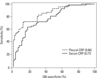
To evaluate whether p- and s-CRP levels could discrimi- nate between MPE and benign PE, cut-off points were de- termined by the maximum sum of sensitivity and specifici- ty. We used cut-off points of 25 mg/L and 82 mg/L for p- and s-CRP, respectively, yielding sensitivity and specificity values of 72% and 83% for p-CRP, and 63% and 73% for s-CRP, respectively. Additionally, p-CRP exhibited elevated diagnostic sensitivity (87%) in lung cancer patients with MPE. The ROC curves of p- and s-CRP for distinguishing lung cancer with MPE from benign PE are shown in Fig. 3.
We found that the AUC of ROC curves (diagnostic accura- cy) of p-CRP (0.86) was superior to that of s-CRP (0.77).
Prognostic implications of p-CRP
We next examined whether the expression status of p-CRP was defined as p<0.05.
RESULTS
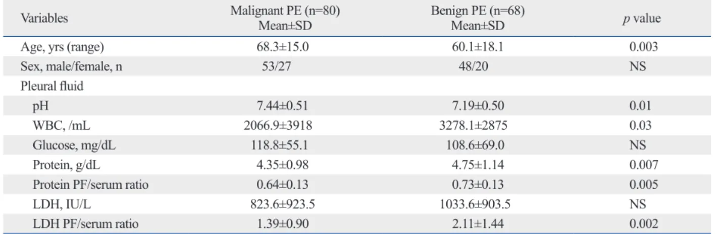
The demographic characteristics of the study subjects are presented in Table 1. There were no significant differences in gender, pleural glucose levels, and pleural lactate dehy- drogenase (LDH) levels between MPE and benign PE pa- tients. However, age and pleural pH were significantly high- er and pleural white blood counts (WBC), protein, protein Light’s ratio and LDH Light’s ratio were significantly lower in MPE patients. Fig. 1 shows significantly positive correla- tion between p- and s-CRP levels (r=0.82, p<0.0001), indi- cating a relationship between p- and s-CRP.
Diagnostic value of p-CRP
As seen in Fig. 2, concentrations of p-CRP were signifi-
Table 1. Demographic and Laboratory Characteristics of the Study Population
Variables Malignant PE (n=80)
Mean±SD Benign PE (n=68)
Mean±SD p value
Age, yrs (range) 68.3±15.0 60.1±18.1 0.003
Sex, male/female, n 53/27 48/20 NS
Pleural fluid
pH 7.44±0.51 7.19±0.50 0.01
WBC, /mL 2066.9±3918 3278.1±2875 0.03
Glucose, mg/dL 118.8±55.1 108.6±69.0 NS
Protein, g/dL 4.35±0.98 4.75±1.14 0.007
Protein PF/serum ratio 0.64±0.13 0.73±0.13 0.005
LDH, IU/L 823.6±923.5 1033.6±903.5 NS
LDH PF/serum ratio 1.39±0.90 2.11±1.44 0.002
PE, pleural effusions; PF, pleural fluid; LDH, lactate dehydrogenase; WBC, white blood counts.
Fig. 1. Correlation between p- and s-CRP in PE; r is the Spearman coeffi- cient of correlation. CRP, C-reactive protein; PE, pleural effusion; p-CRP, pleural CRP; s-CRP, serum CRP.
Fig. 2. Comparison of p-CRP vs. s-CRP levels in MPE and benign PE. CRP, C-reactive protein; MPE, malignant pleural effusion; PE, pleural effusion; p- CRP, pleural CRP; s-CRP, serum CRP.
0 50 100 150 200 250 300
Pleural CRP (mg/L)
0 100 200 300 400 500 600
p<0.0001 r=0.82
Serum CRP (mg/L)
0 20 40 60 80 100 120 140
CRP (mg/L)
Pleural CRP Serum CRP
Malignant Benign p<0.0001
p<0.0001 p<0.0001
ever, the median overall survival was reduced to 2.6 months (95% CI, 1.5-4.7) in patients with high p-CRP expression (Table 2).
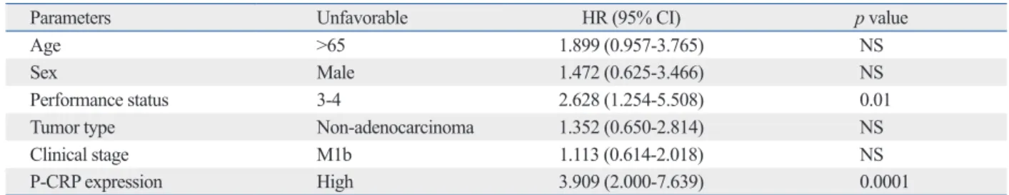
We then conducted multivariate analyses to assess the predictive value of p-CRP expression for overall survival by adjusting other potentially prognostic factors. The re- sults were consistent with unfavorable survival outcome in was correlated with overall survival by using a log-rank sta-
tistic, and found that p-CRP expression was significantly correlated with shorter overall survival (p=0.006). The cu- mulative overall survival curves of patients were significant- ly split by p-CRP expression status (Fig. 4). The median overall survival of patients with low p-CRP expression was 11 months [95% confidence interval (CI), 8.3-13.0]. How-
Table 2. Univariate Analysis for Overall Survival in Lung Cancer with MPE
Parameters No. of patients Overall survival, median (95% CI) p value
Age 0.011
<65 yrs 20 9.7 (7.6-12.9)
>65 yrs 48 5.1 (2.8-7.5)
Sex NS
Female 22 11.3 (5.3-15.0)
Male 46 5.3 (3.1-7.8)
Performance status NS
0-2 47 8.4 (5.7-10.7)
3-4 19 1.7 (1.1-5.1)
Tumor type NS
Adenocarcinoma 49 8.4 (5.4-11.0)
Non-adenocarcinoma 17 5.1 (1.9-5.8)
Clinical stage NS
M1a 26 7.7 (5.4-10.7)
M1b 40 5.1 (1.6-9.3)
S-CRP expression NS
Low 31 11.8 (8.2-13.3)
High 37 3.1 (1.6-5.2)
P-CRP expression 0.006
Low 34 11.0 (8.3-13.0)
High 34 2.6 (1.5-4.7)
MPE, malignant pleural effusion; CRP, C-reactive protein; s-CRP, serum CRP; p-CRP, pleural CRP; CI, confidence interval.
Fig. 3. Comparison of diagnostic accuracies of p-CRP vs. s-CRP in distin- guishing lung cancer with MPE from benign PE, using receiver operating characteristic (ROC) curves. The numbers in parentheses indicate the di- agnostic accuracies (area under the ROC curve). CRP, C-reactive protein;
MPE, malignant pleural effusion; PE, pleural effusion; p-CRP, pleural CRP; s- CRP, serum CRP.
Fig. 4. Overall survival differences between lung cancer with MPE in high and low p-CRP expression, as determined using Kaplan-Meier survival analysis. Patients in the high p-CRP expression group showed reduced overall survival. The p value was obtained from a log-rank test of the differ- ence. CRP, C-reactive protein; MPE, malignant pleural effusion; p-CRP, pleu- ral CRP.
0 0
20 20
40 40
60 60
80 80
100 100
Sensitivity (%) Survival probability (%)
0 20 40 60 80 100 0 5 10 15 20 25 30 35
100-specificity (%) Time (months)
Pleural CRP (0.86) Serum CRP (0.77)
Pleural CRP expression Low pleural CRP High pleural CRP
Log-rank p value=0.006
completely understood. One possible explanation is that, due to cytokine production by tumor tissue, elevated CRP values may indicate a higher tumor burden.30 Scott, et al.31 reported a catabolic effect of acute-phase proteins like CRP on metabolism, and this is associated with an increase in resting energy expenditure and loss of lean tissue in patients with lung cancer, key factors in determining cancer surviv- al. Another reason for elevated CRP may be a cancer-related infection, particularly a post-stenotic pulmonary infection in the case of lung tumors. It is well known that pneumonia may be the first sign that marks lung cancer.32
Multiple risk factors have been identified in advanced NSCLC that are able to discriminate patient groups with a significant difference in survival.33 However, only a few stud- ies have analyzed CRP as a prognostic factor in advanced NSCLC. Moreover, it has been studied in heterogeneous groups of patients without taking treatment into consider- ation.34,35 To our best knowledge, this is the first study ana- lyzing the prognostic value of p-CRP in lung cancer with MPE. In this study, we found by univariate analysis that p- CRP levels ≥25 mg/L and old age were independent predic- tors of survival. Old age, found to be a factor in univariate survival analysis, was no longer significant in multivariate analysis. Both high p-CRP levels and poor performance sta- tus remained associated independently with survival in mul- tivariate analysis. There were no significant differences in WBC and the frequency of bacterial culture growth in PE and sputum samples between high and low p-CRP patients.
These results suggest that infection is not the main stimulus to the increased CRP.
The main limitation of the present study is that it is a ret- rospective study with comparatively small sample size. In spite of relatively high values of statistical significance ob- tained, further research with a larger group of patients is warranted. Further research is also required to identify other sensitive biological markers in PE, in order to define the best combination for marker analysis. Another limitation is that subjects in benign PE were either parapneumonic or tu- patients with high p-CRP expression. In a multivariate Cox
regression analysis, a high p-CRP level was an independent prognostic factor significantly associated with poor survival (p=0.0001). The relative risk of overall survival for patients with high p-CRP was 3.909 (95% CI, 2.000-7.639), regard- less of poor performance status [2.628 (95% CI, 1.254- 5.508)]. The results from the Cox proportional hazards analysis are summarized in Table 3.
DISCUSSION
To the best of our knowledge, this is the first study on pleu- ral effusions in which CRP levels were simultaneously in- vestigated for their diagnostic and prognostic power of lung cancer with MPE. Our findings suggest that p-CRP has a higher diagnostic accuracy than s-CRP for differentiating MPE from benign PE. In multivariate analysis, p-CRP was revealed as a prognostic marker of lung cancer with MPE, together with performance status.
CRP synthesis in hepatocytes is mainly induced by inter- leukin (IL)-6, IL-1, and tumor necrosis factor α. Although several studies have investigated CRP levels in various dis- ease states, measurement of which is relatively inexpensive and easy to quantify in clinical practice, a few studies have focused on its role in patients with pleural effusions.17,27,28 A positive correlation between p- and s-CRP levels was dem- onstrated in the present study, suggesting that p-CRP may result from leakage of s-CRP via inflamed pleura. Further investigation is required to elucidate the exact mechanism.
Botana-Rial, et al.29 reported that the diagnostic accuracies of p- and s-CRP for differentiating MPE from benign PE were 0.752 and 0.667, respectively, as indicated by the area under the ROC curve. In agreement with these results, we demonstrated in the present study that the diagnostic accu- racy of p-CRP for distinguishing lung cancer with MPE from benign PE was 0.86, superior to that of s-CRP (0.77).
The reasons for CRP elevation in cancer patients are not
Table 3. Multivariate Cox Proportional Hazards Analysis for Overall Survival in Lung Cancer with MPE
Parameters Unfavorable HR (95% CI) p value
Age >65 1.899 (0.957-3.765) NS
Sex Male 1.472 (0.625-3.466) NS
Performance status 3-4 2.628 (1.254-5.508) 0.01
Tumor type Non-adenocarcinoma 1.352 (0.650-2.814) NS
Clinical stage M1b 1.113 (0.614-2.018) NS
P-CRP expression High 3.909 (2.000-7.639) 0.0001
HR, hazard ratio; CI, confidence interval; MPE, malignant pleural effusion.
14. Pepys MB, Hirschfield GM. C-reactive protein: a critical update. J Clin Invest 2003;111:1805-12.
15. Korppi M, Heiskanen-Kosma T, Leinonen M. White blood cells, C-reactive protein and erythrocyte sedimentation rate in pneumo- coccal pneumonia in children. Eur Respir J 1997;10:1125-9.
16. Smith RP, Lipworth BJ. C-reactive protein in simple community- acquired pneumonia. Chest 1995;107:1028-31.
17. Castaño Vidriales JL, Amores Antequera C. Use of pleural fluid C-reactive protein in laboratory diagnosis of pleural effusions. Eur J Med 1992;1:201-7.
18. Yilmaz Turay U, Yildirim Z, Türköz Y, Biber C, Erdoğan Y, Keyf AI, et al. Use of pleural fluid C-reactive protein in diagnosis of pleural effusions. Respir Med 2000;94:432-5.
19. Hara M, Matsuzaki Y, Shimuzu T, Tomita M, Ayabe T, Enomoto Y, et al. Preoperative serum C-reactive protein level in non-small cell lung cancer. Anticancer Res 2007;27:3001-4.
20. Pelliniemi TT, Irjala K, Mattila K, Pulkki K, Rajamäki A, Tien- haara A, et al. Immunoreactive interleukin-6 and acute phase pro- teins as prognostic factors in multiple myeloma. Finnish Leuke- mia Group. Blood 1995;85:765-71.
21. Jabs WJ, Busse M, Krüger S, Jocham D, Steinhoff J, Doehn C.
Expression of C-reactive protein by renal cell carcinomas and un- affected surrounding renal tissue. Kidney Int 2005;68:2103-10.
22. Nozoe T, Matsumata T, Kitamura M, Sugimachi K. Significance of preoperative elevation of serum C-reactive protein as an indica- tor for prognosis in colorectal cancer. Am J Surg 1998;176:335-8.
23. Hefler LA, Concin N, Hofstetter G, Marth C, Mustea A, Sehouli J, et al. Serum C-reactive protein as independent prognostic variable in patients with ovarian cancer. Clin Cancer Res 2008;14:710-4.
24. Hashimoto K, Ikeda Y, Korenaga D, Tanoue K, Hamatake M, Ka- wasaki K, et al. The impact of preoperative serum C-reactive pro- tein on the prognosis of patients with hepatocellular carcinoma.
Cancer 2005;103:1856-64.
25. Light RW. Diagnostic principles in pleural disease. Eur Respir J 1997;10:476-81.
26. Papageorgiou E, Kostikas K, Kiropoulos T, Karetsi E, Mpatavanis G, Gourgoulianis KI. Increased oxidative stress in exudative pleu- ral effusions: a new marker for the differentiation between exu- dates and transudates? Chest 2005;128:3291-7.
27. Chierakul N, Kanitsap A, Chaiprasert A, Viriyataveekul R. A sim- ple C-reactive protein measurement for the differentiation between tuberculous and malignant pleural effusion. Respirology 2004;9:
66-9.
28. Kim DY, Lee YS, Ahn S, Chun YH, Lim KS. The usefulness of procalcitonin and C-reactive protein as early diagnostic markers of bacteremia in cancer patients with febrile neutropenia. Cancer Res Treat 2011;43:176-80.
29. Botana-Rial M, Casado-Rey P, Leiro-Fernández V, Andrade-Oliv- ié M, Represas-Represas C, Fernández-Villar A. Validity of pro- calcitonin and C-reactive protein measurement when differentiat- ing between benign and malignant pleural effusion. Clin Lab 2011;57:373-8.
30. Heikkilä K, Ebrahim S, Lawlor DA. A systematic review of the association between circulating concentrations of C reactive pro- tein and cancer. J Epidemiol Community Health 2007;61:824-33.
31. Scott HR, McMillan DC, Forrest LM, Brown DJ, McArdle CS, Milroy R. The systemic inflammatory response, weight loss, per- formance status and survival in patients with inoperable non-small cell lung cancer. Br J Cancer 2002;87:264-7.
32. Søyseth V, Benth JS, Stavem K. The association between hospi-
berculous effusion excluding other causes such as pulmo- nary thromboembolism, connective tissue diseases.
In conclusion, the results of current study indicate that di- agnostic value of p-CRP is superior to that of s-CRP in lung cancer patients with MPE, and that the presence of elevated p-CRP levels predicts poor outcome.
ACKNOWLEDGEMENTS
This study was supported by a grant from Institute of Wonk- wang Medical Science.
REFERENCES
1. Memon A, Zawadzki ZA. Malignant effusions: diagnostic evalua- tion and therapeutic strategy. Curr Probl Cancer 1981;5:1-30.
2. Postmus PE, Brambilla E, Chansky K, Crowley J, Goldstraw P, Patz EF Jr, et al. The IASLC Lung Cancer Staging Project: pro- posals for revision of the M descriptors in the forthcoming (sev- enth) edition of the TNM classification of lung cancer. J Thorac Oncol 2007;2:686-93.
3. Fiegl M, Massoner A, Steurer M, Grünewald K, Krugmann J, Hack R, et al. Improving tumor cell detection in pleural effusions by inter- phase cytogenetics. Cytometry B Clin Cytom 2003;55:60-2.
4. Ferrer J, Villarino MA, Encabo G, Felip E, Bermejo B, Vilà S, et al. Diagnostic utility of CYFRA 21-1, carcinoembryonic antigen, CA 125, neuron specific enolase, and squamous cell antigen level determinations in the serum and pleural fluid of patients with pleu- ral effusions. Cancer 1999;86:1488-95.
5. Miédougé M, Rouzaud P, Salama G, Pujazon MC, Vincent C, Mauduyt MA, et al. Evaluation of seven tumour markers in pleu- ral fluid for the diagnosis of malignant effusions. Br J Cancer 1999;81:1059-65.
6. Gu P, Huang G, Chen Y, Zhu C, Yuan J, Sheng S. Diagnostic utili- ty of pleural fluid carcinoembryonic antigen and CYFRA 21-1 in patients with pleural effusion: a systematic review and meta-anal- ysis. J Clin Lab Anal 2007;21:398-405.
7. Kim HJ, Shin KC, Lee JW, Kim KJ, Hong YH, Chung JH, et al.
TNF-alpha in the pleural fluid for the differential diagnosis of tu- berculous and malignant effusion. Tuberc Respir Dis 2005;59:
625-30.
8. Liang QL, Shi HZ, Qin XJ, Liang XD, Jiang J, Yang HB. Diag- nostic accuracy of tumour markers for malignant pleural effusion:
a meta-analysis. Thorax 2008;63:35-41.
9. Duffy MJ. Role of tumor markers in patients with solid cancers: a critical review. Eur J Intern Med 2007;18:175-84.
10. Coussens LM, Werb Z. Inflammation and cancer. Nature 2002;
420:860-7.
11. Mantovani A, Allavena P, Sica A, Balkwill F. Cancer-related in- flammation. Nature 2008;454:436-44.
12. Chiang AC, Massagué J. Molecular basis of metastasis. N Engl J Med 2008;359:2814-23.
13. Mahmoud FA, Rivera NI. The role of C-reactive protein as a prog- nostic indicator in advanced cancer. Curr Oncol Rep 2002;4:250-5.
temic inflammatory response in patients with inoperable non- small-cell lung cancer. Br J Cancer 2003;89:1028-30.
35. O’Dowd C, McRae LA, McMillan DC, Kirk A, Milroy R. Elevat- ed preoperative C-reactive protein predicts poor cancer specific survival in patients undergoing resection for non-small cell lung cancer. J Thorac Oncol 2010;5:988-92.
talisation for pneumonia and the diagnosis of lung cancer. Lung Cancer 2007;57:152-8.
33. Brundage MD, Davies D, Mackillop WJ. Prognostic factors in non-small cell lung cancer: a decade of progress. Chest 2002;122:
1037-57.
34. Forrest LM, McMillan DC, McArdle CS, Angerson WJ, Dunlop DJ. Evaluation of cumulative prognostic scores based on the sys-