Open Access
Impact of Regional Nodal Irradiation for Breast Cancer Patients with Supraclavicular and/or Internal Mammary Lymph Node Involvement:
A Multicenter, Retrospective Study (KROG 16-14)
Original Article
Purpose
The purpose of this study was to evaluate the treatment outcomes of radiotherapy (RT) for breast cancer with ipsilateral supraclavicular (SCL) and/or internal mammary (IMN) lymph node involvement.
Materials and Methods
A total of 353 patients from 11 institutions were included. One hundred and thirty-six pati- ents had SCL involvement, 148 had IMN involvement, and 69 had both. All patients received neoadjuvant systemic therapy followed by breast-conserving surgery or mastectomy, and postoperative RT to whole breast/chest wall. As for regional lymph node irradiation, SCL RT was given to 344 patients, and IMN RT to 236 patients. The median RT dose was 50.4 Gy.
Results
The median follow-up duration was 61 months (range, 7 to 173 months). In-field progression was present in SCL (n=20) and/or IMN (n=7). The 5-year disease-free survival (DFS) and overall survival rates were 57.8% and 75.1%, respectively. On multivariate analysis, both SCL/IMN involvement, number of axillary lymph node ≥ 4, triple-negative subtype, and mas- tectomy were significant adverse prognosticators for DFS (p=0.022, p=0.001, p=0.001, and p=0.004, respectively). Regarding the impact of regional nodal irradiation, SCL RT dose
≥ 54 Gy was not associated with DFS (5-year rate, 52.9% vs. 50.9%; p=0.696) in SCL- involved patients, and the receipt of IMN RT was not associated with DFS (5-year rate, 56.1%
vs. 78.1%; p=0.099) in IMN-involved patients.
Conclusion
Neoadjuvant chemotherapy followed by surgery and postoperative RT achieved an accept- able in-field regional control rate in patients with SCL and/or IMN involvement. However, a higher RT dose to SCL or IMN RT was not associated with the improved DFS in these pati- ents.
Key words
Breast neoplasms, Supraclavicular lymph node, Internal mammary lymph node, Radiotherapy
Kyubo Kim, MD, PhD
1Yuri Jeong, MD
2Kyung Hwan Shin, MD, PhD
3Jin Ho Kim, MD, PhD
3Seung Do Ahn, MD, PhD
4Su Ssan Kim, MD, PhD
4Chang-Ok Suh, MD, PhD
5Yong Bae Kim, MD, PhD
5Doo Ho Choi, MD, PhD
6Won Park, MD, PhD
6Jihye Cha, MD, PhD
7Mison Chun, MD, PhD
8Dong Soo Lee, MD
9Sun Young Lee, MD, PhD
10Jin Hee Kim, MD, PhD
11Hae Jin Park, MD, PhD
12Wonguen Jung, MD, PhD
1+ + + + + + + + + + + + + + + + + + + + + + + + + + + + + + + + + + + + + + + + + + + + + + + + + + + + + + + + + + + + + + + + + + + + + + + + + + + + + + + + + + + + + + + + + + + + + + + + + + + + + + + + + + + + + + + + + + + + + + + + + + + + + + + + + + + + + + + + + + + + + + + + + + + + + + + + + + + + + + + + + + + + + + + + + + + + + + + + + + + + + + + + + + + + + + + + + + + + + + + + + + + + + + + + + + + + + + + + + + + + + + + + + + + + + + + + + + + + + + + + + + + + + + + + + + + + + + + + + + + + + + + + + + + + + + + + + + + + + + + + + + + + + + + + + + + + + + + + + + + + + + + + + + + + + + + + + + + + + + + + + + + + + + + + + + + + + + + + + + + + + + + + + + + + + + + + + + + + + + + + + + + + + + + + + + + + + + + + + + + + + + + + + + + + + + + + + + + + + + + + + + + + + + + + + + + + + + + + + + + + + + + + + + + + + + + + + + + + + + + + + + + + + + + + + + + + + + + + + + + + + + + + + + + + + + + + + + + + + + + + + + + + + + + + + + + + + + + + + + + +
Correspondence: Kyung Hwan Shin, MD, PhD Department of Radiation Oncology, Seoul National University College of Medicine, 101 Daehak-ro, Jongno-gu, Seoul 03080, Korea Tel: 82-2-2072-2524
Fax: 82-2-765-3317 E-mail: [email protected]
Co-correspondence: Su Ssan Kim, MD, PhD Department of Radiation Oncology, Asan Medical Center, University of Ulsan College of Medicine, 88 Olympic-ro 43-gil, Songpa-gu, Seoul 05505, Korea
Tel: 82-2-3010-5680 Fax: 82-2-3010-6950
E-mail: [email protected]
Received October 20, 2018 Accepted March 13, 2019
*Kyubo Kim and Yuri Jeong contributed equally to this work.
*A list author’s affiliations appears at the end of the paper.
Introduction
Since the recent publication of randomized controlled trials [1,2], elective regional nodal radiotherapy (RT) including supraclavicular lymph nodes (SCL) and internal mammary lymph nodes (IMN) has become the standard treatment in the adjuvant RT for node-positive breast cancer. In the mean- while, neoadjuvant systemic therapy (NST) followed by sur- gery and postoperative RT is a widely accepted treatment for breast cancer with clinically apparent SCL or IMN [3-5]. Ini- tially involved nodal areas are usually included in the RT field, but there are little data on the impact of regional nodal RT in this setting.
Historically, SCL involvement presented as palpable dis- ease, and the IMN involvement was confirmed after surgical excision such as extended radical mastectomy. With the development of imaging technologies such as positron emis- sion tomography–computed tomography (PET-CT), how- ever, extra-axillary regional lymph nodes in locally advanced breast cancer have been increasingly detected [6-8]. Despite the possibility of over-diagnosis, non-palpable SCL and/or clinically detected IMN involvement is becoming more and more common, and more data on the treatment outcome in this setting is needed.
In this study, we evaluated the treatment outcomes of NST followed by surgery and postoperative RT for breast cancer with SCL and/or IMN involvement and investigated the impact of regional nodal RT on the outcomes in these pati- ents.
Materials and Methods
1. Study population
After institutional review board approval, a total of 353 patients with involved SCL and/or IMN but without distant metastases were included from 11 institutions between Jan- uary 2000 and December 2013. SCL and/or IMN metastases were defined as involvement on imaging studies including PET-CT, magnetic resonance imaging (MRI) of the breast, chest CT, and/or breast ultrasonography (US).
Overall, PET-CT was performed in 325 patients (92.1%), MRI in 274 (77.6%), CT in 119 (33.7%), and US in 342 (96.9%).
Among patients with SCL involvement (n=205), SCL was observed in PET-CT (173 of 189), MRI (71 of 141), CT (41 of 76), and US (146 of 198). Meanwhile, IMN was observed in PET-CT (184 of 205), MRI (134 of 187), CT (37 of 69), and US (127 of 213) among 217 patients with IMN involvement.
According to the number of imaging modalities, all four modalities identified SCL in three patients, three modalities in 71, two modalities in 75, and a single modality in 56, while IMN was identified by all four modalities in three patients, three modalities in 95, two modalities in 67, and a single modality in 52. Pathological confirmation was done in 99 patients for SCL and 59 patients for IMN.
2. Treatment
All patients received NST. Chemotherapeutic regimen was adriamycin-based chemotherapy (n=60), taxane-based che- motherapy (n=268), and others (n=18). Seven patients recei- ved hormonal therapy and/or anti–human epidermal grow- th factor receptor 2 (HER2) therapy without cytotoxic che- motherapy.
With regard to surgical treatment for breast, mastectomy was performed in 224 patients, breast-conserving surgery in 127, and no breast surgery in two due to occult breast cancer.
Axillary lymph node dissection was performed in 304 pati- ents and sentinel lymph node biopsy in 49. Fifty-nine pati- ents underwent SCL excision and 14 patients underwent IMN excision.
Postoperative RT was given to breast/chest wall up to a median dose of 50.4 Gy (range, 43.2 to 70 Gy). SCL RT was given in 344 patients, and IMN RT in 236. The median dose to SCL and IMN was 50.4 Gy (range, 45 to 70.4 Gy) and 50.4 Gy (range, 43.2 to 66 Gy), respectively.
One hundred and fifty patients received adjuvant chemo- therapy: adriamycin-based chemotherapy in 22 patients, tax- ane-based chemotherapy in 100, and others in 28. One hund- red and ninety-one patients received hormonal therapy, and 111 patients received anti-HER2 therapy.
3. Statistical analysis
Local recurrences were defined as disease recurrences in the ipsilateral breast or chest wall, and regional recurrences as those in the ipsilateral axillary lymph node, SCL, and/or IMN. Disease recurrences other than local and/or regional recurrences were defined as distant metastases. The time between initial treatment and recurrence or last follow-up was measured as disease-free survival (DFS). Overall sur- vival (OS) was defined from the date of initial treatment to the date of death from any cause or last follow-up. The actu- arial survival rates were calculated by the Kaplan-Meier method and compared using the log-rank test. On multivari- ate analysis, Cox proportional-hazards model was applied.
Factors with p-value < 0.05 on univariate analysis were included in the multivariate analysis. A p < 0.05 was consid- ered statistically significant. Differences in categorical vari- ables were compared using the standard chi-square test or
Fisher exact test. All statistical analyses were performed using SPSS software (release 18.0.1. SPSS Inc., Chicago, IL).
4. Ethical statement
The institutional review board approved this study (appro- val number: H-1612-095-815 at Seoul National University
Hospital), and waived the requirement for obtaining infor- med consent.
Results
1. Characteristics
The patient and tumor characteristics are summarized in Table 1. The median age was 47 years (range, 23 to 84 years).
As for clinical T category, 188 patients had cT1-2, and 161 had cT3-4 disease. One hundred and thirty-six patients had SCL involvement, 148 had IMN involvement, and 69 had both. After NST, pathologic T categories were as follows:
ypT0-is in 66 patients, ypT1-2 in 216, and ypT3-4 in 71. The number of involved axillary lymph nodes was 0 in 103 pati- ents, 1-3 in 98, 4-9 in 90, and ≥ 10 in 62. Regarding the res- ponse of SCL and IMN to NST, each 75.9% of patients show- ed clinical complete remission (CR) on imaging. Hormonal receptor was positive in 53.0% of patients, and HER2 posi- tivity was observed in 37.4% of patients.
2. Outcomes and prognostic factors
The median duration of follow-up was 61 months (range, 7 to 173 months). There were 67 patients with loco-regional recurrences and 140 patients with distant metastases. Among loco-regional recurrences, 20 patients had isolated local recurrences, 23 isolated regional recurrences, and 24 local and regional recurrences. The 5-year DFS and OS were 57.8%
and 75.1%, respectively (Fig. 1).
On univariate analysis, ypT category, extent of SCL and/or IMN involvement, number of involved axillary lymph nodes, histologic grade, lymphovascular invasion, molecular Table 1. Patient and tumor characteristics
Values are presented as median (range) or number (%).
IMN, internal mammary lymph node; SCL, supraclavicu- lar lymph node; HER2, human epidermal growth factor receptor 2; NST, neoadjuvant systemic therapy; CR, com- plete remission.
Variable No. (%) (n=353)
Age (yr) 47 (23-84)
Clinical T category
cT1-2 188 (53.3)
cT3-4 161 (45.6)
cTx 4 (1.1)
Nodal involvement
IMN only 148 (41.9)
SCL only 136 (38.5)
SCL+IMN 69 (19.5)
Pathologic T category
ypT0 47 (13.3)
ypTis 19 (5.4)
ypT1 129 (36.5)
ypT2 87 (24.6)
ypT3-4 71 (20.1)
No. of positive axillary nodes
0 103 (29.2)
1-3 98 (27.8)
4-9 90 (25.5)
≥ 10 62 (17.6)
Hormonal receptor status
Positive 187 (53.0)
Negative 166 (47.0)
HER2 status
Positive 132 (37.4)
Negative 207 (58.6)
Equivocal 14 (4.0)
SCL response to NST
Clinical CR 267 (75.6)
Clinical non-CR 72 (20.4)
Unknown 14 (4.0)
IMN response to NST
Clinical CR 268 (75.9)
Clinical non-CR 66 (18.7)
Unknown 19 (5.4)
Fig. 1. Disease-free and overall survival curves among all the patients (n=353).
Survival rate
1.0
0 0.2 0.4 0.6
0
Time (mo)50 60
10 20 30 40 70 8090100110120 0.8
Disease-free survival rate Overall survival rate
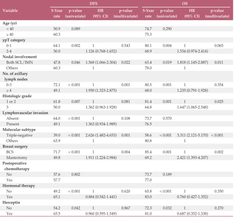
subtype, type of breast surgery, hormonal therapy, and anti- HER2 therapy were significantly associated with DFS. On multivariate analysis, both SCL and IMN involvement (haz- ard ratio [HR], 1.568; 95% confidence interval [CI], 1.066 to 2.304; p=0.022), four or more involved axillary lymph nodes (HR, 1.950; 95% CI, 1.323 to 2.875; p=0.001), triple-negative subtype (HR, 2.626; 95% CI, 1.482 to 4.653; p=0.001), and mas- tectomy (HR, 1.911; 95% CI, 1.224 to 2.984; p=0.004) were
adverse prognostic factors for DFS (Table 2).
With regard to OS, ypT cateogry, extent of SCL and/or IMN involvement, number of involved axillary lymph nodes, histologic grade, molecular subtype, type of breast surgery, hormonal therapy, and anti-HER2 therapy were sig- nificant prognostic factors on univariate analysis. Among these, both SCL and IMN involvement (HR, 1.818; 95% CI, 1.145 to 2.887; p=0.011), histologic grade 3 (HR, 1.647; 95%
DFS OS
Variable 5-Year p-value HR p-value 5-Year p-value HR p-value
rate (univariate) (95% CI) (multivariate) rate (univariate) (95% CI) (multivariate) Age (yr)
< 40 50.9 0.089 74.7 0.290
≥ 40 60.3 75.3
ypT category
0-1 64.1 0.002 1 ( 0.543 80.1 0.004 1 ( 0.065
2-4 50.0 1.126 (0.768-1.652) 68.9 1.534 (0.974-2.414)
Nodal involvement
Both SCL/IMN 47.8 0.046 1.568 (1.066-2.304) 0.022 63.4 0.019 1.818 (1.145-2.887) 0.011
Others 60.3 1 ( 78.0 1 (
No. of axillary lymph nodes
0-3 72.1 < 0.001 1 ( 0.001 80.5 0.001 1 ( 0.354
≥ 4 49.1 1.950 (1.323-2.875) 68.0 1.235 (0.791-1.928)
Histologic grade
1 or 2 61.8 0.007 1 ( 0.081 81.4 0.001 1 ( 0.025
3 50.0 1.362 (0.963-1.928) 64.8 1.647 (1.065-2.548)
Lymphovascular invasion
Absent 64.0 < 0.001 1 ( 0.108 73.7 0.570
Present 49.1 1.363 (0.934-1.989) 76.5
Molecular subtype
Triple-negative 39.0 < 0.001 2.626 (1.482-4.653) 0.001 58.6 < 0.001 3.311 (2.121-5.170) < 0.001
Others 63.9 1 ( 80.8 1 (
Breast surgery
BCS 71.7 < 0.001 1 ( 0.004 85.4 0.001 1 ( 0.002
Mastectomy 49.8 1.911 (1.224-2.984) 69.2 2.421 (1.393-4.207)
Postoperative chemotherapy
No 57.6 0.802 73.7 0.189
Yes 57.7 77.0
Hormonal therapy
No 49.2 < 0.001 1 ( 0.620 65.8 < 0.001 1 ( 0.350
Yes 65.1 0.884 (0.542-1.441) 83.0 0.760 (0.427-1.352)
Herceptin
No 54.2 0.042 1 ( 0.867 72.3 0.032 1 ( 0.270
Yes 65.5 0.960 (0.595-1.549) 81.0 0.687 (0.352-1.338)
DFS, disease-free survival; OS, overall survival; HR, hazard ratio; CI, confidence interval; SCL, supraclavicular lymph node;
IMN, internal mammary lymph node; BCS, breast-conserving surgery.
Table 2. Univariate and multivariate analyses for DFS and OS
CI, 1.065 to 2.548; p=0.025), triple-negative subtype (HR, 3.311; 95% CI, 2.121 to 5.170; p < 0.001), and mastectomy (HR, 2.421; 95% CI, 1.393 to 4.207; p=0.002) were independently correlated with a worse OS on multivariate analysis (Table 2).
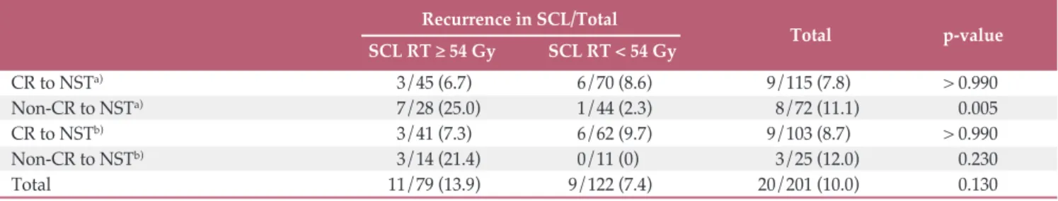
3. Patterns of SCL and IMN failure according to the treat- ments
Among 205 patients with SCL involvement, 201 patients received SCL RT. More patients failed in the SCL with the higher RT dose to SCL ≥ 54 Gy, but the difference was not statistically significant (13.9% vs. 7.4%, p=0.130). According to the response of SCL to NST, significantly more SCL fail- ures were observed with ≥ 54 Gy in patients failing to achieve CR to NST (25.0% vs. 2.3%, p=0.005). When 59 patients undergoing SCL excision were excluded, there were no dif- ferences in the crude SCL failure rates according to the dose to the SCL (Table 3). Among those patients who underwent SCL excision, 12 patients achieved CR to NST and 47 did not, and five patients who failed to achieve CR experienced SCL failures. Regarding four patients who did not receive SCL
RT, they achieved CR to NST and did not undergo SCL exci- sion, and there were no SCL failures during follow-up.
One hundred and eighty-five of 217 patients with IMN involvement received IMN RT. As for IMN failure, there was no significant difference according to the receipt of IMN RT in patients with IMN involvement (3.8% vs. 9.4%, p=0.139).
Patients achieving CR to NST had more IMN failures when IMN RT was not given, but the difference did not reach a sta- tistical significance (13.0% vs. 2.8%, p=0.065). When 14 pati- ents undergoing IMN excision were excluded, a similar finding was observed (Table 4). Among those patients who underwent IMN excision, two patients achieved CR to NST and 12 did not, and no patient experienced IMN failure.
When a log-rank test was performed in 184 patients who did not undergo IMN excision and whose response to NST was available, the 5-year IMN failure-free survival rates were 96.9% and 90.9% in patients with and without IMN RT, res- pectively (p=0.064) (Fig. 2).
Recurrence in SCL/Total
Total p-value
SCL RT ≥ 54 Gy SCL RT < 54 Gy
CR to NSTa) 3/45 (6.7) 6/70 (8.6) 9/115 (7.8) > 0.990
Non-CR to NSTa) 7/28 (25.0) 1/44 (2.3) 8/72 (11.1) 0.005
CR to NSTb) 3/41 (7.3) 6/62 (9.7) 9/103 (8.7) > 0.990
Non-CR to NSTb) 3/14 (21.4) 0/11 (0) 3/25 (12.0) 0.230
Total 11/79 (13.9) 9/122 (7.4) 20/201 (10.0) 0.130
Values are presented as number (%). SCL, supraclavicular lymph node; RT, radiotherapy; CR, complete remission; NST, neoadjuvant systemic therapy. a)14 patients, whose response to NST was unavailable, were excluded, b)73 patients, whose response to NST was unavailable and who underwent SCL excision, were excluded.
Table 3. Patterns of SCL failure among 201 SCL-involved patients receiving SCL RT
Recurrence in IMN/Total
Total p-value
IMN RT (+) IMN RT (–)
CR to NSTa) 3/109 (2.8) 3/23 (13.0) 6/132 (4.5) 0.065
Non-CR to NSTa) 4/60 (6.7) 0/6 (0) 4/66 (6.1) > 0.990
CR to NSTb) 3/107 (2.8) 3/23 (13.0) 6/130 (4.6) 0.068
Non-CR to NSTb) 4/48 (8.3) 0/6 (0) 4/54 (7.4) > 0.990
Total 7/185 (3.8) 3/32 (9.4) 10/217 (4.6) 0.169
Values are presented as number (%). IMN, internal mammary lymph node; RT, radiotherapy; CR, complete remission; NST, neoadjuvant systemic therapy. a)19 patients, whose response to NST was unavailable, were excluded, b)33 patients, whose response to NST was unavailable and who underwent IMN excision, were excluded.
Table 4. Patterns of IMN failure among 217 IMN-involved patients
4. Subgroup analysis according to the involvement of SCL or IMN
Within patients with SCL involvement (n=205), higher ypT category, four or more involved axillary lymph nodes, his- tologic grade 3, triple-negative subtype, clinical non-CR of SCL to NST, mastectomy, and SCL excision were associated with an inferior DFS on univariate analysis. However, the SCL RT dose ≥ 54 Gy did not improve DFS (5-year rate, 52.9%
vs. 50.9%; p=0.696) (Fig. 3). On multivariate analysis, four or more involved axillary lymph nodes (HR, 1.636; 95% CI, 1.020 to 2.621; p=0.041), histologic grade 3 (HR, 1.665; 95%
CI, 1.085 to 2.555; p=0.020), triple-negative subtype (HR, 2.181; 95% CI, 1.369 to 3.473; p=0.001), and mastectomy (HR, 1.867; 95% CI, 1.036 to 3.365; p=0.038) were significant prog-
nosticators predicting a worse DFS.
With regard to those patients with IMN involvement (n=217), additional SCL involvement, four or more involved axillary lymph nodes, lymphovascular invasion, triple-neg- ative subtype, mastectomy, and no hormonal therapy were correlated with an inferior DFS on univariate analysis. How- ever, the receipt of IMN RT did not impact on DFS (5-yr rate, 56.1% with IMN RT vs. 78.1% without IMN RT; p=0.099) (Fig. 4). On multivariate analysis, additional SCL involve- ment (HR, 1.663; 95% CI, 1.070 to 2.587; p=0.024), four or more involved axillary lymph nodes (HR, 1.814; 95% CI, 1.098 to 2.998; p=0.020), triple-negative subtype (HR, 3.995;
95% CI, 2.106 to 7.579; p < 0.001), and mastectomy (HR, 2.409;
95% CI, 1.405 to 4.129; p=0.001) were significant adverse prognosticators for DFS.
5. Impact of SCL or IMN excision on the outcomes
Among 205 patients with SCL involvement, 59 patients underwent SCL excision along with the breast and axillary surgery. Among these, the response to NST was clinical CR in 12 and non-CR in 47. All of them received SCL RT, and the median dose was 50.4 Gy (range, 45 to 66 Gy). Regarding the patterns of failure, there were 10 patients with local recur- rences, nine with regional recurrences including five with SCL recurrences, and 35 with distant metastases. The 5-year DFS was 38.1%, significantly inferior to those without SCL excision (57.5%, p=0.026). As mentioned earlier, however, the significance of SCL excision disappeared on multivariate analysis.
Fourteen of 217 patients with IMN involvement had IMN excision. Among these, the response to NST was clinical CR in two and non-CR in 12. All of them received IMN RT. There were two patients with local recurrences, two with regional Fig. 2. Internal mammary lymph node (IMN) failure-free
survival curves in patients with IMN involvement who did not undergo IMN excision and whose response to neoadjuvant systemic therapy was available (n=184). RT, radiotherapy.
Recurrence-free survival rat
e 1.0
0 0.2 0.4 0.6
0
Time (mo)50 60
10 20 30 40 70 8090100110120 0.8
IMN RT done IMN RT not done
Fig. 3. Disease-free survival curves according to the dose to the supraclavicular lymph node (SCL) among those patients with SCL involvement (n=201). Four patients not receiving radiation to the SCL were excluded.
Disease-free survival rate 1.0
0 0.2 0.4 0.6
0 10 20 30 40Time (mo)50 6070 8090100110120 0.8
< 54 Gy
≥ 54 Gy
Fig. 4. Disease-free survival curves according to the recei- pt of internal mammary lymph node (IMN) irradiation among those patients with IMN involvement (n=217). RT, radiotherapy.
Disease-free survival rate 1.0
0 0.2 0.4 0.6
0
Time (mo)50 60
10 20 30 40 70 8090100110120 0.8
IMN RT done IMN RT not done
recurrences (one in axillary lymph node and the other in the SCL), and five with distant metastases. The 5-year DFS was 56.3%, which was not significantly different from those with- out IMN excision (59.6%, p=0.901).
Discussion
In our study, the outcomes of NST followed by surgery and postoperative RT were analyzed in 353 breast cancer patients with SCL and/or IMN involvement which was detected on imaging studies. The SCL and/or IMN failures were rare, and a higher SCL RT dose as well as the receipt of IMN RT was not associated with a better control rate. DFS and OS were comparable with the contemporary results, but were not improved by a higher dose SCL RT and IMN RT.
As for the treatment for extra-axillary regional lymph nodes in breast cancer, especially IMN, there have been a number of historical reports to date [9]. However, extended radical mastectomy has rarely been performed since several randomized trials noted no survival benefit of such an aggre- ssive treatment [10,11]. With regard to the management for SCL, some institutions reported the outcomes of SCL exci- sion along with breast surgery, but its benefit is not evident until now [12,13]. Therefore, the involved SCL and/or IMN is usually diagnosed clinically and treated by regional nodal RT combined with systemic therapy.
Recently, especially with the development of imaging studies, more data on the multidisciplinary treatment in this setting are becoming available. Among these, Zhang et al. [5]
reviewed 809 patients with cN2-3 patients, and found that 112 patients (13.8%) had IMN involvement on PET-CT, MRI, CT and/or US [5]. They also analyzed the outcomes of NST followed by surgery and postoperative RT in these patients, and 72.1% of IMN-involved patients achieved clinical CR on imaging after NST. In the present study, SCL and/or IMN involvement was determined via the same imaging studies, and each 75.9% of patients with SCL and IMN involvement achieved clinical CR to NST. The treatment outcomes were also very similar, and the 5-year DFS and OS were 56% and 76% in the Zhang et al.’s study [5], and 57.8% and 75.1% in our study, respectively. In addition, Noh et al. [14] also reported the treatment outcomes of SCL- and/or IMN- involved breast cancer which was detected on imaging stud- ies including PET-CT and MRI. When treated with NST fol- lowed by surgery and postoperative RT, the 5-year DFS and OS were 60.2% and 75.5%, respectively. These observations suggest that despite of the advanced nodal disease, favorable DFS and OS can be achieved when multimodality treatments are given. However, these data should be interpreted with
caution, because the involvement of SCL and/or IMN might be overestimated on imaging and those patients with occult distant metastases could be excluded after more meticulous imaging studies such as PET-CT compared with historical controls. Compared with aforementioned studies, Huang et al. [4] reported the relatively inferior DFS and OS, that is, 30%
and 47%, respectively, in SCL-involved breast cancer patients receiving multimodality treatments. In this study, SCL invol- vement was presented as palpable disease in 77.5% of pati- ents, and clinical CR rate to NST was only 49%. These find- ings suggest that their population might have a higher tumor burden. In addition, pathologic confirmation of IMN invol- vement on fine-needle aspiration biopsy was reported to be associated with an inferior progression-free survival com- pared to those of clinically involved IMN on PET-CT [15].
Regarding the impact of regional nodal RT for SCL and/or IMN, Park et al. [3] analyzed 55 patients with PET-detected cN3 breast cancer who were treated with NST followed by surgery and postoperative RT. The 5-year locoregional relapse-free survival, DFS, and OS were 80%, 60%, and 79%, respectively. And, both the higher RT dose to SCL ≥ 55 Gy and the receipt of IMN RT was not associated with better out- comes. We also observed similar findings among 353 patients from 11 institutions, that is, DFS was not improved by a higher dose SCL RT or IMN RT. Although the receipt of IMN RT was associated with a lower IMN failure rate even in pati- ents achieving clinical CR to NST, the difference did not reach statistical significance. Rather, more SCL failures were observed with a higher SCL RT dose in patients failing to achieve clinical CR to NST. The paradoxical dose-response relationship could be explained that a higher RT dose might be prescribed to a larger residual lesion after NST. However, the major patterns of failure were distant metastases, and therefore this finding might not be translated into a mean- ingful DFS difference. In the aforementioned study by Huang et al. [4], a higher SCL RT dose was prescribed to pati- ents with non-CR to NST and was not associated with a bet- ter SCL control. However, non-CR to NST was the indepen- dent prognostic factor associated with an inferior DFS [4].
Considering the increase of treatment failures in those pati- ents with non-CR to NST (S1 and S2 Tables), more intensified systemic therapy should be incorporated to these patients.
The limitations of this study are as follows. First, there might be potential biases which are the typical shortcomings of retrospective studies. Performance of mastectomy was associated with an inferior DFS, which suggests that patients with more advanced disease underwent mastectomy. Sec- ond, the involvement of SCL and/or IMN was determined on imaging studies, and therefore might be overestimated.
In some patients undergoing SCL and/or IMN excision or biopsy, the pathologic confirmation was done, but it was not a routine procedure in most institutions. However, the diag-
nostic accuracy of SCL and/or IMN involvement by PET-CT or US was reported to be about 80%-90% [7,8], and our pati- ents showed positive findings on multiple imaging studies in 72.2% for SCL involvement and 76.0% for IMN involve- ment. Lastly, the extent of SCL and/or IMN involvement was not specified. As mentioned earlier, the paradoxical dose-response relationship between SCL RT dose and SCL failure might be resulted from a higher tumor burden una- vailable from the retrospective chart review. Despite these limitations, our study represents one of the largest series on the outcomes of multidisciplinary treatment for breast cancer patients with SCL and/or IMN involvement.
In conclusion, NST followed by surgery and postoperative RT achieved an acceptable in-field regional control rate in patients with SCL and/or IMN involvement. However, a higher SCL RT dose or IMN RT was not associated with the improved DFS in these patients. SCL and/or IMN excision, which was more frequently performed in patients with non- CR to NST, achieved acceptable local control but did not improve DFS, either. Given the major patterns of failure were distant metastases, more intensified systemic therapy should be incorporated especially in those patients with non-CR to NST.
Electronic Supplementary Material
Supplementary materials are available at Cancer Research and Treatment website (https://www.e-crt.org).
Conflicts of Interest
Conflict of interest relevant to this article was not reported.
Author Details
1Department of Radiation Oncology, Ewha Womans University Col- lege of Medicine, Seoul, 2Department of Radiation Oncology, Won- kwang University Hospital, Wonkwang University School of Medicine, Iksan, 3Department of Radiation Oncology, Seoul Natio- nal University College of Medicine, Seoul, 4Department of Radiation Oncology, Asan Medical Center, University of Ulsan College of Medicine, Seoul, 5Department of Radiation Oncology, Yonsei Cancer Center, Yonsei University College of Medicine, Seoul, 6Department of Radiation Oncology, Samsung Medical Center, Sungkyunkwan University School of Medicine, Seoul, 7Department of Radiation Oncology, Wonju Severance Christian Hospital, Wonju, 8Depart- ment of Radiation Oncology, Ajou University School of Medicine, Suwon, 9Department of Radiation Oncology, Uijeongbu St. Mary’s Hospital, College of Medicine, The Catholic University of Korea, Uijeongbu, 10Department of Radiation Oncology, Chonbuk National University Hospital, Jeonju, 11Department of Radiation Oncology, Keimyung University Dongsan Medical Center, Keimyung Univer- sity School of Medicine, Daegu, 12Department of Radiation Oncol- ogy, Hanyang University College of Medicine, Seoul, Korea
1. Poortmans PM, Collette S, Kirkove C, Van Limbergen E, Budach V, Struikmans H, et al. Internal mammary and medial supraclavicular irradiation in breast cancer. N Engl J Med.
2015;373:317-27.
2. Whelan TJ, Olivotto IA, Parulekar WR, Ackerman I, Chua BH, Nabid A, et al. Regional nodal irradiation in early-stage breast cancer. N Engl J Med. 2015;373:307-16.
3. Park HJ, Shin KH, Cho KH, Park IH, Lee KS, Ro J, et al. Out- comes of positron emission tomography-staged clinical N3 breast cancer treated with neoadjuvant chemotherapy, sur- gery, and radiotherapy. Int J Radiat Oncol Biol Phys. 2011;81:
e689-95.
4. Huang EH, Strom EA, Valero V, Fornage B, Perkins GH, Oh JL, et al. Locoregional treatment outcomes for breast cancer patients with ipsilateral supraclavicular metastases at diagno- sis. Int J Radiat Oncol Biol Phys. 2007;67:490-6.
5. Zhang YJ, Oh JL, Whitman GJ, Iyengar P, Yu TK, Tereffe W, et al. Clinically apparent internal mammary nodal metastasis in patients with advanced breast cancer: incidence and local
control. Int J Radiat Oncol Biol Phys. 2010;77:1113-9.
6. Anderson TL, Glazebrook KN, Murphy BL, Viers LD, Hieken TJ. Cross-sectional imaging to evaluate the extent of regional nodal disease in breast cancer patients undergoing neoadju- vant systemic therapy. Eur J Radiol. 2017;89:163-8.
7. Wang CL, Eissa MJ, Rogers JV, Aravkin AY, Porter BA, Beatty JD. (18)F-FDG PET/CT-positive internal mammary lymph nodes: pathologic correlation by ultrasound-guided fine-nee- dle aspiration and assessment of associated risk factors. AJR Am J Roentgenol. 2013;200:1138-44.
8. Iyengar P, Strom EA, Zhang YJ, Whitman GJ, Smith BD, Woodward WA, et al. The value of ultrasound in detecting extra-axillary regional node involvement in patients with advanced breast cancer. Oncologist. 2012;17:1402-8.
9. Chen RC, Lin NU, Golshan M, Harris JR, Bellon JR. Internal mammary nodes in breast cancer: diagnosis and implications for patient management: a systematic review. J Clin Oncol.
2008;26:4981-9.
10. Lacour J, Le M, Caceres E, Koszarowski T, Veronesi U, Hill C.
References
Radical mastectomy versus radical mastectomy plus internal mammary dissection. Ten year results of an international cooperative trial in breast cancer. Cancer. 1983;51:1941-3.
11. Veronesi U, Marubini E, Mariani L, Valagussa P, Zucali R. The dissection of internal mammary nodes does not improve the survival of breast cancer patients. 30-year results of a ran- domised trial. Eur J Cancer. 1999;35:1320-5.
12. Jung J, Kim SS, Ahn SD, Lee SW, Ahn SH, Son BH, et al. Treat- ment outcome of breast cancer with pathologically proven synchronous ipsilateral supraclavicular lymph node metas- tases. J Breast Cancer. 2015;18:167-72.
13. Chang XZ, Yin J, Sun J, Zhang XH, Cao XC. A retrospective
study of different local treatments in breast cancer patients with synchronous ipsilateral supraclavicular lymph node metastasis. J Cancer Res Ther. 2013;9 Suppl:S158-61.
14. Noh JM, Kim KH, Park W, Suh CO, Huh SJ, Choi DH, et al.
Prognostic significance of nodal involvement region in clinical stage IIIc breast cancer patients who received primary sys- temic treatment, surgery, and radiotherapy. Breast. 2015;24:
637-41.
15. Joo JH, Kim SS, Ahn SD, Choi EK, Jung JH, Jeong Y, et al.
Impact of pathologic diagnosis of internal mammary lymph node metastasis in clinical N2b and N3b breast cancer patients.
Breast Cancer Res Treat. 2017;166:511-8.