ABSTRACT
Purpose: We aimed to analyze the treatment outcomes of ipsilateral cervical lymph node (CLN)-positive breast cancer without other distant metastasis and compare the outcomes with those of supraclavicular lymph node (SCL)-positive breast cancer.
Methods: Seventy-eight patients with breast cancer and ipsilateral CLN metastasis above the supraclavicular fossa (CLN[+] group) were treated at 7 institutions (2000–2014). Seventy- four patients received systemic chemotherapy and breast surgery followed by locoregional radiotherapy. Outcomes of the CLN(+) group were compared with those of the SCL(+) group, which included 183 patients with SCL involvement.
Results: The median follow-up duration was 55.9 months. Twenty-two regional failures were found in 15 patients—axillary lymph node (LN) in 8, SCL in 6, internal mammary LN in 3, previously involved CLN in 4, and previously uninvolved ipsilateral CLN in one patient. The 5-year overall survival (OS), disease-free survival (DFS), locoregional relapse-free survival (LRRFS), and distant metastasis-free survival (DMFS) rates were 68.6%, 46.7%, 68.4%, and 57.0%, respectively. Neck dissection did not improve LRRFS and DFS (p = 0.86 and p = 0.26, respectively). Multivariate analysis showed that hormone receptor negativity and the presence of extracapsular extension were prognostic factors for poor DFS. On comparison with stage IIIC using propensity score matching, survival outcomes of the CLN(+) and SCL(+) groups were not different (5-year OS, p = 0.75; DFS, p = 0.88; LRRFS, p = 0.86; and DMFS, p = 0.45).
Original Article
Jae Sik Kim
1,*, Kyubo Kim
2,*, Kyung Hwan Shin
1, Jin Ho Kim
1, Seung Do Ahn
3, Su Ssan Kim
3, Yong Bae Kim
4, Jee Suk Chang
4, Doo Ho Choi
5, Won Park
5, Tae Hyun Kim
6, Mison Chun
7, Jihye Cha
8, Jin Hee Kim
9, Dong Soo Lee
10, Sun Young Lee
11, Hae Jin Park
121
Department of Radiation Oncology, Seoul National University College of Medicine, Seoul, Korea
2
Department of Radiation Oncology, Ewha Womans University College of Medicine, Seoul, Korea
3
Department of Radiation Oncology, Asan Medical Center, University of Ulsan College of Medicine, Seoul, Korea
4
Department of Radiation Oncology, Yonsei Cancer Center, Yonsei University College of Medicine, Seoul, Korea
5
Department of Radiation Oncology, Samsung Medical Center, Sungkyunkwan University School of Medicine, Seoul, Korea
6
Center for Proton Therapy, Research Institute and Hospital, National Cancer Center, Goyang, Korea
7
Department of Radiation Oncology, Ajou University School of Medicine, Suwon, Korea
8
Department of Radiation Oncology, Wonju Severance Christian Hospital, Wonju, Korea
9
Department of Radiation Oncology, Dongsan Medical Center, Keimyung University School of Medicine, Daegu, Korea
10
Department of Radiation Oncology, Uijeongbu St. Mary's Hospital, The Catholic University of Korea, Uijeongbu, Korea
11
Department of Radiation Oncology, Jeonbuk National University Hospital, Jeonju, Korea
12
Department of Radiation Oncology, Hanyang University College of Medicine, Seoul, Korea
Cervical Lymph Node Involvement above the Supraclavicular Fossa in
Breast Cancer: Comparison with Stage IIIC (KROG 18-02)
Received: Nov 9, 2019 Accepted: Feb 4, 2020 Correspondence to Kyung Hwan Shin
Department of Radiation Oncology, Seoul National University College of Medicine, 101 Daehak-ro, Jongno-gu, Seoul 03080, Korea.
E-mail: [email protected]
*These authors contributed equally to this work.
© 2020 Korean Breast Cancer Society This is an Open Access article distributed under the terms of the Creative Commons Attribution Non-Commercial License (https://
creativecommons.org/licenses/by-nc/4.0/) which permits unrestricted non-commercial use, distribution, and reproduction in any medium, provided the original work is properly cited.
ORCID iDs Jae Sik Kim
https://orcid.org/0000-0002-0039-8667 Kyubo Kim
https://orcid.org/0000-0001-6093-1294 Kyung Hwan Shin
https://orcid.org/0000-0002-5852-7644 Jin Ho Kim
https://orcid.org/0000-0002-7918-1072 Seung Do Ahn
https://orcid.org/0000-0003-4153-4191 Su Ssan Kim
https://orcid.org/0000-0002-8473-302X Yong Bae Kim
https://orcid.org/0000-0001-7573-6862 Jee Suk Chang
https://orcid.org/0000-0001-7685-3382
Doo Ho Choi
https://orcid.org/0000-0002-0524-3883 Won Park
https://orcid.org/0000-0003-4686-2071 Tae Hyun Kim
https://orcid.org/0000-0001-8413-3385 Mison Chun
https://orcid.org/0000-0003-2334-3316 Jihye Cha
https://orcid.org/0000-0001-5926-5685 Jin Hee Kim
https://orcid.org/0000-0001-8608-9250 Dong Soo Lee
https://orcid.org/0000-0001-7880-8147 Sun Young Lee
https://orcid.org/0000-0001-9121-0182 Hae Jin Park
https://orcid.org/0000-0003-3891-8952 Conflict of Interest
The authors declare that they have no competing interests.
Author Contributions
Conceptualization: Kim K, Shin KH; Data curation: Kim JS, Kim K, Shin KH, Kim JH
1, Ahn SD, Kim SS, Kim YB, Chang JS, Choi DH, Park W, Kim TH, Chun M, Cha J, Kim JH
2, Lee DS, Lee SY, Park HJ; Formal analysis: Kim JS, Kim K, Shin KH; Methodology: Kim JS, Kim K, Shin KH; Project administration: Kim K, Shin KH;
Supervision: Kim K, Shin KH; Writing - original draft: Kim JS; Writing - review & editing: Kim JS, Kim K, Shin KH, Kim JH
1, Ahn SD, Kim SS, Kim YB, Chang JS, Choi DH, Park W, Kim TH, Chun M, Cha J, Kim JH
2, Lee DS, Lee SY, Park HJ.
Kim JH
1, Jin Ho Kim; Kim JH
2, Jin Hee Kim.
Conclusion: The comparable clinical outcomes indicate that patients with breast cancer who have ipsilateral CLN metastasis without other distant metastasis may benefit from locoregional treatment of the ipsilateral breast and systemic therapies, as do those with N3c disease.
Keywords: Breast; Carcinoma; Neoplasm metastasis; Survival; Neoplasm staging
INTRODUCTION
The incidence of cervical lymph node (CLN) metastasis from breast cancer is as low as 1%
[1]. Supraclavicular lymph node (SCL) metastasis was considered stage IV disease until 2002 [2]. Brito et al. [3] demonstrated that the survival outcomes of patients with SCL metastasis after combined modality therapy were similar to those with stage IIIB disease and significantly better than patients with visceral stage IV. On the basis of these results [3,4], the International Union Against Cancer/American Joint Committee on Cancer (AJCC) staging system reclassified SCL metastasis from stage IV to stage IIIC in 2003 [5]. However, other CLN metastasis above the SCL is still considered to be M1 in this staging system [6].
Information on CLN metastasis is lacking due to its low incidence. To our knowledge, there are no cohort studies to date on the treatment outcomes of CLN metastasis above the SCL;
thus, there is no consensus on how to manage these patients. The choice of treatment primarily depends on institutional preference [6]. Qin et al. [7] reported a patient with breast cancer and CLN metastasis who underwent modified radical mastectomy and CLN dissection followed by endocrine therapy. In that case report [7], the authors proposed classifying CLN metastasis as a locally advanced stage, which is similar to stage IIIC.
We performed a retrospective multicenter cohort study, Korean Radiation Oncology Group (KROG) 18-02, among patients with breast cancer who had ipsilateral CLN metastasis above the SCL without other distant metastasis. In this study, we evaluated the treatment outcomes and identified the prognostic factors in these patients. We also compared survival outcomes with those of patients with stage IIIC disease.
METHODS
Patients
We retrospectively recruited a total of 78 patients with ipsilateral CLN metastasis above the SCL, without any other distant metastasis at diagnosis (CLN[+] group), from 7 institutions;
the patients were treated between January 2000 and December 2014. All treatments were conducted with curative, not palliative, intent. We excluded patients who had received prior thoracic radiotherapy (RT); those with other cancers except thyroid, cervical, and non- melanoma skin cancer; and those who had not completed the planned treatment.
All patients were staged according to the 8th edition of the AJCC Cancer Staging Manual. Patients
who received neoadjuvant chemotherapy were staged according to the clinical stage; otherwise,
pathologic stage was used for a reasonable comparison. CLN metastasis was identified using the
following imaging modalities: positron emission tomography-computed tomography (PET-CT) in
72 (92.3%) patients; neck ultrasonography in 4 (5.1%) patients; and CT in 2 (2.6%) patients. Thirty-
two patients (41.0%) had a pathologic confirmation of CLN metastasis before initial treatment.
Tumor subtypes were defined as follows. On immunohistochemical staining, tumors with positive hormone receptor (HR[+]: estrogen receptor and/or progesterone receptor) were classified as HR(+)/human epidermal growth factor receptor 2 (HER2)(−) for negative HER2 status and as HR(+)/HER2(+) for positive HER2, regardless of the Ki-67 level. If the hormone receptor was HR(−) and HER2(+), the tumor was defined as the HER2-enriched type.
Otherwise, tumors were categorized as triple negative.
Treatment
Treatments were determined by each institution. All patients except for one received systemic chemotherapy (either neoadjuvant or adjuvant). Sixty patients (76.9%) underwent neoadjuvant chemotherapy. The most frequent regimen was sequential anthracycline plus taxane (A+T) (n
= 28), followed by concurrent A+T (n = 18), and others (n = 14). Adjuvant chemotherapy after breast surgery was administered in 32 patients (41.0%). The adjuvant chemotherapy regimens were sequential A+T (n = 15), concurrent A+T (n = 3), and others (n = 14).
Total mastectomy was performed in 50 (64.1%) patients, and 28 (35.9) patients underwent breast-conserving surgery. Axillary lymph node (LN) dissection was performed in 67 patients (85.9%), and sentinel LN biopsy was performed in only 11 (14.1%). Neck dissection was performed in only 35 patients (44.9%), excision in 25, selective node dissection in 6, and modified radical neck dissection in 4 patients.
Postoperative RT to the whole breast/chest wall and/or the SCL was administered in 75 patients, up to a median dose of 50.4 Gy. Among these, 65 patients also received RT to the CLN. Among 13 patients who did not receive RT to the CLN, only 2 patients underwent neck dissection. In the remaining 11 patients, 10 patients received neoadjuvant chemotherapy with or without adjuvant chemotherapy.
All 40 patients who had an HR(+) tumor received endocrine therapy, and trastuzumab was administered in 31 patients.
Statistical analysis
Overall survival (OS) was defined as the interval from the date of initial treatment (the first day of neoadjuvant chemotherapy or surgery if patients did not receive neoadjuvant chemotherapy) until death from any cause. Disease-free survival (DFS) was defined as the time between initial treatment and any first recurrence or death from any cause. Locoregional relapse-free survival (LRRFS) and distant metastasis-free survival (DMFS) were defined as the interval from initial treatment to locoregional recurrence and distant metastasis, respectively.
In this study, we counted the recurrence of ipsilateral CLN metastasis as regional recurrence, not distant metastasis.
To compare outcomes with patients who had stage IIIC disease, we analyzed the data of 183 patients with SCL metastasis who underwent breast surgery (SCL[+] group) from a previous KROG 16–14 study [8], in which 353 patients were enrolled from 2000 and 2013 to analyze treatment outcomes of regional RT in patients with breast cancer who had SCL and/
or internal mammary LN metastasis. We also considered ipsilateral CLN recurrence in the KROG 16–14 data as regional recurrence.
We used the χ
2test or Fisher's exact test, where appropriate, for categorical variables and
the independent t-test for continuous variables. The survival rates were estimated using the
Kaplan-Meier method and compared using the log-rank test. A Cox proportional hazards model was applied in univariate and multivariate analyses. Propensity score matching of the CLN(+) and SCL(+) groups was performed according to histologic grade, subtypes, neoadjuvant and adjuvant chemotherapy, RT, and trastuzumab. The propensity score was generated with a multivariable logistic regression model. The CLN(+) and SCL(+) groups were matched at a 1:1 ratio using the nearest-neighbor method with a caliper of 0.2. A p-value < 0.05 was considered statistically significant. All statistical analyses were conducted using R version 3.4.2 (https://www.r-project.org/).
Ethical statement
This study was approved by the Institutional Review Board of each institution (Seoul National University Hospital, H-1712-145-909). All procedures performed in studies involving human participants were done in accordance with the ethical standards of the institutional and/or national research committee and the 1964 Declaration of Helsinki and its later amendments or comparable ethical standards.
Because of the retrospective design of the analysis, the requirement for obtaining informed consent of participants included in the study was waived.
RESULTS
Patient characteristics
Patients' baseline characteristics are shown in Table 1. Most patients (n = 73, 93.6%) had invasive ductal carcinoma and 51.3% of patients had a high-grade tumor. Although the most frequent subtype was HR(+)/HER2(−) (n = 22, 28.2%), the distribution of subtypes was relatively even. As for the initial T stage, 46 patients had T1–2 stage and 32 patients had T3–4 stage. The number of pathologically involved axillary LNs was 0 in 21 patients, 1–3 in 17, and
≥ 4 in 40 patients. Among the 21 patients with no axillary LN involvement, all except one received neoadjuvant chemotherapy. Multiple CLN metastases were most common, with 68 cases of level V, followed by 21 of level IV, 12 of level III, 8 of level II, and 1 of level I; the one patient with level I CLN metastases received RT to the neck. Among 8 patients with level II CLN metastases, 4 patients were treated with neck RT. The details of local treatment for CLN are described in Table 2, according to the pathologic confirmation of CLN at diagnosis.
Survival and prognostic factors
The median follow-up duration was 55.9 months and ranged from 10.5 to 142.2 months. Of
38 recurrences, distant metastasis alone (except for ipsilateral CLN) was the most frequent
failure pattern (n = 16, 42.1%). Locoregional recurrence occurred in 7 patients, and 15
patients developed both locoregional and distant failure. Twenty-two cases of regional failure
occurred in 15 patients: with axillary LN in 8 patients, SCL in 6, internal mammary LN in 3,
previously involved CLN in 4, and previously uninvolved ipsilateral CLN in one patient. The
5-year OS, DFS, LRRFS, and DMFS rates were 68.6%, 46.7%, 68.4%, and 57.0%, respectively
(Figure 1). There was a possibility of false positivity in 46 patients owing to the diagnosis of
CLN metastasis using imaging alone, without a biopsy at diagnosis. We compared survival
outcomes according to the presence or absence of a pathologic confirmation of CLN
metastasis at diagnosis. The p-values for OS, DFS, LRRFS, and DMFS were 0.87, 0.77, 0.65,
and 0.49, respectively.
To identify prognostic factors, we performed multivariate analysis using variables with a p-value ≤ 0.10 in univariate analysis (Table 3). HR(+) status (hazard ratio [HR], 0.42; 95%
confidence interval [CI], 0.21–0.83; p = 0.01) and extracapsular extension (ECE) (HR, 2.78;
95% CI, 1.15–6.75; p = 0.02) were identified as independent factors of DFS. There were no prognostic factors identified for LRRFS. Neck dissection and RT to the neck failed to improve LRRFS (p = 0.86 and 1.00, respectively) or DFS (p = 0.26 and 0.22, respectively) in univariate analysis.
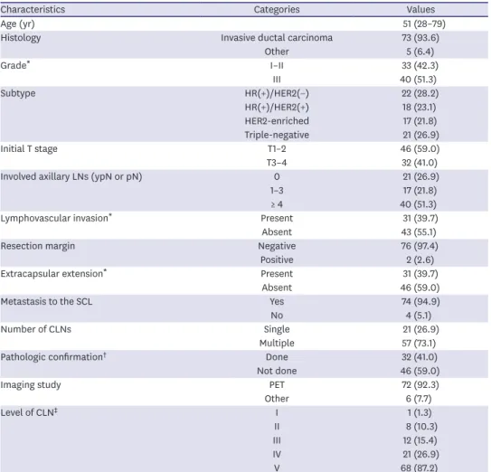
Table 1. Baseline characteristics of 78 patients
Characteristics Categories Values
Age (yr) 51 (28–79)
Histology Invasive ductal carcinoma 73 (93.6)
Other 5 (6.4)
Grade
*I–II 33 (42.3)
III 40 (51.3)
Subtype HR(+)/HER2(−) 22 (28.2)
HR(+)/HER2(+) 18 (23.1)
HER2-enriched 17 (21.8)
Triple-negative 21 (26.9)
Initial T stage T1–2 46 (59.0)
T3–4 32 (41.0)
Involved axillary LNs (ypN or pN) 0 21 (26.9)
1–3 17 (21.8)
≥ 4 40 (51.3)
Lymphovascular invasion
*Present 31 (39.7)
Absent 43 (55.1)
Resection margin Negative 76 (97.4)
Positive 2 (2.6)
Extracapsular extension
*Present 31 (39.7)
Absent 46 (59.0)
Metastasis to the SCL Yes 74 (94.9)
No 4 (5.1)
Number of CLNs Single 21 (26.9)
Multiple 57 (73.1)
Pathologic confirmation
†Done 32 (41.0)
Not done 46 (59.0)
Imaging study PET 72 (92.3)
Other 6 (7.7)
Level of CLN
‡I 1 (1.3)
II 8 (10.3)
III 12 (15.4)
IV 21 (26.9)
V 68 (87.2)
Values are presented as median (range) or number (%).
HR(+) = hormone receptor-positive; HER2 = human epidermal growth factor receptor 2; LN = lymph node; SCL = supraclavicular lymph node; CLN = cervical lymph node; PET = positron emission tomography.
*
Available data only;
†Pathologic confirmation of CLN before initial treatment;
‡Multiple counts.
Table 2. Local treatment for CLN according to the pathologic confirmation
Local treatment for CLN Pathologic confirmation (−) (n = 46) Pathologic confirmation (+) (n = 32)
Neck dissection only 0 2 (6.3)
RT to CLN only 26 (56.5) 6 (18.8)
Both 11 (23.9) 22 (68.8)
None 9
*(19.6) 2
†(6.3)
Values are presented as number (%).
RT = radiotherapy; CLN = cervical lymph node.
*
Eight patients received neoadjuvant chemotherapy;
†All patients received neoadjuvant chemotherapy.
Comparison with stage IIIC
We compared 78 patients (CLN[+] group) with 183 patients who had SCL metastasis (SCL[+]
group) from the KROG 16–14 study. Table 4 summarizes baseline characteristics between the 2 groups before and after propensity score matching. In the SCL(+) group, all patients received neoadjuvant chemotherapy, and 50.8% of patients received adjuvant chemotherapy. Postoperative RT was administered in all SCL(+) patients. There were no differences in treatment outcomes between the 2 groups, even before propensity score matching (OS, p = 0.51; DFS, p = 0.61; LRRFS, p = 0.18; DMFS, p = 0.68). Using the propensity score, 58 patients in the CLN(+) group were successfully matched with 58 patients in the SCL(+) group.
Kaplan–Meier survival curves are shown in Figure 2. The 5-year OS rates in the SCL(+) and CLN(+) groups were 65.8% and 62.6%, respectively (p = 0.75). The 5-year DFS rates were 45.5% in the SCL(+) group and 45.7% in the CLN(+) group (p = 0.88). The respective 5-year Time (mo)
0
OS
0.8 1.0
120 96
60 36
A
0.6
0.4
0.2
108
72 84
12 24 48
No. at risk
78 76 66 57 43 33 24 18 11 6 2
5-yr OS 69%
Time (mo) 0
DFS
0.8 1.0
120 96
60 36
B
0.6
0.4
0.2
108
72 84
12 24 48
No. at risk
78 69 50 40 33 23 19 14 8 3 1
5-yr DFS 47%
Time (mo) 0
LRRF S
0.8 1.0
120 96
60 36
C
0.6
0.4
0.2
108
72 84
12 24 48
No. at risk
78 71 56 46 35 27 21 16 9 4 1
5-yr LRRFS 68%
Time (mo) 0
DMF S
0.8 1.0
120 96
60 36
D
0.6
0.4
0.2
108
72 84
12 24 48
No. at risk
78 73 53 44 37 26 22 16 10 5 2
5-yr DMFS 57%
Figure 1. Kaplan-Meier survival curves of OS (A), DFS (B), LRRFS (C), and DMFS (D) in 78 patients.
OS = overall survival; DFS = disease-free survival; LRRFS = locoregional relapse-free survival; DMFS = distant metastasis-free survival.
LRRFS and DMFS rates were 64.8% and 48.4% in the SCL(+) group and 64.7% and 57.4% in the CLN(+) group (p = 0.86 and 0.45, respectively).
DISCUSSION
The incidence of CLN metastasis from breast cancer is remarkably low [1], although there have been several studies [3,4,9-12] on SCL metastasis. Because no literature exists on guiding treatment and long-term outcome, management of these patients is controversial.
Bisase and Kerawala [6] have reported that the current practice in the United Kingdom varies and there is no unified approach. This is because the question of whether CLN metastasis above the SCL is considered advanced locoregional stage or distant metastasis has not yet been addressed. Prospective trials are needed, but they are difficult to conduct considering the low incidence of CLN metastasis.
To the best of our knowledge, this is the first cohort study to investigate treatment outcomes of patients with ipsilateral CLN metastasis in the absence of other distant metastasis. All but one patient was treated with systemic chemotherapy before and/or after breast surgery, and all patients with an HR(+) tumor received endocrine therapy. RT was administered in all but 3 patients, suggesting that the included patients in this study received locoregional treatment with curative intent, as in patients with stage IIIC disease; in turn, this indicates that clinicians consider CLN metastasis advanced locoregional disease rather than distant metastasis. However, local treatment for CLN itself was not given to all of our patients.
Regarding local treatment for CLN, clinicians in the United Kingdom tend to perform aggressive neck surgery, such as comprehensive neck dissection [6]. Other studies [13,14]
have also suggested that CLN excision should be performed in cases of breast or urogenital tract cancer. However, only 35 patients (44.9) in our study underwent neck surgery, and this was not associated with an improvement in DFS (p = 0.26) or LRRFS (p = 0.86). In contrast to the study by Pergolizzi et al. [10], which highlighted the local control effect of RT on SCL, RT to the CLN did not improve DFS (p = 0.22) or LRRFS (p = 1.00) in the current study.
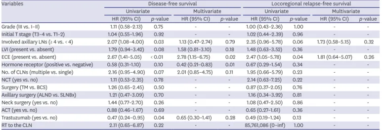
Table 3. Univariate and multivariate analyses of prognostic factors using the Cox proportional hazards model
Variables Disease-free survival Locoregional relapse-free survival
Univariate Multivariate Univariate Multivariate
HR (95% CI) p-value HR (95% CI) p-value HR (95% CI) p-value HR (95% CI) p-value
Grade (III vs. I–II) 1.11 (0.58–2.13) 0.75 - - 1.00 (0.43–2.36) 1.00 - -
Initial T stage (T3–4 vs. T1–2) 1.04 (0.55–1.96) 0.92 - - 1.02 (0.44–2.39) 0.96 - -
Involved axillary LNs (≥ 4 vs. < 4) 2.07 (1.08–4.00) 0.03 1.13 (0.47–2.74) 0.79 2.35 (0.96–5.78) 0.06 1.73 (0.58–5.15) 0.32
LVI (present vs. absent) 1.79 (0.94–3.42) 0.08 1.58 (0.81–3.10) 0.18 1.48 (0.63–3.52) 0.36 - -
ECE (present vs. absent) 2.67 (1.41–5.05) < 0.01 2.78 (1.15–6.75) 0.02 2.47 (1.05–5.78) 0.04 1.81 (0.64–5.07) 0.26 Hormone receptor (positive vs. negative) 0.58 (0.31–1.10) 0.10 0.42 (0.21–0.83) 0.01 0.67 (0.29–1.54) 0.34 - - No. of CLNs (multiple vs. single) 2.16 (0.95–4.90) 0.07 2.01 (0.85–4.75) 0.11 1.95 (0.66–5.79) 0.23 - -
NCT (yes vs. no) 1.11 (0.53–2.35) 0.78 - - 2.14 (0.63–7.25) 0.22 - -
Surgery (TM vs. BCS) 1.26 (0.65–2.45) 0.50 - - 0.87 (0.37–2.05) 0.76 - -
Axillary surgery (ALND vs. SLNBx) 1.21 (0.47–3.09) 0.70 - - 1.16 (0.34–3.92) 0.81 - -
Neck surgery (yes vs. no) 1.44 (0.77–2.70) 0.26 - - 1.08 (0.47–2.50) 0.86 - -
ACT (yes vs. no) 0.88 (0.46–1.67) 0.69 - - 0.65 (0.27–1.61) 0.36 - -
Trastuzumab (yes vs. no) 0.47 (0.24–0.95) 0.04 0.65 (0.30–1.41) 0.28 0.49 (0.19–1.24) 0.13 - -
RT to the CLN 2.11 (0.65–6.87) 0.22 - - 85,761,086 (0–inf) 1.00 - -
HR = hazard ratio; CI = confidence interval; LN = lymph node; LVI = lymphovascular invasion; ECE = extracapsular extension; CLN = cervical lymph node; NCT
= neoadjuvant chemotherapy; TM = total mastectomy; BCS = breast conserving surgery; ALND = axillary lymph node dissection; SLNBx = sentinel lymph node
biopsy; ACT = adjuvant chemotherapy; RT = radiotherapy; Inf = infinity.
According to the literature [15,16], the 5-year OS rate in patients with stage IV disease is 26%–49%. This rate is much lower than the 68.6% 5-year OS rate observed in patients with CLN metastasis of the current study. The 5-year OS rate in our study population was similar to that in patients with stage IIIC disease. There were also no differences in other outcomes between patients with CLN(+) and SCL(+). In the study by Olivotto et al. [4], the OS rate of stage IIIC was intermediate between that of stage IIIB and M1 disease during the first decade.
However, in the present study, the OS curve of the SCL(+) and CLN(+) groups overlapped from the beginning. This finding further supports the idea that the prognosis of CLN metastasis may not differ from that of SCL metastasis, and it could be hypothesized that CLN metastasis is simply an extension of SCL metastasis, that is, a regional LN. However, further studies with a larger number of patients are needed to validate this hypothesis.
We identified that the prognosis of patients with CLN metastasis was similar to that of patients with stage IIIC rather than M1, but the distant metastasis was still a major recurrence Table 4. Baseline characteristics before and after propensity score matching
Characteristics Before PSM After PSM
SCL(+) (n = 183) CLN(+) (n = 78) p-value SCL(+) (n = 58) CLN(+) (n = 58) p-value
Age (yr) 49 (23–84) 51 (28–79) 0.35 48 (27–84) 50 (28–68) 0.34
Histology
IDC 177 (96.7) 73 (93.6) 0.41 56 (96.6) 54 (93.1) 0.68
Other 6 (3.3) 5 (6.4) - 2 (3.4) 4 (6.9) -
Grade
I–II 98 (53.6) 33 (42.3) 0.13 27 (46.6) 27 (46.6) 1.00
III 69 (37.7) 40 (51.3) - 26 (44.8) 26 (44.8) -
Unknown 16 (8.7) 5 (6.4) - 5 (8.6) 5 (8.6) -
Subtype
HR(+)/HER2(−) 68 (37.2) 22 (28.2) 0.55 18 (31.0) 17 (29.3) 0.97
HR(+)/HER2(+) 34 (18.6) 18 (23.1) - 9 (15.5) 11 (19.0) -
HER2-enriched 35 (19.1) 17 (21.8) - 12 (20.7) 11 (19.0) -
Triple-negative 46 (25.1) 21 (26.9) - 19 (32.8) 19 (32.8) -
Initial T stage
T1–2 105 (57.4) 46 (59.0) 0.92 31 (53.4) 29 (50.0) 0.85
T3–4 78 (42.6) 32 (41.0) - 27 (46.6) 29 (50.0) -
Involved axillary LNs
0 39 (21.3) 21 (26.9) 0.60 13 (22.4) 20 (34.5) 0.12
1–3 45 (24.6) 17 (21.8) - 12 (20.7) 16 (27.6) -
≥ 4 99 (54.1) 40 (51.3) - 33 (56.9) 22 (37.9) -
Chemotherapy
NCT and ACT 93 (50.8) 15 (19.2) < 0.01 14 (24.1) 14 (24.1) 1.00
NCT 90 (49.2) 45 (57.7) - 44 (75.9) 44 (75.9) -
ACT 0 (0) 17 (21.8) - 0 (0) 0 (0) -
Not done 0 (0) 1 (1.3) - 0 (0) 0 (0) -
Surgery
BCS 55 (30.1) 28 (35.9) 0.43 17 (29.3) 18 (31.0) 1.00
TM 128 (69.9) 50 (64.1) - 41 (70.7) 40 (69.0) -
Radiotherapy
Done 183 (100.0) 75 (96.2) 0.04 58 (100.0) 58 (100.0) 1.00
Not done 0 (0) 3 (3.8) - 0 (0) 0 (0) -
Trastuzumab
Done 54 (29.5) 31 (39.7) 0.14 22 (37.9) 22 (37.9) 1.00
Not done 129 (70.5) 47 (60.3) - 36 (62.1) 36 (62.1) -
Endocrine therapy
Done 100 (54.6) 40 (51.3) 0.72 27 (46.6) 28 (48.3) 1.00
Not done 83 (45.4) 38 (48.7) - 31 (53.4) 30 (51.7) -
Values are presented as median (range) or number (%).
PSM = propensity score matching; SCL = supraclavicular lymph node; CLN = cervical lymph node; IDC = invasive ductal carcinoma; HR(+) = hormone receptor-positive;
HER2 = human epidermal growth factor receptor 2; LN = lymph node; NCT = neoadjuvant chemotherapy; ACT = adjuvant chemotherapy; BCS = breast conserving
surgery; TM = total mastectomy.
pattern. Thirty-one patients (39.7%) experienced distant metastasis, excluding ipsilateral CLN metastasis during the disease course, although all patients except one underwent systemic chemotherapy. Therefore, proper chemotherapy is emphasized in these patients, and further investigations are needed to overcome distant failures. In addition, we determined that HR(−) and ECE(+) are prognostic factors for poor DFS; previous studies [17,18] have also identified these as risk factors. Treatment intensification may be needed for patients with these risk factors. However, it is difficult to recommend a specific treatment strategy for this population based on the limited number of patients. In this study, we enrolled a selective population treated with curative intent, as in locally advanced breast cancer. However, the role of local treatment for CLN metastasis was not demonstrated. Further research is needed on the optimal local and systemic therapies for patients with CLN metastasis.
Time (mo) 0
OS
0.8 1.0
120 96
60 36
A
0.6
0.4
0.2
108
72 84
12 24 48
CLN (+) SCL (+) No. at risk
58 58
54 55
47 46
37 42
31 31
19 25
17 13
15 8
6 6
5 2
1 0 CLN (+)
SCL (+) 63%
66%
5-yr OS
p = 0.75
Time (mo) 0
DFS
0.8 1.0
120 96
60 36
B
0.6
0.4
0.2
108
72 84
12 24 48
CLN (+) SCL (+) No. at risk
58 58
44 45
31 36
26 29
23 23
15 16
13 8
12 6
4 4
3 1
0 0 CLN (+)
SCL (+) 46%
45%
5-yr DFS
p = 0.88
Time (mo) 0
LRRF S
0.8 1.0
120 96
60 36
C
0.6
0.4
0.2
108
72 84
12 24 48
CLN (+) SCL (+) No. at risk
58 58
47 46
37 39
29 35
24 26
16 19
14 9
13 7
4 5
3 2
0 0 CLN (+)
SCL (+) 65%
65%
5-yr LRRFS
p = 0.86
Time (mo) 0
DMF S
0.8 1.0
120 96
60 36
D
0.6
0.4
0.2
108
72 84
12 24 48
CLN (+) SCL (+) No. at risk
58 58
49 48
34 38
29 32
26 26
18 17
16 8
14 6
6 4
5 1
1 0 CLN (+)
SCL (+) 57%
48%
5-yr DMFS
p = 0.45
Figure 2. Kaplan-Meier survival curves of OS (A), DFS (B), LRRFS (C), and DMFS (D) comparing CLN(+) patients and patients with stage IIIC disease (SCL[+]) after propensity score matching.
OS = overall survival; DFS = disease-free survival; LRRFS = locoregional relapse-free survival; DMFS = distant metastasis-free survival; SCL(+) = supraclavicular
lymph node-positive; CLN(+) = cervical lymph node-positive.
Our study had some limitations. First, this was a retrospective study, although 7 institutions participated. Second, this study only covered a short follow-up period (median, 55.9 months) and the number of patients was small due to the low incidence. Third, 59.0% of patients were diagnosed with CLN metastasis using imaging only. Most patients underwent PET-CT, but it is possible that CLN metastasis was misdiagnosed as false positive, which may have influenced the outcomes. Finally, the indications of local treatment for CLN, including neck dissection and RT, were unclear.
In conclusion, the outcomes of patients with breast cancer who have ipsilateral CLN metastasis without other distant metastasis were comparable to those of patients with stage IIIC breast cancer; the former patients may benefit from locoregional treatment of the ipsilateral breast and systemic therapies with curative intent. The role of local treatment for CLN metastasis, such as neck surgery or RT, was unclear in the present analysis. The prognosis of patients with HR(−) and ECE(+) was poor, and more aggressive treatment may be needed in these patients.
REFERENCES
1. Pedersen AN, Møller S, Steffensen KD, Haahr V, Jensen M, Kempel MM, et al. Supraclavicular recurrence after early breast cancer: a curable condition? Breast Cancer Res Treat 2011;125:815-22.
PUBMED | CROSSREF
2. American Joint Committee on Cancer. Manual for Staging of Cancer, 3rd ed. Philadelphia (PA):
Lippincott; 1998. p.145-50.
3. Brito RA, Valero V, Buzdar AU, Booser DJ, Ames F, Strom E, et al. Long-term results of combined- modality therapy for locally advanced breast cancer with ipsilateral supraclavicular metastases: The University of Texas M.D. Anderson Cancer Center experience. J Clin Oncol 2001;19:628-33.
PUBMED | CROSSREF
4. Olivotto IA, Chua B, Allan SJ, Speers CH, Chia S, Ragaz J. Long-term survival of patients with supraclavicular metastases at diagnosis of breast cancer. J Clin Oncol 2003;21:851-4.
PUBMED | CROSSREF
5. Singletary SE, Allred C, Ashley P, Bassett LW, Berry D, Bland KI, et al. Revision of the American Joint Committee on Cancer staging system for breast cancer. J Clin Oncol 2002;20:3628-36.
PUBMED | CROSSREF
6. Bisase B, Kerawala C. Survey of UK practice for management of breast cancer metastases to the neck. Ann R Coll Surg Engl 2012;94:484-9.
PUBMED | CROSSREF
7. Qin R, Zhang Q, Weng J, Liu W, Zhang B, Lv G, et al. Treatment and prognosis for retrograde cervical lymph node metastases in breast cancer. Contemp Oncol (Pozn) 2015;19:154-6.
PUBMED | CROSSREF
8. Kim K, Jeong Y, Shin KH, Kim JH, Ahn SD, Kim SS, et al. Impact of regional nodal irradiation for breast cancer patients with supraclavicular and/or internal mammary lymph node involvement: a multicenter, retrospective study (KROG 16-14). Cancer Res Treat 2019;51:1500-8.
PUBMED | CROSSREF
9. Chen SC, Chang HK, Lin YC, Leung WM, Tsai CS, Cheung YC, et al. Prognosis of breast cancer after supraclavicular lymph node metastasis: not a distant metastasis. Ann Surg Oncol 2006;13:1457-65.
PUBMED | CROSSREF
10. Pergolizzi S, Settineri N, Santacaterina A, Spadaro P, Maisano R, Caristi N, et al. Ipsilateral supraclavicular lymph nodes metastases from breast cancer as only site of disseminated disease. Chemotherapy alone vs.
induction chemotherapy to radical radiation therapy. Ann Oncol 2001;12:1091-5.
PUBMED | CROSSREF
11. van der Sangen MJ, Coebergh JW, Roumen RM, Rutten HJ, Vreugdenhil G, Voogd AC. Detection, treatment, and outcome of isolated supraclavicular recurrence in 42 patients with invasive breast carcinoma. Cancer 2003;98:11-7.
PUBMED | CROSSREF
12. Jung J, Kim SS, Ahn SD, Lee SW, Ahn SH, Son BH, et al. Treatment outcome of breast cancer with pathologically proven synchronous ipsilateral supraclavicular lymph node metastases. J Breast Cancer 2015;18:167-72.
PUBMED | CROSSREF
13. Sesterhenn AM, Albert US, Barth PJ, Wagner U, Werner JA. The status of neck node metastases in breast cancer--loco-regional or distant? Breast 2006;15:181-6.
PUBMED | CROSSREF
14. Werner JA, Davis RK. Distant metastases. In: Werner JA, Davis RK, editors. Metastasis in Head and Neck Cancer. Heidelberg: Springer; 2004.
15. Siegel RL, Miller KD, Jemal A. Cancer statistics, 2016. CA Cancer J Clin 2016;66:7-30.
PUBMED | CROSSREF
16. Yoo GS, Yu JI, Park W, Huh SJ, Choi DH. Prognostic factors in breast cancer with extracranial oligometastases and the appropriate role of radiation therapy. Radiat Oncol J 2015;33:301-9.
PUBMED | CROSSREF
17. Putti TC, El-Rehim DM, Rakha EA, Paish CE, Lee AH, Pinder SE, et al. Estrogen receptor-negative breast carcinomas: a review of morphology and immunophenotypical analysis. Mod Pathol 2005;18:26-35.
PUBMED | CROSSREF
18. Nottegar A, Veronese N, Senthil M, Roumen RM, Stubbs B, Choi AH, et al. Extra-nodal extension of sentinel lymph node metastasis is a marker of poor prognosis in breast cancer patients: a systematic review and an exploratory meta-analysis. Eur J Surg Oncol 2016;42:919-25.
PUBMED | CROSSREF

![Figure 2. Kaplan-Meier survival curves of OS (A), DFS (B), LRRFS (C), and DMFS (D) comparing CLN(+) patients and patients with stage IIIC disease (SCL[+]) after propensity score matching](https://thumb-ap.123doks.com/thumbv2/123dokinfo/4748539.270619/9.892.66.830.125.789/figure-kaplan-survival-comparing-patients-patients-propensity-matching.webp)
