Purpose: This study was conducted to evaluate prognosis of patients with level I/II axillary lymph node metastases from occult breast cancer (OBC).
Materials and Methods: Data of 53 patients with OBC who received axillary lymph node dissection (ALND) positive/negative (+/–) breast-conserving surgery between 2001 and 2013 were retrospective- ly collected at seven hospitals in Korea. The median number of positive lymph nodes (+LNs) was 2.
Seventeen patients (32.1%) had >3 +LNs. A total of 48 patients (90.6%) received radiotherapy. Ex- tents of radiotherapy were as follows: whole-breast (WB; n = 11), regional lymph node (RLN; n = 2), and WB plus RLN (n = 35).
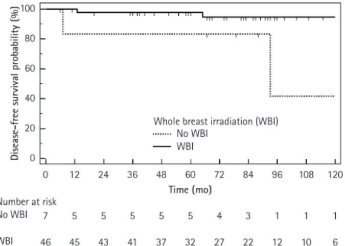
Results: The median follow-up time was 85 months. Recurrence was found in four patients: two in the breast, one in RLN, and one in the breast and RLN. The 5-year and 7-year disease-free survival (DFS) rates were 96.1% and 93.5%, respectively. Molecular subtype and receipt of breast radiotherapy were significantly associated with DFS. Patients with estrogen receptor negative, progesterone recep- tor negative, and human epidermal growth factor receptor 2 negative (ER-/PR-/HER2-) subtype had significantly lower 7-year DFS than those with non-ER-/PR-/HER2- tumor (76.9% vs. 100.0%; p = 0.03). Whole breast irradiation (WBI) was significantly associated with a higher 7-year DFS rate (94.7% for WBI group vs. 83.3% for non-WBI group; p = 0.01). Other factors including patient’s age, number of +LNs, taxane chemotherapy, and RLN irradiation were not associated with DFS.
Conclusion: Patients with OBC achieved favorable outcome after ALND and breast-targeting treat- ment. Molecular subtype and receipt of WBI was significant factors for DFS.
Keywords: Unknown primary neoplasms, Breast neoplasm, Lymph nodes, Radiotherapy
Prognosis of patients with axillary lymph node metastases from occult breast cancer: analysis of multicenter data
Haeyoung Kim 1 , Won Park 1 , Su Ssan Kim 2 , Sung Ja Ahn 3 , Yong Bae Kim 4 , Tae Hyun Kim 5 , Jin Hee Kim 6 , Jin-Hwa Choi 7 , Hae Jin Park 8 , Jee Suk Chang 4 , Doo Ho Choi 1
1
Department of Radiation Oncology, Samsung Medical Center, Sungkyunkwan University School of Medicine, Seoul, Korea
2
Department of Radiation Oncology, Asan Medical Center, University of Ulsan College of Medicine, Seoul, Korea
3
Department of Radiation Oncology, Chonnam National University Medical School, Gwangju, Korea
4
Department of Radiation Oncology, Yonsei Cancer Center, Yonsei University College of Medicine, Seoul, Korea
5
Department of Radiation Oncology, Research Institute and Hospital, National Cancer Center, Goyang, Korea
6
Department of Radiation Oncology, Dongsan Medical Center, Keimyung University School of Medicine, Daegu, Korea
7
Department of Radiation Oncology, Chung-Ang University Hospital, Seoul, Korea
8
Department of Radiation Oncology, Hanyang University College of Medicine, Seoul, Korea
Introduction
Cancer of unknown primary site (CUP) is a rare disease entity in which metastatic cancerous lesions present without any evidence
Radiat Oncol J 2021;39(2):107-112 https://doi.org/10.3857/roj.2021.00241
Received: February 15, 2021 Revised: April 21, 2021 Accepted: April 23, 2021 Corresponding author:
Won Park
Department of Radiation Oncology, Samsung Medical Center, Sungkyunkwan University School of Medicine, 50 Irwon- dong, Gangnam-gu, Seoul 06351, Korea Tel: +82-2-3410-2616
Fax: +82-2-3410-2619
E-mail: [email protected] ORCID:
https://orcid.org/0000-0003-4686-2071 Su Ssan Kim
Department of Radiation Oncology, Asan Medical Center, University of Ulsan College of Medicine, 88 Olympic-ro 43-gil, Songpa-gu, Seoul 05505, Korea Tel: +82-2-3010-5680 Fax: +82-2-3010-6950
E-mail: [email protected] ORCID:
https://orcid.org/0000-0002-8473-302X
of primary tumor. In most patients with CUP, the disease tends to disseminate early and respond poorly to systemic agents [1]. How- ever, there are favorable subsets of patients who have experienced prolonged survival after treatment for putative primary origin [1,2].
Copyright © 2021 The Korean Society for Radiation Oncology
This is an Open Access article distributed under the terms of the Creative Commons Attribution Non-Commercial License (http://creativecommons.org/licenses/by-
nc/4.0/) which permits unrestricted non-commercial use, distribution, and reproduction in any medium, provided the original work is properly cited.
Cases of axillary lymph node adenocarcinoma with unknown pri- mary site (AxCUP) which is detected in females are one of the fa- vorable subsets of CUP [1]. AxCUP in females is generally regarded as a presentation of occult breast cancer (OBC). Hence, it has been recommended that AxCUP in females needs to be managed as per the treatment for primary breast cancer [3].
OBC accounts for only 0.1%–1.0% of all breast cancer cases [4,5]; therefore, there was little evidence regarding optimal treat- ment strategies for OBC. Recent studies reported that axillary lymph node dissection (ALND) along with breast-targeting treat- ment such as mastectomy or breast-conserving surgery (BCS) re- sulted in favorable survival among patients with OBC [5-7]. The addition of postoperative radiotherapy to surgical treatment was associated with improved survival when compared to surgery alone [5,8]. Nonetheless, there is little consensus regarding which area should be irradiated for patients with OBC. Given that patients with OBC have no cancerous lesion in the ipsilateral breast even after detailed imaging studies, it is questionable whether the breast needs to be irradiated or not. Moreover, it is unknown whether prophylactic radiotherapy to uninvolved regional lymph nodes (RLNs), such as supraclavicular lymph nodes (SCN) or internal mammary lymph nodes (IMN), has prognostic impact in patients with OBC with axillary lymph node involvement.
In this study, we evaluated prognosis and patterns of failure in patients with axillary lymph node metastasis from OBC.
Materials and Methods
1. Patients and treatments
Females who received breast-conserving treatment (BCT) including ALND and/or BCS for OBC between January 2001 and December 2013 were included in this study. OBC was defined as adenocarci- noma or poorly differentiated carcinoma in axillary lymph nodes without an evidence of primary breast tumor on physical examina- tion and imaging studies including mammography, breast ultraso- nography (US), magnetic resonance imaging (MRI) of the breast, chest computed tomography (CT), or positron emission tomogra- phy-computed tomography (PET-CT). Patients were ineligible for inclusion in this study if they had cancerous lesions in other organs other than the axillary lymph nodes, previous history of other can- cer, or previous radiotherapy. Patients who had mastectomy with subsequent identification of primary breast tumor on pathologic evaluation were excluded from this study. Seven hospitals that are members of the Korean Radiation Oncology Group provided data of 53 patients who met the inclusion criteria of this study. The Insti- tutional Review Board of each hospital approved this study. The in- formed consent was waived.
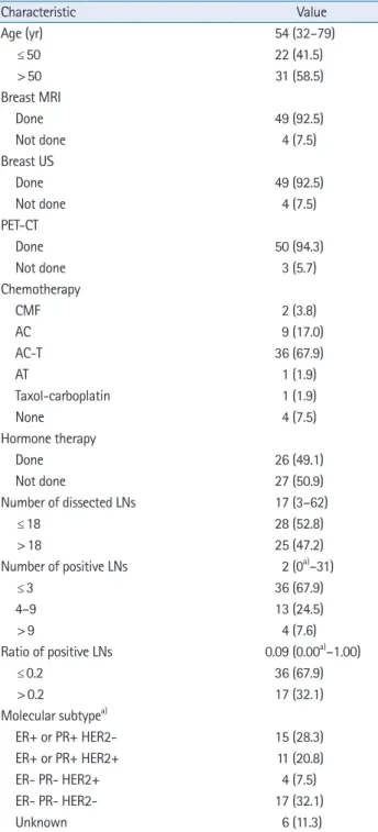
Mammography and either breast US or breast MRI were per- formed in all patients. All four patients who did not undergo breast MRI were evaluated with breast US and PET-CT. ALND and/or BCS was administered to all patients. Blind upper outer quadrantecto- my was performed in 11 patients, while 42 patients underwent no breast surgery. The median number of dissected lymph nodes was 17 (range, 3 to 62). Immunohistochemical staining for estrogen re- ceptor (ER), progesterone receptor (PR), and human epidermal growth factor receptor 2 (HER2) of tumor in lymph nodes was per- formed. ER/PR positivity was defined as an Allred score of 3–8 by immunohistochemistry (IHC). HER2 positivity was defined as either staining 3+ by IHC or 2+ by IHC with positive fluorescence in situ hybridization (FISH) or silver in situ hybridization (SISH). Tax- ane-based chemotherapy was provided to 71.7% of patients. Neo- adjuvant systemic treatment was administered before ALND in five patients (9.4%). Among 15 patients with HER2-positive tumor, eight patients (53.3%) underwent anti-HER2 agents. Patient’s characteristics are shown in Table 1. Radiotherapy was provided to all but five patients. Fields and doses of radiotherapy were decided according to each institutional policy. Whole breast or RLNs was treated with a total dose of 40.05–50.4 Gy at 1.8–2.67 Gy per frac- tion. Three patients received intensity-modulated radiotherapy, while others underwent three-dimensional conformal radiotherapy.
Details of radiation fields are depicted in Table 2.
2. Statistical analysis
Overall survival (OS), disease-free survival (DFS), and breast can- cer-free survival (BCFS) were defined as the interval from the date of ALND or the first day of neoadjuvant systemic treatment to death, cancer recurrence, and ipsilateral breast cancer occurrence, respectively. Survival probability was estimated using the Ka- plan-Meier method and the log-rank test was used to compare survival between groups with different variables. Factors with a significance at p < 0.05 on univariate analysis were included in a multivariate Cox stepwise regression analysis. Statistical signifi- cance was calculated at 95% confidence level (p < 0.05). Statisti- cal analyses were performed using SPSS version 22.0 for Windows (IBM Corp., Armonk, NY, USA).
Results
The median follow-up time was 85 months (range, 7 to 178
months). Recurrence was found in four patients (7.5%): two (3.7%)
in the ipsilateral breast, one (1.9%) in RLN, and one (1.9%) in the
ipsilateral breast and RLN (Table 3). No patient showed distant me-
tastases. Cancer in the ipsilateral breast occurred in three patients
(5.6%) within 7 to 93 months after the completion of treatment
for OBC. Of the three patients who developed breast cancer, one did not have breast irradiation while two received radiotherapy to the ipsilateral breast for the treatment of OBC.
The 5-year DFS, BCFS, and OS of all patients were 96.1%, 98.0%, and 96.0%, respectively. The 7-year DFS, BCFS, and OS of all pa- tients were 93.5%, 95.4%, and 96.0%, respectively. In the univari- ate analyses, molecular subtype and receipt of breast radiotherapy were significant factors for DFS. Patients with ER-/PR-/HER2- sub- type had significantly lower 7-year DFS than those with non-ER-/
PR-/HER2- tumor (76.9% vs. 100.0%; p = 0.03). In addition, whole breast irradiation (WBI) was significantly associated with a higher 7-year DFS rate (94.7% for WBI group vs. 83.3% for non-WBI group; p = 0.01) (Fig. 1). However, in multivariate analyses, there were no factors significantly associated with DFS. Other factors such as patient’s age, number of metastatic lymph nodes, ratio of positive lymph nodes, types of breast surgery, RLN irradiation, and taxane chemotherapy were not related to patient’s DFS (Table 4).
Discussion and Conclusion
In this study, we found that patients with OBC presenting as Ax- CUP achieved favorable outcome after ALND and BCT including WBI and systemic treatment. Tumor subtype of non-ER-/PR-/
HER2- and administration of WBI was significantly associated with Table 1. Patient’s characteristics
Characteristic Value
Age (yr) 54 (32–79)
≤ 50 22 (41.5)
> 50 31 (58.5)
Breast MRI
Done 49 (92.5)
Not done 4 (7.5)
Breast US
Done 49 (92.5)
Not done 4 (7.5)
PET-CT
Done 50 (94.3)
Not done 3 (5.7)
Chemotherapy
CMF 2 (3.8)
AC 9 (17.0)
AC-T 36 (67.9)
AT 1 (1.9)
Taxol-carboplatin 1 (1.9)
None 4 (7.5)
Hormone therapy
Done 26 (49.1)
Not done 27 (50.9)
Number of dissected LNs 17 (3–62)
≤ 18 28 (52.8)
> 18 25 (47.2)
Number of positive LNs 2 (0
a)–31)
≤ 3 36 (67.9)
4–9 13 (24.5)
> 9 4 (7.6)
Ratio of positive LNs 0.09 (0.00
a)–1.00)
≤ 0.2 36 (67.9)
> 0.2 17 (32.1)
Molecular subtype
a)ER+ or PR+ HER2- 15 (28.3)
ER+ or PR+ HER2+ 11 (20.8)
ER- PR- HER2+ 4 (7.5)
ER- PR- HER2- 17 (32.1)
Unknown 6 (11.3)
Values are presented as median (range) or number (%).
LNs, lymph nodes; MRI, magnetic resonance imaging; US, ultrasonogra- phy; PET-CT, positron emission tomography-computed tomography;
CMF, cyclophosphamide, methotrexate, and fluorouracil; AC, doxorubi- cin and cyclophosphamide; AC-T, doxorubicin and cyclophosphamide followed by paclitaxel or docetaxel; AT, doxorubicin and paclitaxel or docetaxel; ER, estrogen receptor; PR, progesterone receptor; HER2, hu- man epidermal growth factor receptor 2.
a)