© 2016 Korean Breast Cancer Society. All rights reserved. http://ejbc.kr | pISSN 1738-6756
INTRODUCTION
In 2008, the Korean Radiation Oncology Group (KROG) initiated a prospective phase III randomized trial (KROG 08-
06) designed to investigate the effect of internal mammary node irradiation (IMNI) on disease-free survival (DFS) and toxicity in breast cancer patients. Until that time, no consen- sus existed regarding the use of IMNI in postmastectomy ra- diotherapy or radiotherapy after breast-conserving surgery (BCS). Inclusion of the internal mammary nodes (IMNs) in breast cancer radiotherapy is mainly dependent on the prefer- ences of the treating radiation oncologists. A Korean pattern- of-care study showed that approximately 50% of patients re- ceived IMNI during postmastectomy radiotherapy [1]. Vari- able patterns of clinical practice regarding IMNI, which were culture-driven not evidence-based, have been reported [2,3].
Because IMNI may increase radiation exposure to critical
Radiation Pneumonitis in Association with Internal Mammary Node Irradiation in Breast Cancer Patients: An Ancillary Result from the KROG 08-06 Study
Jinhyun Choi, Yong Bae Kim, Kyung Hwan Shin1, Sung-Ja Ahn2, Hyung-Sik Lee3, Won Park4, Su Ssan Kim5, Jin Hee Kim6, Kyu Chan Lee7, Dong Won Kim8, Hyun Suk Suh9, Kyung Ran Park9, Hyun Soo Shin10, Chang-Ok Suh
Department of Radiation Oncology, Yonsei Cancer Center, Yonsei University College of Medicine, Seoul; 1Department of Radiation Oncology, Proton Therapy Center, Research Institute and Hospital, National Cancer Center, Goyang; 2Department of Radiation Oncology, Chonnam National University Hwasun Hospital, Hwasun; 3Department of Radiation Oncology, Dong-A University Hospital, Dong-A University College of Medicine, Busan; 4Department of Radiation Oncology, Samsung Medical Center, Sungkyunkwan University School of Medicine, Seoul; 5Department of Radiation Oncology, Asan Medical Center, University of Ulsan College of Medicine, Seoul; 6Department of Radiation Oncology, Dongsan Medical Center, Keimyung University School of Medicine, Daegu; 7Department of Radiation Oncology, Gachon University Gil Medical Center, Incheon; 8Department of Radiation Oncology, Pusan National University Hospital, Pusan National University School of Medicine, Busan; 9Department of Radiation Oncology, Ewha Womans University Mokdong Hospital, Seoul; 10Department of Radiation Oncology, CHA Bundang Hospital, CHA University College of Medicine, Seongnam, Korea
ORIGINAL ARTICLE
Purpose: The aim of this study is to present the incidence of ra- diation pneumonitis (RP) reported within 6 months after treat- ment for breast cancer with or without internal mammary node irradiation (IMNI). Methods: In the Korean Radiation Oncology Group (KROG) 08-06 phase III randomized trial, patients who were node-positive after surgery were randomly assigned to re- ceive radiotherapy either with or without IMNI. A total of 747 pa- tients were enrolled, and three-dimensional treatment planning with computed tomography simulation was performed for all pa- tients. Of the 747 patients, 722 underwent chest X-rays before and within 6 months after radiotherapy. These 722 patients un- derwent evaluation, and RP was diagnosed on the basis of chest radiography findings and clinical symptoms. The relation- ship between the incidence of RP and clinical/dosimetric para- meters was analyzed. Results: RP developed in 35 patients (4.8%),
including grade 1 RP in 26 patients (3.6%), grade 2 RP in nine patients (1.2%); there was no incidence of grade 3 or higher RP.
Grade 2 RP cases were observed in only the IMNI group. The risk of developing RP was influenced by IMNI treatment; pneu- monitis occurred in 6.5% of patients (n=23/356) who underwent IMNI and in 3.3% of patients (n=12/366) who did not (p=0.047).
The differences in lung dosimetric parameters (mean lung dose, V10–40) were statistically significant between the two groups.
Conclusion: IMNI treatment resulted in increased radiation expo- sure to the lung and a higher rate of RP, but the incidence and severity of RP was minimal and acceptable. This minor impact on morbidity should be balanced with the impact on survival outcome in future analyses.
Key Words: Breast neoplasms, Lymphatic irradiation, Radiation pneumonitis
Correspondence to: Chang-Ok Suh
Department of Radiation Oncology, Yonsei Cancer Center, Yonsei University College of Medicine, 50-1 Yonsei-ro, Seodaemun-gu, Seoul 03722, Korea Tel: +82-2-2228-8095, Fax: +82-2-2227-7823
E-mail: [email protected]
This work was supported by a grant from the National R&D Program for Cancer Control, Ministry for Health, Welfare, and Family Affairs, Republic of Korea (0820010).
Received: June 28, 2016 Accepted: August 18, 2016
Cancer
organs, new studies must determine whether the expected benefits of elective IMNI is worth the risk of late toxicity to critical organs, such as the lungs and heart [4]. In the European Organisation for Research and Treatment of Cancer (EORTC) 22922/10925 trial, instances of pulmonary toxicity were sig- nificantly higher in the IM-MS (internal mammary and medial supraclavicular chain) treatment group than in the control group [5]. The National Cancer Institute of Canada Clinical Trial Group MA.20 trial demonstrated improved DFS in patients with one to three positive nodes with the addition of regional nodal irradiation, including IMNI. However, this additional irradiation was associated with an increase in the incidence of grade 2 or higher pneumonitis (1.3% vs. 0.2%) [6].
Upon its completion in February 2013, KROG 08-06 had enrolled a total of 747 patients. In this study, we reviewed col- lected data from the evaluable 722 patients and present the in- cidence of radiation pneumonitis (RP) reported within 6 months after radiotherapy with or without IMNI.
METHODS
Randomization and patient characteristics
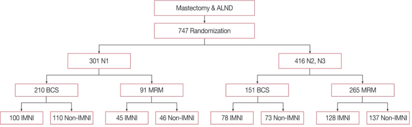
Eligible patients were pathologically confirmed to have axil- lary node-positive breast cancer after surgery consisting of ei- ther modified radical mastectomy (MRM) or BCS, regardless of histologic type. All patients underwent axillary dissection, in which eight or more lymph nodes were identified. Patients were stratified according to N stage (N1 vs. N2 or N3) and type of surgery (breast conservation vs. mastectomy), and then were randomly assigned to receive radiotherapy either with or without IMNI (Figure 1). Three hundred and one pa- tients were diagnosed with pathologic stage N1 disease, and 416 patients were diagnosed with N2 or N3 disease. Three hundred and sixty-one patients were treated with BCS, and
the remaining 356 patients were treated with MRM. Among the patients with BCS, 178 were randomly assigned to the IMNI group, and 183 were randomly assigned to the non- IMNI group. Among the patients with MRM, 173 were ran- domly assigned to the IMNI group, and 183 patients were as- signed to the non-IMNI group. All patients had unilateral in- vasive breast cancer and were eligible for adjuvant chemother- apy with or without hormonal therapy. Patients who received neoadjuvant systemic therapy or had a previous history of cancer or distant metastasis were excluded.
Between November 2008 and February 2013, we enrolled 747 patients from the 12 participating institutions in Korea.
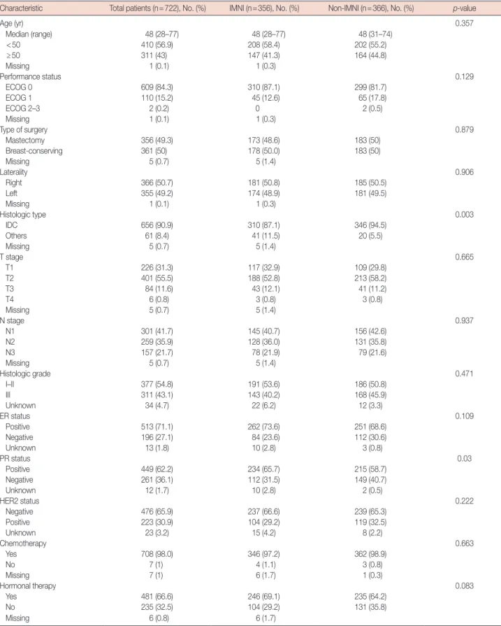
The study protocol was approved by the institutional review board (IRB approval number: 4-2008-0263), and all patients provided written informed consent. Twenty-five patients (3.3%) who had not undergone chest X-ray within 6 months of radiotherapy completion were excluded from the analysis, leaving 722 analyzable patients. The characteristics of the pa- tients in this study are presented in Table 1. The median pa- tient age was 48 years (range, 28–77 years) in both groups.
The majority of the patients enrolled (99.6%) had Eastern Co- operative Oncology Group performance scores of 0 or 1. The non-IMNI group included a significantly higher percentage of patients with ductal carcinoma and a progesterone receptor- negative status compared with the IMNI group (p=0.003 and p=0.030, respectively). Other patient characteristics were not significantly different between the two groups.
Radiation treatment
Radiation was administered once per day at a dose of 1.8–2 Gy, up to a total dose of 45–50.4 Gy; additionally, 381 patients (52.8%) received boost radiotherapy to the primary tumor bed with a median dose of 10 Gy (range, 9–16 Gy) in five fractions.
All patients received supraclavicular irradiation, as routinely
Mastectomy & ALND
747 Randomization
210 BCS 91 MRM 151 BCS 265 MRM
78 IMNI 73 Non-IMNI 128 IMNI 137 Non-IMNI 46 Non-IMNI
45 IMNI 110 Non-IMNI
100 IMNI
301 N1 416 N2, N3
Figure 1. KROG-08-06 trial diagram.
KROG=Korean Radiation Oncology Group; ALND=axillary lymph node dissection; BCS=breast-conserving surgery; MRM=modified radical mas- tectomy; IMNI=internal mammary node irradiation.
Table 1. Patients and tumor characteristics
Characteristic Total patients (n=722), No. (%) IMNI (n=356), No. (%) Non-IMNI (n=366), No. (%) p-value
Age (yr) 0.357
Median (range) 48 (28–77) 48 (28–77) 48 (31–74)
<50 410 (56.9) 208 (58.4) 202 (55.2)
≥50 311 (43) 147 (41.3) 164 (44.8)
Missing 1 (0.1) 1 (0.3)
Performance status 0.129
ECOG 0 609 (84.3) 310 (87.1) 299 (81.7)
ECOG 1 110 (15.2) 45 (12.6) 65 (17.8)
ECOG 2–3 2 (0.2) 0 2 (0.5)
Missing 1 (0.1) 1 (0.3)
Type of surgery 0.879
Mastectomy 356 (49.3) 173 (48.6) 183 (50)
Breast-conserving 361 (50) 178 (50.0) 183 (50)
Missing 5 (0.7) 5 (1.4)
Laterality 0.906
Right 366 (50.7) 181 (50.8) 185 (50.5)
Left 355 (49.2) 174 (48.9) 181 (49.5)
Missing 1 (0.1) 1 (0.3)
Histologic type 0.003
IDC 656 (90.9) 310 (87.1) 346 (94.5)
Others 61 (8.4) 41 (11.5) 20 (5.5)
Missing 5 (0.7) 5 (1.4)
T stage 0.665
T1 226 (31.3) 117 (32.9) 109 (29.8)
T2 401 (55.5) 188 (52.8) 213 (58.2)
T3 84 (11.6) 43 (12.1) 41 (11.2)
T4 6 (0.8) 3 (0.8) 3 (0.8)
Missing 5 (0.7) 5 (1.4)
N stage 0.937
N1 301 (41.7) 145 (40.7) 156 (42.6)
N2 259 (35.9) 128 (36.0) 131 (35.8)
N3 157 (21.7) 78 (21.9) 79 (21.6)
Missing 5 (0.7) 5 (1.4)
Histologic grade 0.471
I–II 377 (54.8) 191 (53.6) 186 (50.8)
III 311 (43.1) 143 (40.2) 168 (45.9)
Unknown 34 (4.7) 22 (6.2) 12 (3.3)
ER status 0.109
Positive 513 (71.1) 262 (73.6) 251 (68.6)
Negative 196 (27.1) 84 (23.6) 112 (30.6)
Unknown 13 (1.8) 10 (2.8) 3 (0.8)
PR status 0.03
Positive 449 (62.2) 234 (65.7) 215 (58.7)
Negative 261 (36.1) 112 (31.5) 149 (40.7)
Unknown 12 (1.7) 10 (2.8) 2 (0.5)
HER2 status 0.222
Negative 476 (65.9) 237 (66.6) 239 (65.3)
Positive 223 (30.9) 104 (29.2) 119 (32.5)
Unknown 23 (3.2) 15 (4.2) 8 (2.2)
Chemotherapy 0.663
Yes 708 (98.0) 346 (97.2) 362 (98.9)
No 7 (1) 4 (1.1) 3 (0.8)
Missing 7 (1) 6 (1.7) 1 (0.3)
Hormonal therapy 0.083
Yes 481 (66.6) 246 (69.1) 235 (64.2)
No 235 (32.5) 104 (29.2) 131 (35.8)
Missing 6 (0.8) 6 (1.7)
IMNI =internal mammary node irradiation; ECOG =Eastern Cooperative Oncology Group; IDC =invasive ductal carcinoma; ER =estrogen receptor;
PR=progesterone receptor; HER2=human epidermal growth factor receptor 2.
performed for node-positive disease. Each patient underwent computed tomography (CT)-based simulation, and structures were manually contoured on CT scan slices. This protocol con- tained no strict guidelines on radiotherapy technique; the techniques were determined at the discretion of the physician, and included the reverse hockey stick, standard tangent, par- tial wide tangent, and photon/electron combination tech- niques. A detailed distribution of the patients according to ra- diotherapy technique is shown in Table 2. In the MRM-IMNI group, the partial wide tangent (n=84, 48.5%) was the most commonly used technique, followed by the reverse hockey stick method (n=51, 29.4%). In the BCS group, the partial wide tangent method (n=101, 58.7%) and the photon/elec- tron combination method (n=71, 41.2%) were used for IMN irradiation. In the non-IMNI group, the most commonly used radiotherapy technique was the standard tangent method for both MRM patients (n=132, 72.1%) and BCS patients (n=
182, 100%).
Radiation pneumonitis assessment
After completion of radiotherapy, follow-up examinations including chest X-rays and physical examinations were ob- tained within 6 months. Chest X-rays obtained at baseline (before radiotherapy) were compared with those obtained within 6 months after treatment to determine RP levels. RP- related symptoms, such as cough, dyspnea, and the incidence of steroid treatment, were also identified and recorded. RP grade was scored on a scale of 0–5, based on the Radiation Therapy Oncology Group/EORTC toxicity criteria as follows:
grade 0=no change over baseline; 1=asymptomatic or mild symptoms (dry cough), slight radiographic appearances;
2=moderate symptomatic fibrosis or pneumonitis (severe
cough), low-grade fever, patch radiographic appearances;
3=severe symptomatic fibrosis or pneumonitis, dense radio- graphic appearance; 4=severe respiratory insufficiency, con- tinuous O2, assisted ventilation; 5=death. To avoid any inter- observer variation between the 12 participating institutions, two radiation oncologists on-site visited and reviewed all ab- normal chest X-ray findings and assessed them together.
Dosimetric analysis
To identify predictive factors associated with RP develop- ment, clinical variables and dosimetric parameters were ana- lyzed via univariate analysis using the Pearson chi-square test.
Dosimetric parameters such as mean lung dose (MLD), V10, V20, V30, and V40 were included in the analysis, and the cor- relation with RP was analyzed using the Student t-test. Addi- tionally, the significance of the association between treatment assignment and patient characteristics was assessed using the chi-square test. Statistical analysis was carried out using SPSS version 18.0 (SPSS Inc., Chicago, USA). A p-value ≤0.05 was considered statistically significant.
RESULTS
Incidence of radiation pneumonitis
The incidence of RP was higher in the IMNI group (Table 3). Of the 722 patients, RP developed in 35 patients (4.8%), including 26 patients (3.6%) with grade 1 RP and nine pa- tients (1.2%) with grade 2 RP. No cases of grade 3 or higher RP were found. All grade 2 RP cases developed in the IMNI group. RP occurred in 6.5% (n=23/356) of patients who were treated with IMNI and 3.3% (n=12/366) of those who were treated without IMNI (p=0.047). However, most RP cases were asymptomatic minimal pulmonary radiologic changes defined as grade 1. Of the 26 patients with grade 1 RP, only six patients experienced mild dry cough, which improved spon- taneously. In all grades of RP, most radiologic changes devel- oped 2 to 3 months into the follow-up period after radiation treatment.
Table 2. Radiotherapy techniques used in each treatment group and incidence of radiation pneumonitis
RT technique IMNI
No. (%)
Non-IMNI No. (%)
No. of RP (IMNI/non-IMNI) MRM (n=356)
Reverse hockey stick 51 (29.4) 50 (27.3) 10 (7/3) Standard tangent 0 132 (72.1) 5 (0/5) Partial wide tangent 84 (48.5) 0 9 (9/0) Photon/electron
combination
38 (21.9) 1 (0.5) 2 (2/0) BCS (n=354)
Standard tangent 0 182 (100.0) 4 (0/4) Partial wide tangent 101 (58.7) 0 4 (4/0) Photon/electron
combination 71 (41.2) 0 1 (1/0)
RT =radiation therapy; IMNI =internal mammary node irradiation; RP = radiation pneumonitis; MRM=modified radical mastectomy; BCS=breast- conserving surgery.
Table 3. Incidence of radiation pneumonitis by treatment groups Total patients
(n=722) No. (%)
IMNI (n=356) No. (%)
Non-IMNI (n=366)
No. (%)
No RP 687 (95.2) 333 (93.5) 354 (96.7)
RP 35 (4.8) 23 (6.5) 12 (3.3)
G1 26 (3.6) 14 (3.9) 12 (3.3)
G2 9 (1.2) 9 (2.5) 0
G3 or higher 0 0 0
IMNI=internal mammary node irradiation; RP=radiation pneumonitis.
With respect to radiotherapy techniques, the patterns of RP incidence differed between the two study arms (Table 2).
Among the 23 patients who developed RP in the IMNI group, 13 patients (n=13/185, 7.0%) developed RP after undergoing treatment with the partial wide tangent method, and seven patients (n=7/51, 13.7%) developed RP after the reverse hockey stick method. The others (n=3/109, 2.8%) developed RP after the photon/electron combination method. In the non-IMNI group, 12 patients developed RP, including nine patients (n=9/314, 2.9%) after the standard tangent method and three patients (n=3/50, 6.0%) after the reverse hockey stick method. The overall incidence of RP after the partial wide tangent method (13 patients) was similar to that after the reverse hockey stick method (10 patients), but the percentage of RP patients treated with each technique were 7.0% (n=
13/185) and 9.9% (n=10/101), respectively.
Dosimetric parameters
All dosimetric parameters were significantly different be- tween treatment groups (Figure 2). The MLD was 17.66±5.33 Gy with IMNI and 13.29±4.37 Gy without IMNI (p<0.001).
The V10 and V20 with IMNI were 45.68%±18.19% and 34.94% ±12.15%, respectively, and 31.71% ±12.87% and 25.49%±10.53, respectively, without IMNI. The V30 and V40 with IMNI were 27.48%±8.86% and 19.40%±6.92%, respec- tively, and 21.07%±8.81% and 14.65%±6.67%, respectively, without IMNI.
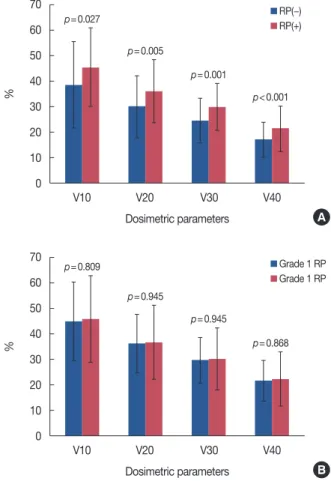
Univariate analysis of dosimetric parameters for predicting the development of RP showed that all dosimetric parameters were significantly different between the RP and non-RP groups (Figure 3A). In the RP group, the MLD was 17.88±5.75 Gy, and the V10, V20, V30, and V40 were 45.31%±15.69%, 36.20%±
11.96%, 29.47%±9.73%, and 21.54%±8.67%, respectively. In the non-RP group, the MLD was 15.38±5.30 Gy, and the V10, V20, V30, and V40 were 38.38%±17.25%, 29.93%±12.26%, 24.02%±9.30%, and 16.80%±7.04%, respectively. However, dosimetric parameters were not significantly different between grade 1 and grade 2 RP patients (Figure 3B).
DISCUSSION
The use of IMNI has been debated, but several reports have provided evidence that it improves survival in patients with breast cancer [7,8]. A previous retrospective study from our institution reported a long-term DFS advantage following IMNI in postoperative patients [9]. Although more studies are needed to clearly define the role of IMNI in long-term surviv- al and toxicity, the findings of this retrospective study showed that IMNI was obviously effective in patients with N2 disease and patients with inner/central tumors. However, there is no consensus regarding whether IMNs should be treated, be- Figure 2. Dosimetric parameters of both internal mammary node irradi-
ation (IMNI) and non-IMNI groups. All dosimetric parameters (V10–40) were significantly different between treatment groups. Data are the mean±SD.
70 60 50 40 30 20 10 0
V10 V20w V30 V40 Dosimetric parameters
%
IMNI Non-IMNI All p<0.001
Figure 3. Dosimetric parameters in the patients with radiation pneumo- nitis. All dosimetric parameters were significantly different between the radiation pneumonitis (RP) and non-RP groups (A), not significantly dif- ferent between grade 1 and grade 2 RP (B). Data are the mean±SD.
70 60 50 40 30 20 10 0
V10 V20 V30 V40
Dosimetric parameters
%
A RP(−) RP(+) p=0.027
p=0.005
p=0.001
p<0.001
70 60 50 40 30 20 10 0
V10 V20 V30 V40
Dosimetric parameters
%
B Grade 1 RP Grade 1 RP p=0.809
p=0.945
p=0.945
p=0.868
cause of the possible increase in the risk for late toxicity. As a result of the anatomic location of IMNs, IMNI increases the exposure of critical organs, such as the lungs and heart, to ra- diation.
In this study, we investigated lung toxicity, which may lead to deterioration of the patient’s performance status or quality of life. We specifically examined RP occurring within 6 months of treatment and its association with IMNI. RP is a common type of toxicity caused by radiation exposure to the lung and usually appears within 6 months of the completion of radiotherapy. Clinical symptoms, including cough and low- grade fever, occur following completion of the radiotherapy course, and can also be seen as nonspecific infiltration on chest X-rays. The rate of pneumonitis may be also influenced by systemic therapy [10,11]. Various techniques to irradiate the IMN while minimizing normal tissue irradiation have been suggested [12]. We previously reported that the partial wide tangent method is the best technique for patients under- going BCS because of the IMN coverage involved with this method and because of the reduced dose to the lungs and heart. However, the photon/electron combination method showed better isodose distribution in some patients [13]. The developments in radiotherapy techniques and the availability of three-dimensional (3D) treatment planning have allowed us to more precisely and safely irradiate IMN. In our country, 3D treatment planning with CT simulation has been used since the mid-2000s; therefore, all patients enrolled in this study had undergone CT simulation and 3D treatment plan- ning. In this study, various radiotherapy techniques were al- lowed at the discretion of the treating radiation oncologists. In the IMNI treatment group, the most commonly used tech- nique was the partial wide tangent method for both MRM and BCS. The reverse hockey stick method was exclusively used for MRM cases, both in the IMNI and non-IMNI groups.
The incidence of symptomatic RP (grade ≥2) was reported as 2.3% after whole breast and supraclavicular lymph node treatment without IMNI, and 3% after breast irradiation using the partial wide tangent technique, which includes the first three IMNs, in single-institution studies [14,15]. As a multi- institutional study, we showed that the incidence of RP, when patients were treated without IMNI, was 3.3% (n=12/366), with the BCS group accounting for 2.2% and the MRM group accounting for 4.3%. In addition, the incidence rates of grade 1 and 2 RP after using the partial wide tangent method were 4.9% (n=9/185) and 2.2% (n=4/185), respectively. In the MRM group, the risk of RP can be reduced by using the re- verse hockey stick method, in which the anterior chest wall is irradiated with an electron beam using an individualized step-
bolus. However, 13.7% of patients experienced RP, which may have resulted from improper administration of the step-bolus.
The results of the current study are in line with previous re- ports on pulmonary toxicity associated with breast radiother- apy [5,16,17]. Although the incidence of RP, including asymp- tomatic radiologic changes, was significantly increased with IMNI, the clinical impact was minimal. In the EORTC 22922/10925 trial, researchers found no significant difference between the deterioration of the performance status and in- creased lung toxicity [5]. Thus, we suggest that IMNI can be applied without any significantly increased risk.
The incidence of RP correlates with the irradiated lung vol- ume and radiation dose. A previous study suggested that if the ipsilateral lung irradiation volume is less than 12%, then the risk of pneumonitis is minimal, even with coverage of the su- praclavicular area [18]. In general, the MLD and V20 are relat- ed to RP, and the ipsilateral V20 can predict the risk of pulmo- nary toxicity [19]. In the current study, each patient underwent CT-based simulation; therefore, we obtained and analyzed the relationship between the dosimetric parameters and the inci- dence of RP with or without IMNI. MLD and V20 (17.66±
5.33 Gy and 34.94%±12.15%, respectively) in the IMNI group were higher than in the non-IMNI group (13.29±4.37 Gy and 25.49%±10.53%, respectively). Other lung dosimetric para- meters such as V10, V30, and V40 also exhibited statistically sig- nificant differences between the two groups. A previous single- institution study showed that the incidence of RP was higher in patients with MLD ≥20.5 Gy or a normal tissue complica- tion probability ≥23% [15]. Through this large prospective trial, we confirmed that the incidence of RP, as evaluated using chest X-ray, increased with higher doses of radiation to the lung, which was associated with IMNI. However, clinically meaningful grade 2 RP was not predictable on the basis of do- simetric parameters. Other patient factors that increase the risk of RP can also be considered. It has been reported that RP is more likely to occur when certain chemotherapy drugs are administered along with radiation. However, because all the patients in our trial received chemotherapy, we could not eval- uate the effect of chemotherapy.
One drawback of this study is that the chest X-ray follow- up visit could occur at any time within 6 months after RT.
Considering that most radiologic changes in this study were found at 2 or 3 months after RT, the heterogeneity of the fol- low-up time among patients may have caused an underesti- mation of asymptomatic grade 1 RP. Furthermore, we did not assess the change in performance status between the enroll- ment and post-RT periods, which may have helped to evalu- ate the effect of IMNI on quality-of-life. In this study, we fo- cused only on short-term RP incidence; we did not plan to in-
vestigate radiation-related cardiac disease, because late cardiac toxicity often appears 10 to 15 years after radiotherapy, mean- ing long-term follow-up is required [20-22]. Nilsson et al. [23]
reported that radiation to the supraclavicular lymph nodes and IMNs increased the risk of stroke. The EORTC trial 22922/10925 assessed the impact of elective internal mamma- ry and medical supraclavicular lymph node irradiation on the well-known toxicities of breast cancer radiotherapy, including lung, skin, and heart toxicity [5]. In contrast, they found that increased lung toxicity with IMNI was the only statistically significant factor between the two treatment groups at 3 years.
With newer treatment techniques, such as the breath-hold technique, intensity-modulated RT, particle therapy, and volu- metric-modulated arc therapy, IMNI can be delivered at even lower doses to the organ at risk, especially in left-sided breast cancer [24-26]. Consequently, the incidence of RP and dose parameters with IMNI estimated in this study can be further decreased by including these newer methods.
In conclusion, results from this large data collection study clearly showed that treatment of IMNs resulted in increased radiation to the lungs and a higher rate of RP, but the inci- dence and severity of RP was minimal. Therefore, we suggest that IMNI is well tolerated with a very low risk of symptomat- ic RP; however, future analyses should assess whether this mi- nor impact on morbidity could affect long-term survival out- comes.
CONFLICT OF INTEREST
The authors declare that they have no conflict of interests.
REFERENCES
1. Keum KC, Shim SJ, Lee IJ, Park W, Lee SW, Shin HS, et al. The 1998, 1999 patterns of care study for breast irradiation after mastectomy in Korea. J Korean Soc Ther Radiol Oncol 2007;25:7-15.
2. Chargari C, Castadot P, Macdermed D, Vandekerkhove C, Bourgois N, Van Houtte P, et al. Internal mammary lymph node irradiation contrib- utes to heart dose in breast cancer. Med Dosim 2010;35:163-8.
3. Taghian A, Jagsi R, Makris A, Goldberg S, Ceilley E, Grignon L, et al.
Results of a survey regarding irradiation of internal mammary chain in patients with breast cancer: practice is culture driven rather than evi- dence based. Int J Radiat Oncol Biol Phys 2004;60:706-14.
4. Freedman GM, Fowble BL, Nicolaou N, Sigurdson ER, Torosian MH, Boraas MC, et al. Should internal mammary lymph nodes in breast cancer be a target for the radiation oncologist? Int J Radiat Oncol Biol Phys 2000;46:805-14.
5. Matzinger O, Heimsoth I, Poortmans P, Collette L, Struikmans H, Van Den Bogaert W, et al. Toxicity at three years with and without irradia- tion of the internal mammary and medial supraclavicular lymph node chain in stage I to III breast cancer (EORTC trial 22922/10925). Acta
Oncol 2010;49:24-34.
6. Whelan TJ, Olivotto I, Ackerman I, Chapman JW, Chua B, Nabid A, et al. NCIC-CTG MA. 20: an intergroup trial of regional nodal irradiation in early breast cancer. J Clin Oncol 2011;29(15 Suppl):LBA1003.
7. Overgaard M, Hansen PS, Overgaard J, Rose C, Andersson M, Bach F, et al. Postoperative radiotherapy in high-risk premenopausal women with breast cancer who receive adjuvant chemotherapy: Danish Breast Cancer Cooperative Group 82b Trial. N Engl J Med 1997;337:949-55.
8. Overgaard M. Postoperative radiotherapy in high-risk postmenopausal breast cancer: authors’ reply. Lancet 1999;354:866.
9. Chang JS, Park W, Kim YB, Lee IJ, Keum KC, Lee CG, et al. Long-term survival outcomes following internal mammary node irradiation in stage II-III breast cancer: results of a large retrospective study with 12- year follow-up. Int J Radiat Oncol Biol Phys 2013;86:867-72.
10. Kubo A, Osaki K, Kawanaka T, Furutani S, Ikushima H, Nishitani H.
Risk factors for radiation pneumonitis caused by whole breast irradia- tion following breast-conserving surgery. J Med Invest 2009;56:99-110.
11. Lingos TI, Recht A, Vicini F, Abner A, Silver B, Harris JR. Radiation pneumonitis in breast cancer patients treated with conservative surgery and radiation therapy. Int J Radiat Oncol Biol Phys 1991;21:355-60.
12. Arthur DW, Arnfield MR, Warwicke LA, Morris MM, Zwicker RD. In- ternal mammary node coverage: an investigation of presently accepted techniques. Int J Radiat Oncol Biol Phys 2000;48:139-46.
13. Jeong K, Shim SJ, You SH, Kim YB, Keum KC, Kim JD, et al. A study of the radiotherapy techniques for the breast including internal mammary lymph nodes. J Korean Soc Ther Radiol Oncol 2009;27:35-41.
14. Kim HJ, Jang WI, Kim TJ, Kim JH, Kim SW, Moon SH, et al. Radiation- induced pulmonary toxicity and related risk factors in breast cancer. J Breast Cancer 2009;12:67-72.
15. Chung Y, Yoon HI, Kim YB, Ahn SK, Keum KC, Suh CO. Radiation pneumonitis in breast cancer patients who received radiotherapy using the partially wide tangent technique after breast conserving surgery. J Breast Cancer 2012;15:337-43.
16. Krengli M, Sacco M, Loi G, Masini L, Ferrante D, Gambaro G, et al.
Pulmonary changes after radiotherapy for conservative treatment of breast cancer: a prospective study. Int J Radiat Oncol Biol Phys 2008;70:
1460-7.
17. Muren LP, Maurstad G, Hafslund R, Anker G, Dahl O. Cardiac and pulmonary doses and complication probabilities in standard and con- formal tangential irradiation in conservative management of breast cancer. Radiother Oncol 2002;62:173-83.
18. Das IJ, Cheng EC, Freedman G, Fowble B. Lung and heart dose volume analyses with CT simulator in radiation treatment of breast cancer. Int J Radiat Oncol Biol Phys 1998;42:11-9.
19. Lind PA, Wennberg B, Gagliardi G, Rosfors S, Blom-Goldman U, Lideståhl A, et al. ROC curves and evaluation of radiation-induced pul- monary toxicity in breast cancer. Int J Radiat Oncol Biol Phys 2006;64:
765-70.
20. Prosnitz RG, Chen YH, Marks LB. Cardiac toxicity following thoracic radiation. Semin Oncol 2005;32(2 Suppl 3):S71-80.
21. Prosnitz RG, Hubbs JL, Evans ES, Zhou SM, Yu X, Blazing MA, et al.
Prospective assessment of radiotherapy-associated cardiac toxicity in breast cancer patients: analysis of data 3 to 6 years after treatment. Can- cer 2007;110:1840-50.
22. Harris EE. Cardiac mortality and morbidity after breast cancer treat-
ment. Cancer Control 2008;15:120-9.
23. Nilsson G, Holmberg L, Garmo H, Terent A, Blomqvist C. Radiation to supraclavicular and internal mammary lymph nodes in breast cancer increases the risk of stroke. Br J Cancer 2009;100:811-6.
24. Bartlett FR, Colgan RM, Carr K, Donovan EM, McNair HA, Locke I, et al. The UK HeartSpare Study: randomised evaluation of voluntary deep-inspiratory breath-hold in women undergoing breast radiothera- py. Radiother Oncol 2013;108:242-7.
25. Mast ME, van Kempen-Harteveld L, Heijenbrok MW, Kalidien Y, Rozema H, Jansen WP, et al. Left-sided breast cancer radiotherapy with and without breath-hold: does IMRT reduce the cardiac dose even further?
Radiother Oncol 2013;108:248-53.
26. Lee HY, Chang JS, Lee IJ, Park K, Kim YB, Suh CO, et al. The deep inspi- ration breath hold technique using Abches reduces cardiac dose in pa- tients undergoing left-sided breast irradiation. Radiat Oncol J 2013;31:
239-46.