R E S E A R C H
Open Access
Evaluation of optimal treatment planning
for radiotherapy of synchronous bilateral
breast cancer including regional lymph
node irradiation
Yeona Cho, Yoon Jin Cho, Won Suk Chang, Jun Won Kim, Won Hoon Choi and Ik Jae Lee
*Abstract
Background: We evaluated the optimal radiotherapy (RT) plan for synchronous bilateral breast cancer (SBBC), especially treatment plans including the regional lymph node (LN) area.
Methods: This study was conducted using 15 patients with SBBC (5 with small breasts, 5 with large breasts, and 5 who underwent a left total mastectomy). The clinical target volume (CTV) was defined as the volume enveloping the bilateral whole breasts/chest wall and left regional LN area. We established the following plans: 1) volumetric-modulated arc therapy (VMAT)-the only plan using two pairs of partial arcs for the whole target volume, 2) VMAT using one partial arc for the left CTV followed by a 3D tangential technique for the right breast (primary hybrid plan), and 3) VMAT for the left CTV followed by a tangential technique using an automatically calculated
prescription dose for the right breast, considering the background dose from the left breast VMAT plan (modified hybrid plan). The Tukey test and one-way analysis of variance were used to compare the target coverage and doses to organs at risk (OARs) of the three techniques.
Results: For target coverage, the VMAT-only and modified hybrid plans showed comparable target coverage in terms of Dmean(50.33 Gy vs. 50.53 Gy,p = 0.106). The primary hybrid plan showed the largest distribution of the
high-dose volume, with V105%of 25.69% and V110%of 6.37% for the planning target volume (PTV) (p < 0.001). For
OARs including the lungs, heart, and left anterior descending artery, the percentages of volume at various doses (V5Gy, V10Gy, V20Gy, V30Gy) and Dmeanwere significantly lower in both the primary and modified hybrid plans
compared to those of the VMAT-only plan. These results were consistent in subgroup analyses of breast size and morphological variation.
Conclusions: The modified hybrid plan, using an automatically calculated prescription dose for the right breast and taking into consideration the background dose from the left breast VMAT plan, showed comparable target
coverage to that of the VMAT-only plan, and was superior for saving OARs. However, considering that VMAT can be adjusted according to the physician’s intention, further evaluation is needed for developing a better plan.
Keywords: Radiotherapy dosage, Breast Cancer, Volumetric-modulated arc therapy, Organs at risk
© The Author(s). 2019 Open Access This article is distributed under the terms of the Creative Commons Attribution 4.0 International License (http://creativecommons.org/licenses/by/4.0/), which permits unrestricted use, distribution, and reproduction in any medium, provided you give appropriate credit to the original author(s) and the source, provide a link to the Creative Commons license, and indicate if changes were made. The Creative Commons Public Domain Dedication waiver (http://creativecommons.org/publicdomain/zero/1.0/) applies to the data made available in this article, unless otherwise stated.
* Correspondence:[email protected]
Department of Radiation Oncology, Gangnam Severance Hospital, Yonsei University College of Medicine, 211 Eonju-ro, Gangnam-gu, Seoul 06273, Korea
Background
Breast cancer is the most common cancer in women, and 1–2% of these patients are diagnosed with syn-chronous bilateral breast cancer (SBBC) [1]. While the incidence of SBBC is low, it presents significantly poorer overall survival than that of unilateral breast cancer [2,
3]. There is no standard guideline for treating SBBC,
and owing to an increasing demand for
breast-conserving treatment in many cases, synchronous bilateral breast irradiation is commonly required.
Radiotherapy (RT) for unilateral breast cancer has used a tangential field with two-dimensional (2D) or three-dimensional conformal radiotherapy (3D-CRT). However, when the traditional field is applied to SBBC, overlapping of the RT fields could be inevitable, thereby compromising the target coverage. There is also a prob-lem with the daily set-up of the patients, especially in the case of RT including regional node irradiation (RNI). Multiple isocenters are used in this method, so setting up the patient’s posture may often be inaccurate. More-over, a larger treatment volume is required for SBBC, so the radiation dose to organs at risk (OARs), like the lung and heart, is considerable.
For treating such a complex target volume, recent trends have shown that intensity-modulated radiation
therapy (IMRT) using helical tomotherapy or
volumetric-modulated arc therapy (VMAT) are applied. When IMRT is used for SBBC, problems associated with the isocenter and junction can be solved. Because the ir-radiation beam in IMRT emits in many directions, a large volume of the lung and heart may be irradiated un-necessarily during bilateral breast treatment. Therefore, physicians have tried to reduce the dose to OARs by using hybrid-VMAT, limiting the beam direction, or using a static angle only in tomotherapy [4–6].
However, there is still no standard for the best plan, and in most previous studies, only RT for bilateral breasts without RNI was considered. In addition, the op-timal RT plan considering the morphologic variation of patients (e.g. large/small breasts or a funnel-like chest) has not been reported. In this study, we evaluated the optimal RT plan for SBBC, especially treatment plans in-cluding RNI, and analyzed the strategies considering the patients’ morphologic variations.
Methods
Patients selection and planning objectives
In order to consider various breast sizes and types of breast surgery, we included 10 patients who received bi-lateral partial mastectomy (5 with small breasts and 5 with large breasts) and 5 patients who received a right breast partial mastectomy and a left breast total mastec-tomy. A small breast was defined as one where the depth from the nipple to the chest wall was < 3 cm in an
axial computed tomography (CT) image, whereas a large breast was defined as one where the depth was > 3 cm (Additional file1: Figure S1). The patients were immobi-lized in a supine position on a breast board with both arms raised, and planning CT was conducted.
The clinical target volume (CTV) for the breast, chest wall, and lymph node (LN) were contoured with refer-ence to the Radiation Therapy Oncology Group con-touring atlas [7]. To closely examine the irradiation dose to the heart, treatment of LN area was set to the left side. Thus, the CTV was defined as the volume that enveloped the bilateral whole breasts (or the right breast and left chest wall) and the left regional LN area, includ-ing the internal mammary, axillary level I, II, III, and supraclavicular LNs. The planning target volumes (PTVs) were obtained by 5 mm expansion in all direc-tions from the CTVs and were also restricted to have a skin gap (trim) of 3 mm from the surface in the case of breast CTV. However, for patients who received a total mastectomy, we included the skin in the CTV without a gap. The whole lung, heart, and left anterior descending artery (LAD) were considered OARs.
The prescribed dose was 50.4 Gy at 1.8 Gy per fraction to the PTVs. Radiation boost to the tumor bed was not included in this study. The primary aim in RT planning was to deliver 95% of the prescribed dose in 95% of the PTV. For planning approval, PTV D95%≥ 47.8 Gy, a near-maximum dose (D2%)≤ 55.4 Gy to the PTV, and a minimum dose to 95% of both PTVs that is not lower than 95% of their respective prescribed doses were re-quired. As no distinct treatment protocol has been sug-gested for bilateral breast cancer, the OAR dose constraints guideline was established based on a study of several patients with unilateral breast cancer [8] and pre-vious SBBC research [9]. The irradiation dose to the OARs was restricted as follows: the planning objectives were a mean lung dose of 15 Gy with V20Gy< 30% (no more than 30% of the OAR volume receiving 20 Gy) for the lungs and V25Gy< 20% for the heart. For the LAD, an objective of 25 Gy for the maximum dose (Dmax) was retained. A bolus was not used for any chest wall planning.
Planning techniques
The RT plans were generated using RayStation software version 5.2 (RaySearch Laboratories, Sweden) and all plans were made for treatment on a Versa HD radiother-apy system (Elekta). The energy of 6 MV photon was used in all VMAT and 3D CRT plans.
Using only VMAT for the whole PTV (VMAT-only plan)
For the VMAT-only plan, a single isocenter was used for the whole PTV. The plans used two pairs of partial arcs with a total length of 240° per arc (clockwise and
counterclockwise), which consisted of a rotating beam on each breast. The starting gantry angle was 240° and the stop gantry angle was 120° for all patients. A fixed collimator angle is generally recommended to avoid an excessive inter-leaf leakage dose to the patient during VMAT. The impact is different depending on certain factors such as MLC type. With reference to previous papers on VMAT plans for the breast, the collimator and couch angles in this study were set at zero, and only a coplanar beam was used [4, 10, 11]. No monitor unit constraint was used in order to allow maximum OAR sparing.
VMAT planning for the left breast followed by
conventional 3D-tangential technique for the right breast (primary hybrid plan)
To cover the left PTV, one partial arc was used with a total length of 195°, a starting gantry angle between 300° and 305°, and a stop angle between 140° and 145°. After completing the VMAT plan for the left PTV, the right breast PTV was planned with a conventional tangential field, without considering the background dose of the previous VMAT plan. A total dose of 50.4 Gy with 28 fractions was prescribed at the prescription point of the right breast PTV.
VMAT planning for the left breast followed by
conventional tangential technique for the right breast, considering the background dose of previous VMAT planning (modified hybrid plan)
The VMAT plans for the left PTV in this method were the same as those of the primary hybrid plan. However, owing to the out-of-field dose distribution of the left VMAT plan in the right breast (Additional file2: Figure S2), following the 3D plan may result in a high dose area if we use the prescribed dose of 50.4 Gy as is. Thus, we set the dose distribution of the previous left VMAT plan as a background dose when setting the new beam for the right breast. The multiple beam set planning in RayStation enables the optimization of the summed dose distribution for both VMAT and the 3D plan simultan-eously. Objective functions can be assigned to each beam set dose, or to their sum. In this plan, the RaySta-tion system calculated the background dose distribuRaySta-tions in the right PTV, and automatically adjusted the dose of the prescription point to a final dose of 50.4 Gy.
Analysis of plans
The plans were evaluated by dose-volume histogram (DVH) analysis. For PTV, the mean doses and values of V105%and V110%(the percentage of the PTV receiving at least 105 and 110% of the prescribed dose, respectively), Dmax, and Dmean (mean dose) were reported. The D98% and D2%(minimum dose to 98 and 2% of the PTV) were
also reported. The conformity index (CI) was measured by BV95% (defined below), and the dose homogeneity index (HI) was measured by D5%/D95% [12]. CI and HI were calculated using the definitions below, and the closer the CI and HI values are to 1, the better the con-formal coverage:
CI¼ BV95%=PTV volume
(BV95%= body volume of the isodose of 95% of the prescribed dose)
HI¼ D5%=D95%
(D5%= minimum dose to 5% of the PTV, D95%= mini-mum dose to 95%of the PTV)
To evaluate the irradiated dose to OARs, the analysis included the mean dose and VXGy(OAR volume receiv-ing X Gy), dependreceiv-ing upon the organ. For the lung, inci-dence of radiation pneumonitis (RP) was < 20% when the mean lung dose was less than approximately 20 Gy. With regard to Vdose threshold models, the risk of RP was < 20% for V20 < 30–35% or V5 < 60% with conven-tional fractionation [13–15]. The V30Gy, V25Gy, and Dmean for the heart were compared; V20Gy, V10Gy, Dmax, and Dmeanfor LAD were also compared. The Tukey test and one-way ANOVA were used to compare the PTV and OAR values of the three techniques. Statistical analyses were performed using SPSS software version 23 (IBM Corp, Armonk, NY). Differences were reported to be statistically significant atp < 0.05.
Results
Target dose distribution
The median pathologic tumor size of the 15 patients was 1.7 cm (range, 0.4–3.8 cm) for the left breast and 0.8 cm (range, 0.1–2.8 cm) for the right breast. Mean volume of the whole PTV was 988.6 ± 327.6 ml; the range of PTV volume was from 410.0 ml to 1765.5 ml. In the modified hybrid plan, the mean prescribed dose to the right PTV was 48.09 Gy (± 0.93 Gy) at the prescription point. The characteristics of the 15 patients in this study are pre-sented in Additional file3: Table S1.
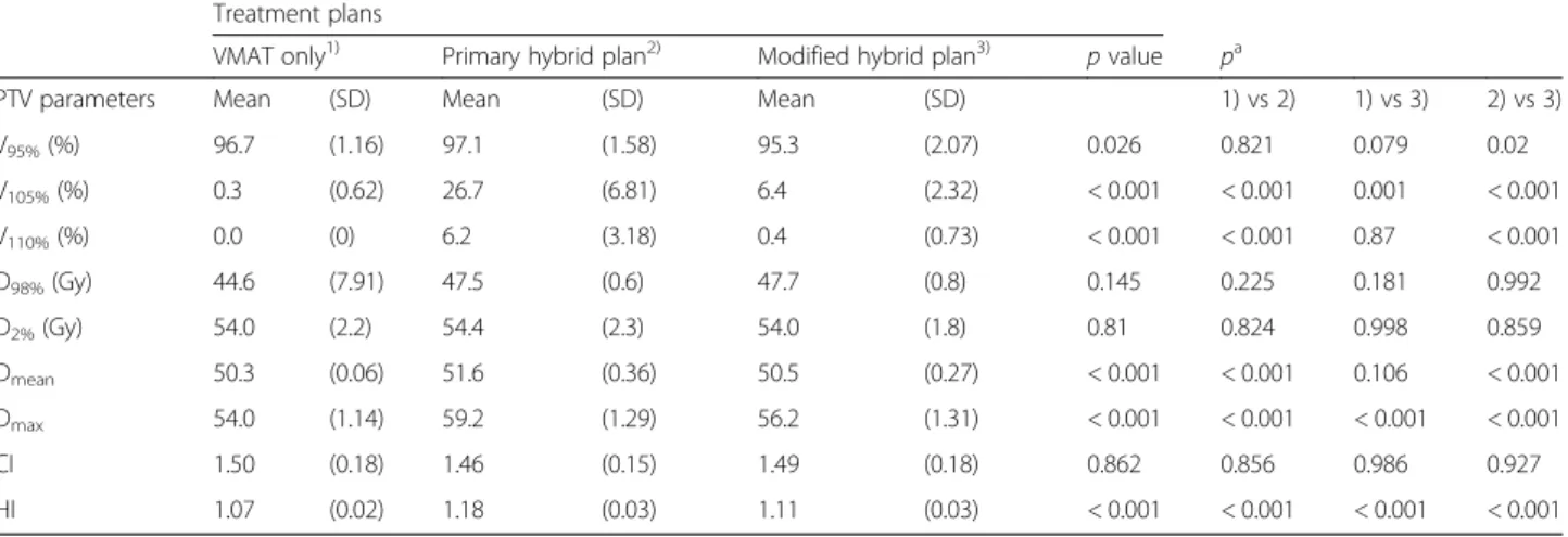
Table 1shows the Dn%(dose (Gy) to n% of the PTV),
Vm% (percentage of the PTV receiving ≥m% of
pre-scribed dose), Dmean, and Dmax. The primary hybrid plan showed the largest distribution of high-dose volume with a V105% of 25.69% and V110% of 6.37% in the PTV (p < 0.001). This plan also showed the highest Dmax (59.21 ± 1.29 Gy), followed by the modified hybrid (56.2 ± 1.31 Gy) and VMAT-only plans (54 ± 1.14 Gy). The modified hybrid plan showed higher V105% than the VMAT-only plan, but V110% did not differ between the two plans. Figure 1 shows the isodose distribution of one patient treated with various radiotherapy plans.
The VMAT-only plan and modified hybrid plan showed comparable target coverage according to Dmean with mean values of 50.33 Gy and 50.53 Gy, respectively (p = 0.106), whereas the primary hybrid plan showed higher Dmean. Although D98% and D2% were not signifi-cantly different between the 3 RT plans, D98% for the VMAT-only plan was the lowest, with a large standard deviation, indicating that the D98% of the VMAT-only plan is distributed widely around the mean value. There was also no significant difference in CI. However, the
VMAT-only plan showed the best HI value (1.07 ± 0.02), followed by the modified hybrid (1.11 ± 0.03) and pri-mary hybrid plans (1.18 ± 0.03) (p < 0.001).
Among the 10 patients who received bilateral partial mastectomy, 5 had small breasts and the other 5 had large breasts. In the subgroup analysis for patients with small breasts, the VMAT-only and modified hybrid plans showed equivalent target coverage in terms of Dmean (50.3 Gy vs 50.6 Gy, p = 0.057), whereas the primary hy-brid plan showed a high Dmeanvalue (51.6 Gy, p < 0.001)
Table 1 The comparison of planning target volume (PTV) coverage for VMAT only, primary hybrid, and modified hybrid plan using dosimetric parameters
Treatment plans
VMAT only1) Primary hybrid plan2) Modified hybrid plan3) p value pa
PTV parameters Mean (SD) Mean (SD) Mean (SD) 1) vs 2) 1) vs 3) 2) vs 3) V95%(%) 96.7 (1.16) 97.1 (1.58) 95.3 (2.07) 0.026 0.821 0.079 0.02 V105%(%) 0.3 (0.62) 26.7 (6.81) 6.4 (2.32) < 0.001 < 0.001 0.001 < 0.001 V110%(%) 0.0 (0) 6.2 (3.18) 0.4 (0.73) < 0.001 < 0.001 0.87 < 0.001 D98%(Gy) 44.6 (7.91) 47.5 (0.6) 47.7 (0.8) 0.145 0.225 0.181 0.992 D2%(Gy) 54.0 (2.2) 54.4 (2.3) 54.0 (1.8) 0.81 0.824 0.998 0.859 Dmean 50.3 (0.06) 51.6 (0.36) 50.5 (0.27) < 0.001 < 0.001 0.106 < 0.001 Dmax 54.0 (1.14) 59.2 (1.29) 56.2 (1.31) < 0.001 < 0.001 < 0.001 < 0.001 CI 1.50 (0.18) 1.46 (0.15) 1.49 (0.18) 0.862 0.856 0.986 0.927 HI 1.07 (0.02) 1.18 (0.03) 1.11 (0.03) < 0.001 < 0.001 < 0.001 < 0.001
pa: Post-Hoc Analysis Using Tukey’s HSD test
Fig. 1 Isodose distribution of individual patients treated with various radiotherapy plans. The VMAT-only (a), primary hybrid (b), and modified hybrid plans (c) are shown. High dose areas (107% of the prescribed dose) are represented with yellow color
(Additional file3: Table S2). HI (1.1 ± 0.01 p < 0.001) was best in the VMAT-only plan, while D98%, D2%, and CI showed no significant differences between the three plans.
For patients with large breasts, the VMAT-only and modified hybrid plans also showed comparable target volume coverage regarding Dmean. Moreover, HI was not significantly different between the VMAT-only plan and modified hybrid plan. Additional analysis for those who received a left breast total mastectomy showed results consistent with those of the patients with large breasts. All three subgroup analyses by breast shape (small, large, and total mastectomy) revealed that Dmax, V105%, and V110% were significantly higher in the primary hybrid plan than the other two plans (Additional file 3: Table S3).
OAR dose distribution
Regarding the whole lung, the average Dmeanof all cases was 12.6 ± 1.6 Gy, while V5Gy, V10Gy, and V20Gy were 60.5 ± 9.1%, 36.1 ± 7%, and 21.9 ± 4.6%, respectively. Table 2 shows the statistical comparison of the OAR dose distribution for each treatment technique. All dosi-metric parameters for the whole lung were significantly higher in the VMAT-only plans than in the two hybrid plans, while there was no significant difference between the hybrid plans.
The average heart dose in all cases was 9.7 ± 3.5 Gy. The irradiation dose to the heart was also highest in the VMAT-only plan, resulting in Dmeanof 13.2 ± 2.3 Gy and V25Gyof 11.5 ± 6.4%. Like in the whole lung, the primary and modified hybrid plans showed almost the same
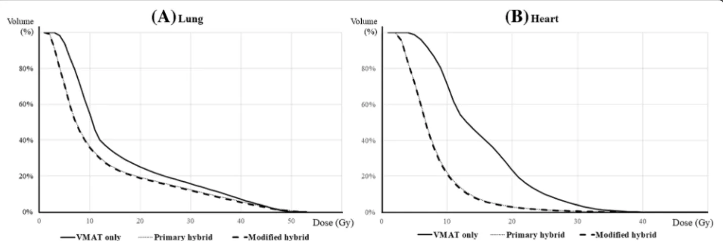
results for the heart dose. Compared to the VMAT-only plan, the hybrid plans had an approximately 5 Gy lower mean heart dose. No cases reported the heart volume re-ceiving ≥40 Gy. Figure2 shows the typical dose volume histogram for the whole lung (A) and heart (B). In both
OARs, the VMAT-only plan showed a relatively
high-volume distribution in both low- and high-dose regions.
For LAD, both the primary hybrid and modified hy-brid plans showed lower Dmean than VMAT-only plans (14.7 Gy vs 21.8 Gy, p < 0.001). V10Gy and V20Gy were also significantly larger in VMAT-only plans. Dmax showed no significance between the three RT plans.
In every subgroup analysis according to the surgery and breast shape, the VMAT-only plan showed signifi-cantly higher dose distribution in terms of mean lung dose, V20Gy of lung, mean heart dose, and mean LAD dose (Additional file3: Table S3).
Three patients in this study had funnel-like chests with convex lungs whose central lung distances (CLDs, dis-tance from the chest wall to the edge of the field at the central axis) were larger than 2.5 cm, resulting in larger lung volume in the RT field in the conventional tangen-tial plan (Additional file4: Figure S3). We evaluated the lung dose distribution in these patients, and found that the V5Gy, V10Gy and V20Gy in the VMAT-only plan were greater than those in the two hybrid plans, as was Dmean (15.2 Gy, 11.9 Gy, and 12.0 Gy, respectively).
Treatment efficiency
The VMAT-only plans resulted in a longer beam on time than those of the hybrid plans (115.3 s, 73.5 s, and 77.5 s,
Table 2 The comparison of the organs at risk (OAR) dose using dosimetric parameters
Treatment plans
VMAT only1) Primary hybrid plan2) Modified hybrid plan3) pa
Mean (SD) Mean (SD) Mean (SD) p value 1) vs 2) 1) vs 3) 2) vs 3) Whole Lung Dmean (Gy) 14.4 (0.67) 11.8 (1.19) 11.6 (1.18) < 0.001 < 0.001 < 0.001 0.935 V5Gy (%) 67.9 (9.63) 57.0 (6.34) 56.7 (6.38) < 0.001 0.001 0.001 0.993 V10Gy (%) 41.1 (7.9) 33.2 (4.39) 34.0 (5.6) < 0.001 0.003 0.008 0.926 V20Gy (%) 27.5 (2.02) 19.2 (2.42) 19.0 (2.4) < 0.001 < 0.001 < 0.001 0.971 Heart Dmean (Gy) 13.2 (2.33) 8.0 (2.61) 8.0 (2.6) < 0.001 < 0.001 < 0.001 1 V25Gy (%) 11.5 (6.41) 4.0 (4.71) 4.0 (4.71) 0.001 0.001 0.001 1 V30Gy (%) 6.4 (4.86) 2.4 (3.49) 2.4 (3.49) 0.005 0.024 0.023 1 LAD Dmean (Gy) 21.8 (3.73) 14.7 (5.25) 14.7 (5.27) < 0.001 0.001 0.001 0.999 Dmax (Gy) 33.7 (2.65) 33.0 (7.19) 32.9 (7.15) 0.566 0.947 0.933 0.999 V10Gy (%) 92.7 (12.88) 56.7 (23.19) 54.9 (23.61) < 0.001 < 0.001 < 0.001 0.97 V20Gy (%) 59.8 (21.3) 29.2 (22.38) 29.0 (22.46) 0.001 0.001 0.001 1
respectively, p < 0.001) (Table 3). The VMAT-only plans required 50% more time to deliver the radiation beam than the modified hybrid plan (115.3 ± 11.6 s vs. 7.5 ± 8.8 s,p < 0.001). All plans had similar MU values with no statistically significant differences.
Discussion
In this study, the optimal RT plan for SBBC, especially when including regional LN, was investigated. The PTV is very large for treating the bilateral breasts and re-gional LN area. Thus, radiation exposure to OARs, like the lungs, increases compared to the PTV of unilateral breast cancer. A modified prescribed dose in a 3D plan for the right breast, considering the low dose distribu-tion of the previous VMAT plan for the left breast and regional LN (i.e., the modified hybrid plan), was the best way to reduce the OAR dose while delivering the appro-priate target volume coverage.
VMAT for the left breast and regional LN area im-proves target volume coverage and reduces treatment time and radiation dose to the lung, heart, and even LAD compared to the 3D plan [11, 16, 17]. However, despite these dosimetric advantages of VMAT, treatment for SBBC is more complicated. When using the arc in multiple directions to treat both breasts, a larger volume of the bilateral lungs is inevitably exposed to irradiation. To solve this problem, Subramanian et al. proposed the
hybrid–VMAT (h-VMAT) technique [5]. The h-VMAT
planning involves two steps. First, a field-in-field (FIF) forward planning setup with 80% of the prescription dose was planned for both breasts. The heart and lungs were spared using a high definition multi-leaf collimator. Second, the remaining 20% prescription dose for both the breasts was optimized using VMAT with three con-tinuous arcs (arc length: 150°–210°) by keeping the dose delivered in a FIF arrangement as the base dose plan. With this method, the radiation dose for the lung was significantly decreased compared to that in the conven-tional VMAT plan. However, this strategy could be ap-plied only for SBBC, but not including regional LN irradiation, and additional evaluation is needed.
The need to control and minimize the impact of breathing motion during IMRT for breast cancer has been investigated by several groups, and the breath-hold technique or breathing gating technique was identified as the most convenient and safe approach. As multileaf collimators without fully-opening jaws are used for VMAT, several set-up or positional errors could occur. Efforts are needed to reduce this gap, and Nicolini et al. suggested optimal VMAT planning for breast cancer [18]. Alternative images were generated with an artificial expansion of 10 mm from the body in the breast region and additional PTV was contoured to this image. The two treatment plans, which are performed on original and alternative images respectively, were optimized using original and alternative images. The proposed Fig. 2 Comparison of dose volume for the planning target volume of the three radiotherapy plans. Histograms are shown for the (a) whole lung and (b) heart. The solid line represents the VMAT-only plan, the dotted line represents the primary hybrid plan, and the dashed line represents the modified hybrid plan (the two hybrid plans show almost the same dose distribution to the whole lung and heart)
Table 3 The comparison of the delivery parameters for VMAT only, primary hybrid, and modified hybrid plan
VMAT only Primary hybrid plan Modified hybrid plan p value Mean (SD) Mean (SD) Mean (SD)
Beam on time (s) 115.3 (11.6) 73.5 (8.8) 77.5 (8.8) < 0.001 MU segment 795.1 (131) 827.5 (59.2) 811.7 (67.3) 0.424
planning strategy could represent a robust approach that could account for moderate changes in target or body volume during the course of breast radiotherapy and also account for residual intrafractional respiratory mo-tion in VMAT.
Here, we conducted a comparative study for the opti-mal RT plan for SBBC including regional LN irradiation. Among the previous studies on SBBC, Seppälä et al. and Boman et al. reported RT for SBBC including RNI [19,
20]. However, they only included axillary LNs and/or in-ternal mammary LNs, whereas we included supraclavi-cular LNs; this resulted in a significant increase in lung dose. We implemented a hybrid plan using both VMAT and 3D-CRT instead of VMAT for this large target vol-ume. Specifically, we used a 3D-tangential plan consider-ing the widespread but low-dose area generated by previous VMAT plans on the left side. The dose cover-age for the PTV was comparable to that of the VMAT-only plan, and the dose to OAR was significantly reduced. Especially for the lung, VMAT plans generally produced a larger volume in which a low dose of radi-ation is distributed, compared to that of a tangential field [11]. Considering the mean lung dose (MLD) of 6– 16 Gy in previous SBBC studies [4,9,19,21], our results were comparable to previous reports, despite the inclu-sion of the regional LN area such as the internal mam-mary chain and supraclavicular area.
In breast cancer, evidence is accumulating that RT can increase the risk of heart disease [22], and an increased risk of stenosis in the LAD for left-sided RT compared to right-sided RT has been reported [23]. Contemporary techniques usually deliver lower mean doses to the heart than they did in the past, but some parts of the heart may still receive high doses including the LAD, which is located near the left breast and may receive a high dose in RT for left breast cancer. Although Darby et al. sug-gested that increasing the mean heart dose could in-crease the incidence of ischemic heart disease [22, 24], specific thresholds have been defined for neither the dose in the heart nor the dose in the LAD, according to several authors [25]. Since no threshold doses for the heart and LAD are available and the clinical effect of low doses is not completely clear, we think the best clin-ical practice would be to keep the dose in the heart and LAD as low as achievable. In this study, the mean dose and VxGy of the heart and LAD were also highest in the bilateral VMAT plan. Hybrid techniques using VMAT +3D-CRT can reduce the heart dose, and additional methods such as deep inspiration breath holding can re-duce the radiation exposure to cardiac structures.
Although the hybrid plans showed shorter beam on time than that of the VMAT-only plan, hybrid plans would consume more treatment time due to the move-ment of the isocenter. Moreover, the setup during
treatment could result in some errors involving factors such as quality of treatment and patients’ satisfaction. However, previous studies reported the feasibility of the two-isocenter technique in treating SBBC [20]. The two-isocenter technique induced 2–5 mm of errors but no clinically significant change of dose coverage. The studies also emphasized that the two-isocenter VMAT technique could reduce the mean dose for the lung and heart better than the single isocenter technique.
There are several limitations in this study. First, we only considered the regional LN in the left side. For bi-lateral regional LN, the VMAT-plan may be the best, as the AP field for supraclavicular LN in 3D CRT generates a large amount of upper lung dose. To counter this issue further evaluation is needed based on the two-isocenter VMAT suggested by Boman et al. [18]. Second, this study was conducted using an Asian population, and the criterion for classifying large breasts was extremely small when compared to those for Caucasian populations; therefore, further studies based on Caucasian popula-tions are needed. In addition, only the beam delivery time was calculated, and it was the longest when the VMAT-only plan was performed. In actual patient treat-ment, however, two isocenters are used in the hybrid plan, which may lead to a longer patient set-up time. Therefore, further evaluation is needed to confirm whether hybrid plans are more efficient than the VMAT-only plan in clinical use. Moreover, since VMAT plans can be adjusted according to the physician’s intention, better plans could be generated by modifying the geometry of the beams.
Conclusions
The modified hybrid plan using VMAT + modified 3D-CRT is best when considering both PTV coverage and protection of OARs. To identify the clinical efficacy of the modified hybrid plan in terms of oncologic out-comes and treatment toxicities, advanced long-term follow-up studies with a large number of patients are needed. In addition, there is no standard guideline for the RT of SBBC including RNI, so it is also necessary to determine whether there are additional RT strategies be-yond the method presented in this study.
To reach a compromise between dosimetric and thera-peutic efficiency, and to improve the treatment of pa-tients with SBBC, further studies of the optimal RT planning method, with a larger number of participants, should be performed.
Additional files
Additional file 1:Figure S1. A large breast was defined as the one where the deputh was > 3 cm in an axial computed tomography (CT) image (A), whereas a small breast was defined as one where the depth
from the nipple to the chest wall was < 3 cm (B). (C) is the CT image of patient who received left total mastectomy. (TIF 341 kb)
Additional file 2:Figure S2. Isodose distribution of VMAT plan of left PTV. Out-field dose distribution of the left VMAT plan is observed in right breast. (TIF 495 kb)
Additional file 3:Table S1. Patients and tumor characteristics. Table S2. The comparison of planning target volume (PTV) coverage for VMAT only, primary hybrid, and modified hybrid plans according to the patients’ morphologic variations using dosimetric parameters. Table S3. The comparison of the organs at risk dose according to the patients’ morphologic variations using dosimetric parameters (DOCX 28 kb) Additional file 4:Figure S3. A computed tomography image of a patients whose central lung distance (CLDs, distance from the chest wall to the edge of the field at the central axis) is larger than 2.5 cm. (TIF 318 kb)
Abbreviations
2D:two-dimensional; 3D: three-dimensional; BV95%: body volume of the isodose of 95% of the prescribed dose; CI: conformity index; CRT: conformal radiotherapy; CT: computed tomography; CTV: clinical target volume; Dmax: maximum dose; Dmean: mean dose; HI: homogeneity index;
IMRT: intensity-modulated radiation therapy; LAD: left anterior descending artery; LN: lymph node; OAR: organ at risk; PTV: planning target volume; RNI: regional node irradiation; RT: radiotherapy; SBBC: synchronous bilateral breast cancer; VMAT: volumetric-modulated arc therapy; VX%: percentage of
the PTV receiving at least X% of the prescribed dose; VXGy: OAR volume
receiving a dose of X Gy Acknowledgements
This work was supported by the National Research Foundation of Korea Grant funded by the Korean Government (number
NRF-2016R1D1A1B03930229. Funding
None.
Availability of data and materials
The data and materials related to patients’ personal information are not readily available; readers who require access to data can contact the authors. Authors’ contributions
Study concepts: YC, IJL. Study design: YC, JWK, IJL. Radiotherapy planning: YJC, WSC. Data acquisition: YC. Data analysis and interpretation: YC, WHC, JWK. Manuscript preparation, editing and review: YC, JWK, IJL. All authors read and approved the final manuscript.
Ethics approval and consent to participate
This study was approved by the institutional review board of the Gangnam Severance Hospital (Protocol number: 3–2018-0196). Because we used the CT images of patients who already had finished their treatment in our institution, patient informed consent was not required for this study. Consent for publication
Not applicable. Competing interests
The authors declare that they have no competing interests.
Publisher’s Note
Springer Nature remains neutral with regard to jurisdictional claims in published maps and institutional affiliations.
Received: 9 August 2018 Accepted: 20 March 2019
References
1. Jobsen JJ, van der Palen J, Ong F, et al. Synchronous, bilateral breast cancer: prognostic value and incidence. Breast. 2003;12(2):83–8.
2. Jobsen JJ, van der Palen J, Ong F, et al. Bilateral breast cancer, synchronous and metachronous; differences and outcome. Breast Cancer Res Treat. 2015; 153(2):277–83.
3. Kheirelseid EA, Jumustafa H, Miller N, et al. Bilateral breast cancer: analysis of incidence, outcome, survival and disease characteristics. Breast Cancer Res Treat. 2011;126(1):131–40.
4. Kim SJ, Lee MJ, Youn SM. Radiation therapy of synchronous bilateral breast carcinoma (SBBC) using multiple techniques. Med Dosim. 2018;43(1):55–68. 5. Sitaraman Balaji Subramanian KB, Thirunavukarasu M, Premkumar S. Bilateral
breast irradiation using hybrid volumetric modulated arc therapy (h-VMAT) technique: a planning case report. Cureus. 2016;8(12):e914.
6. Wadasadawala T, Visariya B, Sarin R, Upreti RR, Paul S, Phurailatpam R. Use of tomotherapy in treatment of synchronous bilateral breast cancer: dosimetric comparison study. British J Radiol. 2015;2015(88):20140612.
7. (RTOG) RTOG. Breast cancer atlas for radiation therapy planning: consensus definitions. 2017.
8. Rudra S, Al-Hallaq HA, Feng C, et al. Effect of RTOG breast/chest wall guidelines on dose-volume histogram parameters. J Appl Clin Med Phys. 2014;15(2):4547.
9. Nicolini G, Clivio A, Fogliata A, et al. Simultaneous integrated boost radiotherapy for bilateral breast: a treatment planning and dosimetric comparison for volumetric modulated arc and fixed field intensity modulated therapy. Radiat Oncol. 2009;4:27.
10. Lin JF, Yeh DC, Yeh HL, et al. Dosimetric comparison of hybrid volumetric-modulated arc therapy, volumetric-volumetric-modulated arc therapy, and intensity-modulated radiation therapy for left-sided early breast cancer. Med Dosim. 2015; 40(3):262–7.
11. Popescu CC, Olivotto IA, Beckham WA, et al. Volumetric modulated arc therapy improves dosimetry and reduces treatment time compared to conventional intensity-modulated radiotherapy for locoregional radiotherapy of left-sided breast cancer and internal mammary nodes. Int J Radiat Oncol Biol Phys. 2010;76(1):287–95.
12. Wang X, Zhang X, Dong L, et al. Effectiveness of noncoplanar IMRT planning using a parallelized multiresolution beam angle optimization method for paranasal sinus carcinoma. Int J Radiat Oncol Biol Phys. 2005;63(2):594–601. 13. Marks LB, Ten Haken RK, Martel MK. Guest editor's introduction to
QUANTEC: a users guide. Int J Radiat Oncol Biol Phys. 2010;76(3 Suppl):S1–2. 14. Bentzen SM, Constine LS, Deasy JO, et al. Quantitative analyses of Normal
tissue effects in the clinic (QUANTEC): an introduction to the scientific issues. Int J Radiat Oncol Biol Phys. 2010;76(3 Suppl):S3–9.
15. Marks LB, Bentzen SM, Deasy JO, et al. Radiation dose-volume effects in the lung. Int J Radiat Oncol Biol Phys. 2010;76(3 Suppl):S70–6.
16. Pasler M, Georg D, Bartelt S, et al. Node-positive left-sided breast cancer: does VMAT improve treatment plan quality with respect to IMRT? Strahlenther Onkol. 2013;189(5):380–6.
17. Tyran M, Mailleux H, Tallet A, et al. Volumetric-modulated arc therapy for left-sided breast cancer and all regional nodes improves target volumes coverage and reduces treatment time and doses to the heart and left coronary artery, compared with a field-in-field technique. J Radiat Res. 2015;56(6):927–37. 18. Giorgia N, Antonella F, Alessandro C, et al. Planning strategies in volumetric
modulated are therapy for breast. Med Phys. 2011;38(7):4025–31. 19. Seppala J, Heikkila J, Myllyoja K, et al. Volumetric modulated arc therapy for
synchronous bilateral whole breast irradiation - a case study. Rep Pract Oncol Radiother. 2015;20(5):398–402.
20. Boman E, Rossi M, Kapanen M. The robustness of dual isocenter VMAT radiation therapy for bilateral lymph node positive breast cancer. Phys Med. 2017;44:11–7. 21. Fiorentino A, Mazzola R, Naccarato S, et al. Synchronous bilateral breast cancer
irradiation: clinical and dosimetrical issues using volumetric modulated arc therapy and simultaneous integrated boost. Radiol Med. 2017;122(6):464–71. 22. Darby SC, Ewertz M, McGale P, et al. Risk of ischemic heart disease in women
after radiotherapy for breast cancer. N Engl J Med. 2013;368(11):987–98. 23. Mast ME, van Kempen-Harteveld L, Heijenbrok MW, et al. Left-sided breast
cancer radiotherapy with and without breath-hold: does IMRT reduce the cardiac dose even further? Radiother Oncol. 2013;108(2):248–53. 24. Gagliardi G, Lax I, Ottolenghi A, et al. Long-term cardiac mortality after
radiotherapy of breast cancer--application of the relative seriality model. Br J Radiol. 1996;69(825):839–46.
25. Sardaro A, Petruzzelli MF, D'Errico MP, et al. Radiation-induced cardiac damage in early left breast cancer patients: risk factors, biological mechanisms, radiobiology, and dosimetric constraints. Radiother Oncol. 2012;103(2):133–42.