397
Background : Mucin (MUC)1 and MUC4 (MUC1, 4) are high molecular weight glycopro- teins expressed in normal and malignant epithelial cells, and these expressions are related to the prognosis of some carcinomas. In non-small cell lung carcinoma (NSCLC), the relation- ship between MUC1, 4 expressions and their prognostic significance is not well known. We evaluated these relationships in a series of NSCLC: 1) between MUC1, 4 expression levels and histologic subtypes, and 2) between high expression of MUC1, 4 and their prognostic significance. Methods : We performed immunohistochemical staining for MUC1, 4 in paraf- fin-embedded tissues from 165 NSCLC cases arranged in a tissue microarray. Results : We found a significant correlation between MUC1, 4 expressions and NSCLC histologic sub- types (p < 0.05). High MUC1 expression was characteristic of adenocarcinoma. Low MUC1, 4 expressions were characteristic of squamous cell carcinoma. In adenocarcinoma, we found significant association between diffuse MUC1 expression and short patient survival (p = 0.005).
In squamous cell carcinoma, diffuse MUC4 expression showed long patient survival trend (p = 0.128). Conclusions : MUC1, 4 expression levels were significantly correlated with NSCLC histologic subtypes. Diffuse MUC1 expression was significantly associated with shortened survival in NSCLC patients, especially in adenocarcinoma.
Key Words : Mucin-1; MUC4 protein, human; Carcinoma, non-small cell; Prognosis
Ji Min Jeon Hye Won Lee Ji Young Park Hye Ra Jung Ilseon Hwang Sun Young Kwon Mi Sun Choe Yu Na Kang Sang Pyo Kim Sang Sook Lee Won Il Choi1 Kun Young Kwon
397
Expression of MUC1 and MUC4 and Its Prognostic Significance in Non-Small Cell Lung Carcinoma
397 397 Corresponding Author
Kun Young Kwon, M.D.
Department of Pathology, Keimyung University School of Medicine, 194 Dongsan-dong, Jung-gu, Daegu 700-712, Korea
Tel: 82-53-580-3812 Fax: 82-53-580-3823 E-mail: [email protected]
*The present research has been conducted by the Bisa Research Grant of Keimyung University in 2005.
Departments of Pathology and 1Internal Medicine, Keimyung University School of Medicine, Daegu, Korea
Received : March 17, 2010 Accepted : April 1, 2010
Lung cancer is one of the most common malignancies in the world, and generally has a poor prognosis. Even after complete resection of stage I tumors, approximately 30% of patients will experience disease recurrence and eventually die from the dis- ease.1Therefore, it is important to evaluate the malignant poten- tial of tumor cells for more precise prediction of prognosis in patients with early-stage lung cancer. Until recently, few stud- ies have addressed the prognostic implication of mucin (MUC) and MUC-associated antigens in lung cancer.2,3
Epithelial MUCs are major glycoprotein components of mu- cus, and have an established protective role in epithelial tissues.
In addition, MUCs have a proposed involvement in the renew- al and differentiation of the epithelium, modulation of cell adhe- sion and cell signaling.4Twenty MUC-encoding genes have been characterized.5,6MUC gene expression is organ-, tissue-, and cell type-specific. Moreover, quantitative and qualitative structural alterations and changes in the expression levels of MUCs have been observed in neoplastic lesions. These MUC-associated chan-
ges have been proposed as molecular markers of malignant trans- formation in several organs and tissues. Furthermore, the expres- sion levels of MUCs were related to the prognosis of breast can- cer and colorectal carcinoma.7,8
MUC1 is expressed in epithelial tissues. Its expression is up- regulated in breast, pancreatic, and colorectal carcinomas.6,9,10 Previous studies have reported that, high MUC1 mRNA expres- sion in adenocarcinoma is associated with poor prognosis.2,9,10
MUC4 is expressed in normal epithelial tissues including tracheobronchial mucosa, colon, stomach, cervix and lung.4,7 Abnormal expression of MUC4 has been reported in pancreat- ic, colon, pulmonary and gastric carcinomas.3,8,11MUC4 over- expression was associated with aggressiveness and metastasis in breast cancer, extrahepatic bile duct carcinoma, and cholangio- carcinoma.12,13In contrast, improved patient survival is associ- ated with MUC4 expression in ovarian cancer, mucoepidermoid carcinoma of the salivary glands and squamous cell carcinoma of the lung.3,14
To evaluate the prognostic significance and expression char- acteristics of MUC1 and MUC4 proteins, we attempted to de- termine MUC1 and MUC4 expressions semi-quantitatively by high throughput tissue microarray in a series of 165 resected non-small cell lung carcinoma (NSCLC) specimens.
MATERIALS AND METHODS
Archival tissue specimens of 243 lung cancer patients were obtained from curative excision at the Keimyung University Dongsan Hospital. Histopathologic diagnosis was performed according to the World Health Organization classification (2004) for lung cancer.15Cases with histopathologic diagnosis of squa- mous cell carcinoma, adenocarcinoma, large cell carcinoma, and adenosquamous carcinoma were included in the study. Cases with mixed non-small cell and small cell lung carcinoma, giant cell carcinoma, mucoepidermoid carcinoma, sarcomatoid carci- noma, and basaloid carcinoma were excluded. Of all 243 cases, 216 cases had sufficient tissue for microarray slide analysis. Of these 216 cases, adequate clinical information was available in 165 cases.
Retrospective chart review was performed to obtain patient clinical data, including age, gender, and survival status. The tumor stage was assigned using the International Staging Sys- tem for Lung Cancer.16The median follow up period was 111 months.
Construction of high-throughput tissue microarrays
Tissue microarrays were constructed from archival formalin- fixed paraffin-embedded tissues from the selected 165 patients with NSCLCs. Light microscopic examination of hematoxylin and eosin-stained sections was utilized to identify areas rich in viable tumor cells for use in tissue microarrays. A core measur- ing 0.5 cm in diameter was taken from the donor paraffin tis- sue blocks of each case, and arranged in a recipient paraffin tis- sue array block using a manual tissue arrayer (MTA-1, Beecher Instruments Inc., Sun Prairie, WI, USA).
Immunohistochemistry and interpretation criteria
Five mm sections were obtained from the tissue microarray blocks and incubated in an oven at 60℃overnight. Sections were dewaxed in xylene for 10 minutes, and rehydrated through a graded series of ethanol solutions to water. Endogenous per-
oxidases were blocked with 3% H2O2. The sections were steamed for 25 minutes in citrate buffered saline (pH 6.0) prior to incu- bation with mouse monoclonal anti-MUC1 and anti-MUC4 (1 : 750, Zymed Laboratories Inc., San Francisco, CA, USA) anti- bodies. Detection was performed using the Envision Plus labeled polymer system (DakoCytomation, Carpenteria, CA, USA). Non- neoplastic lung tissues were used as positive and negative con- trol specimens. MUC1 and MUC4 expressions were assessed in a blind fashion, regardless of the patient’s identity and clinical history. The extent and intensity of stained tumor cells were assessed by a semi-quantitative scale in each sample.
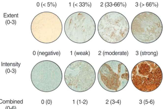
The extent scores of MUC1 and MUC4 staining in tumor cells were graded as follows: extent score 0 (< 5% immunore- active), extent score 1 (< 33% immunoreactive), extent score 2 (33-66% immunoreactive), extent score 3 (> 66% immunore- active). The intensity scores of MUC1 and MUC4 staining in tumor cells were graded using the following scale: score 0 (neg- ative), score 1 (weak), score 2 (moderate), score 3 (strong) (Fig.
1). A combined score (CS) was calculated by adding the extent scores and intensity scores of staining, using the following scale:
CS 0 = 0, CS 1 = 1-2, CS 2 = 3-4, CS 3 = 5-6.
Statistical analysis
Statistical analysis was performed using SPSS ver. 12.0 (SPSS Inc., Chicago, IL, USA). The chi-square test was used to evalu- ate the association between MUC1 or MUC4 expression and the clinicopathological parameters. A p-value less than 0.05 was considered statistically significant. We performed Kaplan-Meier analysis to investigate the potential relationship between MUC1 or MUC4 expression and patient survival.
0 (< 5%) Extent
(0-3)
Intensity (0-3)
Combined (0-6)
1 (< 33%) 2 (33-66%) 3 (> 66%)
0 (negative)
0 (0) 1 (1-2) 2 (3-4) 3 (5-6)
1 (weak) 2 (moderate) 3 (strong)
Fig. 1. The scoring criteria of the positive expression levels of mucin (MUC)1 and MUC4 immunoreactivity using tissue microarray in non-small cell lung carcinoma.
RESULTS Clinicopathological characteristics
The patient population consisted of 129 men (78%) and 36 women (22%). The mean patient age at the time of diagnosis was 60 years (range, 29 to 77 years). The most common histo- pathologic type was squamous cell carcinoma (59%) followed by adenocarcinoma (33%), large cell carcinoma (6%), and ade- nosquamous carcinoma (2%). The cases were classified into the following pathological stages: stage I (77, 47%), II (48, 29%), III (40, 24%), and IV (0).
The mean survival for each histologic type were as follows:
squamous cell carcinoma (54 months), adenocarcinoma (38 mon- ths), large cell carcinoma (21 months), and adenosquamous car- cinoma (22 months).
Immunohistochemical expression of MUC1 and MUC4 proteins in NSCLC
In general, MUC1 expression was observed more frequently and at a higher intensity than MUC4 expression throughout
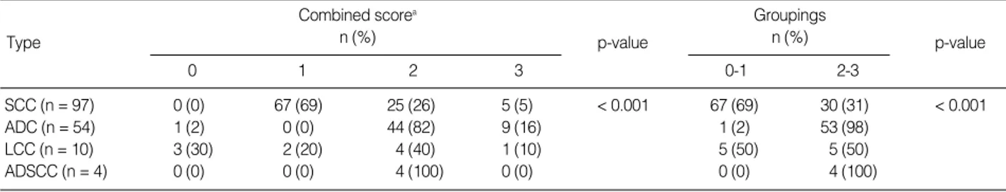
the series. Among NSCLC, adenocarcinoma showed high expres- sion for MUC1 (98%) and low expression for MUC4 (29%) on CS 2-3 (Tables 1, 2).
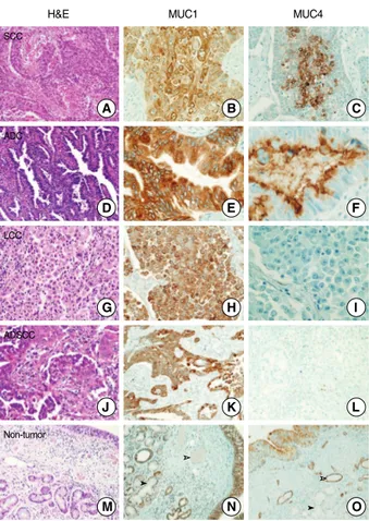
Squamous cell carcinoma showed diffuse MUC1 expression in the tumor cells (Fig. 2B), but focal MUC4 expression in the central portion of tumor cell nests (Fig. 2C). Adenocarcinoma cases showed diffuse positive MUC1 staining in the luminal portion and cytoplasm of tumor cells, whereas MUC4 staining was observed mainly in the apical and luminal portions of the tumor glands (Fig. 2E, F). Large cell carcinoma demonstrated diffuse positive expression of MUC1, but no expression of MUC4 (Fig. 2H, I). Adenosquamous carcinoma showed positive MUC1 expression in the tumor cells of the adenocarcinoma and squa- mous cell carcinoma. However, MUC4 expression was negative in the tumor cells (Fig. 2K, L). In the non-tumorous portions, MUC1 showed positive expression in the alveolar and bronchial epithelial cells, and in the submucosal glands, but was negative in the vascular endothelial cells (Fig. 2N). In contrast, MUC4 showed positive expression in the alveolar and bronchial epithe- lial cells, and in the vascular endothelial cells, but was negative in the submucosal glands (Fig. 2O).
aDegree of immunohistochemical expression using the combination of extent and intensity of the staining; combined score 0 (0), combined score 1 (1-2), combined score 2 (3-4), combined score 3 (5-6).
MUC, mucin; NSCLC, non-small cell lung carcinoma; SCC, squamous cell carcinoma; ADC, adenocarcinoma; LCC, large cell carcinoma; ADSCC, adenosquamous carcinoma.
Type
Combined scorea n (%)
0 1 2 3 0-1 2-3
p-value p-value
Groupings n (%)
SCC (n = 97) 0 (0) 67 (69) 25 (26) 5 (5) < 0.001 67 (69) 30 (31) < 0.001
ADC (n = 54) 1 (2) 0 (0) 44 (82) 9 (16) 1 (2) 53 (98)
LCC (n = 10) 3 (30) 2 (20) 4 (40) 1 (10) 5 (50) 5 (50)
ADSCC (n = 4) 0 (0) 0 (0) 4 (100) 0 (0) 0 (0) 4 (100)
Table 1. Comparison of MUC1 expression between each combined score and groupings of combined scores in NSCLC (n = 165)
aDegree of immunohistochemical expression using the combination of extent and intensity of the staining; combined score 0 (0), combined score 1 (1-2), combined score 2 (3-4), combined score 3 (5-6).
MUC, mucin; NSCLC, non-small cell lung carcinoma; SCC, squamous cell carcinoma; ADC, adenocarcinoma; LCC, large cell carcinoma; ADSCC, adenosquamous carcinoma.
Type
Combined scorea n (%)
0 1 2 3 0-1 2-3
p-value p-value
Groupings n (%)
SCC (n = 97) 36 (37) 54 (56) 6 (6) 1 (1) < 0.05 90 (93) 7 (7) < 0.01
ADC (n = 54) 15 (28) 23 (43) 12 (22) 4 (7) 38 (71) 16 (29)
LCC (n = 10) 5 (50) 4 (40) 0 (0) 1 (10) 9 (90) 1 (10)
ADSCC (n = 4) 1 (25) 3 (75) 0 (0) 0 (0) 4 (100) 0 (0)
Table 2. Comparison of MUC4 expression between each combined score and groupings of combined scores in NSCLC (n = 165)
Statistical analysis of MUC1 and MUC4 expression in NSCLC
There were no statistically significant associations between MUC1 or MUC4 expression levels and patient gender, age at the time of diagnosis and pathological stage. However, MUC1 and MUC4 expression levels were significantly correlated with
the histologic subtype of NSCLC. High expression of MUC1 was characteristic in adenocarcinoma, with combined scores of 2 and 3 (98%, p < 0.001) (Table 1). In contrast, low expression levels of MUC1 and MUC4 were characteristic of squamous cell carcinoma, with combined scores of 0 and 1 (69% and 93%, res- pectively) (Tables 1, 2). Statistical analyses of MUC1 and MUC4 expression levels were not available for large cell carcinoma and adenosquamous carcinoma due to insufficient numbers.
Prognostic significance of MUC1 and MUC4 expression in NSCLC
We performed survival analysis in patients with squamous cell carcinoma, adenocarcinoma, and other NSCLCs. There were no prognostic significances according to the combined scores of MUC1 and MUC4. However, there were some prognostic sig- nificance according to the extent scores of MUC1 and MUC4.
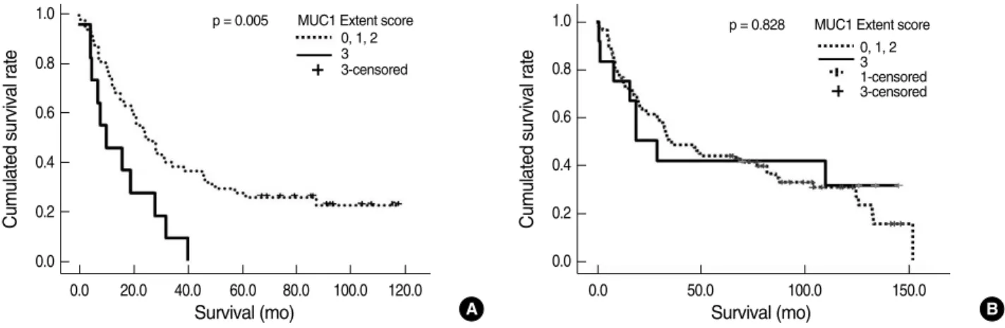
Survival analysis showed that diffuse MUC1 expression (extent score 3) was significantly associated with shortened survival in NSCLC patients, excluding cases of squamous cell carcinoma (p = 0.005) (Fig. 3A). There was no significant correlation be- tween diffuse MUC1 expression and shortened patient survival in squamous cell carcinoma (Fig. 3B). There was a trend of longer patient survival in NSCLC patients with diffuse MUC4 expres- sion (extent score 2 and 3), excluding squamous cell carcinoma (p = 0.128) (Fig. 4A). The survival analysis for diffuse MUC4 expression (extent score 2 and 3) showed no significant associa- tion in squamous cell carcinoma (Fig. 4B), which was similar to the result for MUC1 expression. No significant correlation was detected between high MUC1 and MUC4 expression lev- els and patient survival in NSCLC, adenocarcinoma, and large cell carcinoma (p > 0.05). After controlling for disease stage, there was no significant association between high MUC1 and MUC4 expressions and patient survival in patients with stage I and II tumors.
DISCUSSION
This study demonstrated high expression levels of MUC1 and MUC4 in NSCLC, with more frequent detection of MUC1 than MUC4. Our results support a previous study by Seregni et al.17 demonstrated by northern blot analysis that MUC1 was the highest expressed MUC gene in most lung carcinomas, followed by MUC4.
MUCs are high-molecular weight glycoconjugates (154 to
H&E MUC1 MUC4
Fig. 2. Mucin (MUC)1 and MUC4 proteins show various expres- sion patterns in different non-small cell lung carcinoma histologic subtype. (A-C) Squamous cell carcinoma (SCC): MUC1 shows diffuse expression in the tumor cells (B), but MUC4 shows well differentiated tumor cells, especially the central portion of the tumor nest (C). (D-F) Adenocarcinoma (ADC): MUC1 shows diffuse cy- toplasmic expression (E), but MUC4 shows expression in the api- cal and luminal portion of the tumor gland (F). (G-I) Large cell car- cinoma (LCC): MUC1 shows diffuse expression in the tumor cells (H), but MUC4 shows no expression (I). (J-L) Adenosquamous carcinoma (ADSCC): MUC1 shows positive expression in the tu- mor cells of both SCC and ADC (K), but MUC4 shows no expres- sion in the tumor cells (L). (M-O) Non-tumor: MUC1 shows posi- tive expression in the bronchial epithelial cells and submucosal glands, but no expression in the vascular endothelial cells (N). In contrast MUC4 shows positive expression in the bronchial epithe- lial cells and vascular endothelial cells, but no expression in the submucosal glands (O). White arrow head, vascular structure;
Black arrow head, submucosal glands; H&E, hematoxylin and eosin.
SCC
ADC
LCC
ADSCC
Non-tumor
A
D
G
J
M
B
E
H
K
N
C
F
I
L
O
over 7,000 kDa) with hundreds of oligosaccharide chains in O- glycosidic linkages to a protein backbone. MUC1 is a trans- membrane-bound MUC with a transmembrane segment, fol- lowed by a 69-amino acid cytoplasmic tail at the amino termi- nal end. Maeshima et al.18reported high frequency of MUC1 expression in adenocarcinoma compared to other types of lung cancer. This study showed that high MUC1 expression was char- acteristic of adenocarcinoma, whereas low expression of MUC1 and MUC4 was characteristic of squamous cell carcinoma. Mae- shima et al.18reported that high MUC1 expression was associ- ated with poor survival in patients with NSCLC. Tsuda et al.19 reported that high MUC1 gene expression was correlated with poor survival in 38 patients with NSCLC. Several studies sug- gested that MUC1 expression was an important component of biochemical events that enabled metastasis in cancer cells.20-22
Wesseling et al.23showed that MUC1 inhibited E-cadherin mediated the cell-cell adhesion system, and inhibition is depen- dent on the length of MUC1 ectodomain. High MUC1 levels also reduced integrin mediated cell adhesion to the extracellu- lar matrix.24MUC1 expressing cells are less susceptible to T cell mediated lysis, which may contribute to the ability of cancer cells to escape from immune surveillance.25
MUC4 is a heterodimeric glycoprotein complex consists of a MUC subunit, MUC4 alpha (ASGP-1 in the rat), tightly bound to a transmembrane subunit, MUC4 beta (ASGP-2 in the rat).26 The relationship between MUC4 expression and tumor behav- ior appears to be tissue-dependent.12,13,26-28MUC4 over-expres- sion was associated with aggressiveness and metastasis in breast cancer, extrahepatic bile duct carcinoma, and cholangiocarcino- ma.12,13,27Conversely, improved patient survival was associated
Cumulated survival rate
1.0
0.8
0.6
0.4
0.2
0.0
0.0 20.0 40.0 60.0 80.0 100.0 120.0 Survival (mo)
p = 0.005 MUC1 Extent score 0, 1, 2 3 3-censored
Cumulated survival rate
1.0
0.8
0.6
0.4
0.2
0.0
0.0 50.0 100.0 150.0
Survival (mo)
p = 0.828 MUC1 Extent score 0, 1, 2 3 1-censored 3-censored
A B
Fig. 3. Kaplan-Meier survival curves for mucin (MUC)1 expression according to non-small cell lung carcinoma (NSCLC) histologic subtype.
(A) In NSCLC, excluding cases of squamous cell carcinoma, there is a significant correlation between diffuse MUC1 expression (extent score 3) and shortened patient survival (p = 0.005). (B) In squamous cell carcinoma, there is no significant association between diffuse MUC1 expression (extent score 3) and patient survival (p = 0.828).
Cumulated survival rate
1.0
0.8
0.6
0.4
0.2
0.0
0.0 20.0 40.0 60.0 80.0 100.0 120.0 Survival (mo)
p = 0.128 MUC4 Extent score 0 + 1 2 + 3 0 + 1-censored 2 + 3-censored
Cumulated survival rate
1.0
0.8
0.6
0.4
0.2
0.0
0.0 50.0 100.0 150.0
Survival (mo)
p = 0.986 MUC4 Extent score 0 + 1 2 + 3 0 + 1-censored 2 + 3-censored
A B
Fig. 4. Kaplan-Meier survival curves for mucin (MUC)4 expression according to non-small cell lung carcinoma (NSCLC) histologic sub- type. (A) In NSCLC, excluding squamous cell carcinoma, Kaplan-Meier analysis reveals a trend toward improved survival in association with increased MUC4 expression (extent score 2 and 3) (p = 0.128). (B) In squamous cell carcinoma, there is no significant association between increased MUC4 expression (extent score 2 and 3) and patient survival (p = 0.986).
with high MUC4 expression in ovarian cancer, mucoepidermoid carcinoma of the salivary glands, and squamous cell carcinoma of the upper aerodigestive tract.26,28These divergent clinical be- haviors may result from tissue and tumor-specific differences in the relative importance of differentiation versus proliferative signals mediated by MUC4. Animal studies elucidating the functional relationship between MUC4 and ErbB2 suggested that these two membrane molecules associate as ligand and re- ceptor under various conditions, mediating different down- stream signaling pathways.29Two pathways of MUC4-mediat- ed ErbB2 tyrosine phosphorylation have been demonstrated.
Phosphorylation of the MUC4-ErbB2 complex can occur in the absence of heterodimerization with other receptors of the ErbB family, and in the absence of other known soluble lig- ands.30The heterodimers formed by MUC4-activated ErbB2 and neuregulin-activated ErbB3 are characterized by potentiat- ed phosphorylation of each of the 2 ErbB receptors, compared with MUC4-ErbB2 or ErbB2-ErbB3-neuregulin complex.
This study investigated the correlation between the expres- sion of both membrane-bound MUCs (MUC1 and MUC4) and NSCLC prognosis. We found that diffuse MUC1 expression sig- nificantly correlated with shortened survival in NSCLC, exclud- ing squamous cell carcinoma. However, diffuse MUC4 expres- sion was associated with longer survival in NSCLC, excluding squamous cell carcinoma. In adenocarcinoma, our results on the relationship between diffuse expression of MUC1, and MUC4 and survival were not consistent with previous reports. It remains unclear whether the expression of MUC1 and MUC4 proteins in lung carcinoma cells correlates with mRNA expression. We speculated that increased expression of MUC1 and MUC4 pro- teins was either due to increased mRNA levels, or to alterations of MUC1 and MUC4 glycosylation. Future studies in our labo- ratory will focus on the correlation between mRNA expression and the protein expression of MUC1 and MUC4 in NSCLC.
In conclusion, the expression levels of MUC1 and MUC4 were significantly correlated with the NSCLC histologic sub- type. Diffuse MUC1 expression was significantly correlated with shortened survival in NSCLC, excluding squamous cell carcinoma. In addition, diffuse MUC4 expression showed longer survival trend in NSCLC, excluding squamous cell carcinom.
REFERENCES
1. Kelsey CR, Boyd JA, Hubbs JL, et al. Local/regional recurrence fol- lowing surgery for early-stage lung cancer: a 10-year experience
with 975 patients. J Clin Oncol 2008; 26(15 Suppl): 7542.
2. Liu XQ, Ren HX, Wu ZP. Detecting MUC-1 mRNA for diagnosing peripheral blood micro-metastasis in non-small cell lung cancer pa- tients. Ai Zheng 2008; 27: 1267-70.
3. Komatsu M, Tatum L, Altman NH, Carothers Carraway CA, Car- raway KL. Potentiation of metastasis by cell surface sialomucin com- plex (rat MUC4), a multifunctional anti-adhesive glycoprotein. Int J Cancer 2000; 87: 480-6.
4. Moniaux N, Escande F, Porchet N, Aubert JP, Batra SK. Structural organization and classification of the human mucin genes. Front Biosci 2001; 6: D1192-206.
5. Senapati S, Sharma P, Bafna S, Roy HK, Batra SK. The MUC gene family: their role in the diagnosis and prognosis of gastric cancer.
Histol Histopathol 2008; 23: 1541-52.
6. Nakamori S, Ota DM, Cleary KR, Shirotani K, Irimura T. MUC1 mu- cin expression as a marker of progression and metastasis of human colorectal carcinoma. Gastroenterology 1994; 106: 353-61.
7. Ho SB, Niehans GA, Lyftogt C, et al. Heterogeneity of mucin gene expression in normal and neoplastic tissues. Cancer Res 1993; 53:
641-51.
8. Okabe S, Ishikawa T, Tanami S, et al. Clinicopathological study of mucous pooling referred to as mucinous component (MUC) in col- orectal submucosal invasive carcinomas. J Med Dent Sci 2001; 48:
69-78.
9. Monges GM, Mathoulin-Portier MP, Acres RB, et al. Differential MUC 1 expression in normal and neoplastic human pancreatic tis- sue: an immunohistochemical study of 60 samples. Am J Clin Pa- thol 1999; 112: 635-40.
10. Khodarev NN, Pitroda SP, Beckett MA, et al. MUC1-induced tran- scriptional programs associated with tumorigenesis predict outcome in breast and lung cancer. Cancer Res 2009; 69: 2833-7.
11. Andrianifahanana M, Moniaux N, Schmied BM, et al. Mucin (MUC) gene expression in human pancreatic adenocarcinoma and chronic pancreatitis: a potential role of MUC4 as a tumor marker of diag- nostic significance. Clin Cancer Res 2001; 7: 4033-40.
12. Carraway KL, Price-Schiavi SA, Komatsu M, Jepson S, Perez A, Car- raway CA. Muc4/sialomucin complex in the mammary gland and breast cancer. J Mammary Gland Biol Neoplasia 2001; 6: 323-37.
13. Shibahara H, Tamada S, Higashi M, et al. MUC4 is a novel prognos- tic factor of intrahepatic cholangiocarcinoma-mass forming type.
Hepatology 2004; 39: 220-9.
14. Hanaoka J, Kontani K, Sawai S, et al. Analysis of MUC4 mucin ex- pression in lung carcinoma cells and its immunogenicity. Cancer 2001; 92: 2148-57.
15. Travis WD, Brambilla E, Muller-Hermelink HK, Harris CC. World Health Organization classification of tumours: pathology and genet-
ics of tumours of the lungs, pleura, thymus and heart. Lyon: IARC Press, 2004.
16. Goldstraw P, Crowley J, Chansky K, et al. The IASLC Lung Cancer Staging Project: proposals for the revision of the TNM stage group- ings in the forthcoming (seventh) edition of the TNM classification of malignant tumours. J Thorac Oncol 2007; 2: 706-14.
17. Seregni E, Botti C, Lombardo C, et al. Pattern of mucin gene expres- sion in normal and neoplastic lung tissues. Anticancer Res 1996; 16:
2209-13.
18. Maeshima A, Miyagi A, Hirai T, Nakajima T. Mucin-producing adenocarcinoma of the lung, with special reference to goblet cell type adenocarcinoma: immunohistochemical observation and Ki- ras gene mutation. Pathol Int 1997; 47: 454-60.
19. Tsuda T, Mitsudomi T, Ohgami A, et al. Mucin gene expression is associated with the poor prognosis in human non-small cell lung cancer. Proc Am Assoc Cancer Res 1995; 36: 214.
20. Zhao Q, Guo X, Nash GB, et al. Circulating galectin-3 promotes me- tastasis by modifying MUC1 localization on cancer cell surface. Can- cer Res 2009; 69: 6799-806.
21. Nogi H, Takeyama H, Uchida K, Agata T, Horiguchi-Yamada J, Yamada H. Detection of MUC1 and keratin 19 mRNAs in the bone marrow by quantitative RT-PCR predicts the risk of distant metas- tasis in breast cancer patients. Breast Cancer 2003; 10: 74-81.
22. Nitta T, Sugihara K, Tsuyama S, Murata F. Immunohistochemical study of MUC1 mucin in premalignant oral lesions and oral squa- mous cell carcinoma: association with disease progression, mode of invasion, and lymph node metastasis. Cancer 2000; 88: 245-54.
23. Wesseling J, van der Valk SW, Hilkens J. A mechanism for inhibi-
tion of E-cadherin-mediated cell-cell adhesion by the membrane- associated mucin episialin/MUC1. Mol Biol Cell 1996; 7: 565-77.
24. Wesseling J, van der Valk SW, Vos HL, Sonnenberg A, Hilkens J.
Episialin (MUC1) overexpression inhibits integrin-mediated cell adhesion to extracellular matrix components. J Cell Biol 1995; 129:
255-65.
25. van de Wiel-van Kemenade E, Ligtenberg MJ, de Boer AJ, et al. Epi- sialin (MUC1) inhibits cytotoxic lymphocyte-target cell interaction.
J Immunol 1993; 151: 767-76.
26. Weed DT, Gomez-Fernandez C, Yasin M, et al. MUC4 and ErbB2 expression in squamous cell carcinoma of the upper aerodigestive tract: correlation with clinical outcomes. Laryngoscope 2004; 114: 1- 32.
27. Takao S, Uchikura K, Yonezawa S, Shinchi H, Aikou T. Mucin core protein expression in extrahepatic bile duct carcinoma is associated with metastases to the liver and poor prognosis. Cancer 1999; 86:
1966-75.
28. Weed DT, Gomez-Fernandez C, Pacheco J, et al. MUC4 and ERBB2 expression in major and minor salivary gland mucoepidermoid car- cinoma. Head Neck 2004; 26: 353-64.
29. Theodoropoulos G, Carraway CA, Carraway KL. MUC4 involve- ment in ErbB2/ErbB3 phosphorylation and signaling in response to airway cell mechanical injury. J Cell Biochem 2009; 107: 112-22.
30. Ramsauer VP, Pino V, Farooq A, Carothers Carraway CA, Salas PJ, Carraway KL. Muc4-ErbB2 complex formation and signaling in pol- arized CACO-2 epithelial cells indicate that Muc4 acts as an unor- thodox ligand for ErbB2. Mol Biol Cell 2006; 17: 2931-41.