https://doi.org/10.5468/ogs.2018.61.6.662 pISSN 2287-8572 · eISSN 2287-8580
Introduction
Cervical cancer is the leading cause of cancer-related mor- tality in women worldwide, and 80% of cases occur in developing countries [1]. Although the incidence rates of
Risk factors for cytological progression in HPV 16 infected women with ASC-US or LSIL: The Korean HPV cohort
Kyeong A So
1, Seon Ah Kim
1, Yoo Kyung Lee
1, In Ho Lee
1, Ki Heon Lee
1, Jee Eun Rhee
2, Mee Kyung Kee
2, Chi Heum Cho
3, Sung Ran Hong
4, Chang Sun Hwang
5, Mi Seon Jeong
6, Ki Tae Kim
7, Moran Ki
8,
Soo Young Hur
9, Jong Sup Park
9, Tae Jin Kim
11Department of Obstetrics and Gynecology, Cheil General Hospital and Women's Healthcare Center, College of Medicine, Dankook University, Seoul;
2Division of Viral Disease Research Center for Infectious Disease Research, National Institute of Health, Korea Centers for Disease Control and Prevention, Cheongju; 3Department of Obstetrics and Gynecology, Keimyung University Dongsan Medical Center, Daegu; 4Department of Pathology, Cheil General Hospital and Women's Healthcare Center, Dankook University College of Medicine, Seoul; 5Human Resource Biobank, Cheil General Hospital and Women's Healthcare Center, Dankook University College of Medicine, Seoul; 6Laboratory of R&D for Genomics, Cheil General Hospital and Women's Healthcare Center, College of Medicine, Dankook University, Seoul; 7Department of Obstetrics and Gynecology, Busan Paik Hospital, Inje University College of Medicine, Busan; 8Department of Cancer Control and Policy, Graduate School of Cancer Science and Policy, National Cancer Center, Goyang; 9Department of Obstetrics and Gynecology, Seoul St. Mary's Hospital, The Catholic University, Seoul, Korea
Objective
This study was to identify the risk factors for cytological progression in women with atypical squamous cells of undetermined significance (ASC-US) or low-grade squamous intraepithelial lesions (LSIL).
Methods
We analyzed data from women infected with the human papillomavirus (HPV) who participated in the Korean HPV cohort study. The cohort recruited women aged 20–60 years with abnormal cervical cytology (ASC-US or LSIL) from April 2010. All women were followed-up at every 6-month intervals with cervical cytology and HPV DNA testing.
Results
Of the 1,158 women included, 654 (56.5%) and 504 (43.5%) women showed ASC-US and LSIL, respectively. At the time of enrollment, 143 women tested positive for HPV 16 (85 single and 58 multiple infections). Cervical cytology performed in the HPV 16-positive women showed progression in 27%, no change in 23%, and regression in 50% of the women at the six-month follow-up. The progression rate associated with HPV 16 infection was higher than that with infection caused by other HPV types (relative risk [RR], 1.75; 95% confidence interval [CI], 1.08–2.84; P=0.028).
The cytological progression rate in women with persistent HPV 16 infection was higher than that in women with incidental or cleared infections (P <0.001). Logistic
regression analysis showed a significant relationship between cigarette smoking and cytological progression (RR, 4.15; 95% CI, 1.01–17.00).
Conclusion
The cytological progression rate in HPV 16-positive women with ASC-US or LSIL is higher than that in women infected with other HPV types. Additionally, cigarette smoking may play a role in cytological progression.
Keywords: Papillomaviridae; Smoking; Epidemiology
Articles published in Obstet Gynecol Sci are open-access, distributed under the terms of the Creative Commons Attribution Non-Commercial License (http://creativecommons.
org/licenses/by-nc/3.0/) which permits unrestricted non-commercial use, distribution, and reproduction in any medium, provided the original work is properly cited.
Copyright © 2018 Korean Society of Obstetrics and Gynecology
Received: 2017.11.18. Revised: 2018.02.12. Accepted: 2018.03.08.
Corresponding author: Tae Jin Kim
Department of Obstetrics and Gynecology, Cheil General Hospital and Women's Healthcare Center, College of Medicine, Dankook University, 17 Seoae-ro 1-gil, Jung-gu, Seoul 04619, Korea E-mail: [email protected]
https://orcid.org/0000-0002-5322-2745 Co-corresponding author: Jong Sup Park
Department of Obstetrics and Gynecology, Seoul St. Mary's Hospital, The Catholic University of Korea College of Medicine, 222 Banpo-daero, Seocho-gu, Seoul 06591, Korea
E-mail: [email protected]
https://orcid.org/0000-0003-4086-4885
cervical cancer have consistently declined in Koreans, it was identified as the third most common cancer in women aged between 15 and 34 years [2,3]. There are 3,500 newly di- agnosed cervical cancer cases with 960 deaths in Korea [3].
Human papillomavirus (HPV) infection has been definitively implicated in the pathogenesis of cervical intraepithelial neo- plasia (CIN) and cervical cancer [4,5]. Most HPV infections are cleared by the immune system within one to two years [6].
However, persistent infection with high-risk HPV can trigger the development of high-grade squamous intraepithelial le- sions (HSIL) and cervical cancer [7]. Among the 170 known HPV types, a few have been classified as a high-risk variety because of their high rate of detection in women with cervi- cal cancers [8]. HPV 16 and 18 are the most prevalent high- risk types that account for more than 70% of all invasive cervical cancers [9]. Additionally, the ten-year cumulative incidence of HSIL or cancer in women with HPV 16 infection (17%) is significantly higher than that in women infected by other high-risk genotypes (3%) [10]. Thus, the detection of HPV 16 infection in cervical samples is useful to identify women at high risk of developing HSIL or cancer.
Although women with both HPV infection and abnormal Pap results are commonly observed in clinical practice, most of them do not develop cervical cancer. Previous studies have reported that the risk of development of subsequent HSIL or cancer following low-grade lesions such as atypical squamous cells of undetermined significance (ASC-US) and low-grade squamous intraepithelial lesions (LSIL) of the cervix ranges between only 1.9% and 13% [11-13]. The clinical significance of low-grade cervical lesions remains unclear. An interaction between demographic, immunological, and envi- ronmental factors can affect the persistence of HPV infection and the development of high-grade lesions [14]. Therefore, a population-based study is warranted to evaluate the risk factors determining the progression of low-grade cervical le- sions and the subsequent management of these women. We evaluated the cytological prognosis and epidemiological risk factors for cytological progression in HPV 16-positive women with ASC-US or LSIL.
Materials and methods
We analyzed data obtained as part of the Korean HPV co- hort study, a large, prospective, multicenter study includ-
ing 5 university medical centers nationwide, funded by the Korea Centers for Disease Control and Prevention [15]. The Korean HPV cohort study is an ongoing research project with long-term follow-up that aims to establish the infrastructure for the optimal management of HPV infection. This study enrolled a total of 1,158 women between April 2010 and October 2014 and included women aged 20–60 years who were HPV-positive (ASC-US or LSIL on cytological examina- tion), regardless of previous HPV infection. Exclusion criteria were pregnancy, malignant disease including cervical cancer diagnosed at the time of enrollment, actively treated psycho- logical disease, and a history of hysterectomy or CIN treated within six months of enrollment. Institutional Review Board approval (CGH-IRB-2010-13) was obtained at each study center, and all women provided written informed consent.
Upon enrollment, all women completed a self-administered questionnaire comprising the following categories of data:
sociodemographic status, health-related lifestyle, women- specific conditions, and sexual habits. Additionally, women were followed-up with cervical cytology and HPV DNA test- ing at six-month intervals. For cervical cytological examina- tion, both conventional and liquid-based Pap smears were acceptable as baseline evaluation tests; however, only the liquid-based Pap smear using the Cervex-Brush (Rovers Medi- cal Devices, Oss, Netherlands) was used during follow-up examinations. The cytological results were reported at Cheil General Hospital. HPV genotyping was performed using a DNA microarray technique based on a polymerase chain re- action method using a Cheil HPV DNA chip kit (Cheil General Hospital, Seoul, Korea). Samples were tested for the pres- ence of 19 high-risk HPV types (16, 18, 31, 33, 35, 39, 45, 51, 52, 53, 56, 58, 59, 66, 67, 68a, 68b, 69, and 82) and 17 low-risk HPV types (6, 11, 30, 32, 40, 42, 43, 44, 54, 55, 62, 70, 72, 81, 84, 90, and 91).
We categorized the cytological results obtained at follow- up as progression, regression, or no change. Progression was defined as atypical squamous cells, and the inability to exclude a high-grade squamous intraepithelial lesion (ASC- H), HSIL or LSIL from ASC-US, and HSIL from LSIL. Regression was defined as a change to normal cytological features, to ASC-US from LSIL, or to normal cytological features from ASC-US. HPV test results observed during follow-up were classified into three groups as follows: persistence, incidence, and clearance. Persistence was defined as positivity for both the same HPV type as that observed at the time of enroll-
ment or the same HPV type concomitant with other types.
Incidence was defined as positivity only for types different from HPV types at enrollment. Clearance was defined as a negative HPV test.
1. Statistical analysis
Baseline data are presented as mean±standard deviation for continuous variables or frequency (%) for categorical variables. Patients’ general and clinical characteristics were compared using the Pearson’s χ2 test. The Kruskal-Wallis test was used to analyze prognosis based on the HPV status. A logistic regression model was used to estimate relative risk (RR) and 95% confidence intervals (CIs) to characterize the associations between HPV infection and epidemiological characteristics. All P-values <0.05 were considered statisti- cally significant. The IBM SPSS Statistics software, version 20.0 (SPSS Inc., Chicago, IL, USA) was used for all data analyses.
Results
The Korean HPV cohort enrolled 654 (56.5%) and 504 (43.5%) women with ASC-US and LSIL, respectively. How- ever, 359 women did not undergo any six-month follow-up tests. All 799 women included in the study were followed-up at least once. Six-month follow-up examination showed that ASC-US regressed to normal cytological features in 46.3% of the women, persisted in 31.0% women, and progressed to LSIL, ASC-H, or HSIL in 7.2%, 6.8%, and 8.7% women, re- spectively. Similarly, LSIL showed regression to normal cytol- ogy in 42.5% or to ASC-US in 29.9% and persisted as LSIL in 14.4%. Progression to HSIL occurred in only 45 women (13.2%).
Among the 1,158 enrolled patients, the 10 most common- ly identified HPV types were all high-risk types. In the order of decreasing prevalence these were HPV 16, 58, 56, 53, 52, 39, 18, 51, 68, and 66. HPV 16 showed the highest preva- lence (12.3%). Among the 799 women who underwent six- month follow-up tests, 72 women infected with unknown
Table 1. Changes in cervical cytology and human papillomavirus infection status in women initially diagnosed with atypical squamous cells of undetermined significance/low-grade squamous intraepithelial lesion
Category 6-month, No. (%)
P-value 12-month, No. (%)
P-value
HPV 16 Other HPVs HPV 16 Other HPVs
Cytology
Progression 27 (27.0) 122 (17.5) 0.028 13 (20.3) 79 (16.0) 0.373
No change 23 (23.0) 168 (24.0) 0.901 13 (20.3) 98 (19.8) 0.869
Regression 50 (50.0) 409 (58.5) 0.130 38 (59.4) 318 (64.2) 0.490
Total 100 (100) 699 (100) 64 (100) 495 (100)
HPV status
Persistent infection 54 (54.0) 291 (46.4) 0.163 25 (39.1) 148 (33.3) 0.398
Incidental infection 25 (25.0) 159 (25.4) 1.000 22 (34.4) 136 (30.6) 0.565
Clearance 21 (21.0) 177 (28.2) 0.147 17 (26.6) 160 (36.0) 0.161
Total 100 (100) 627 (100) 64 (100) 444 (100)
HPV, human papillomavirus.
Table 2. The association between human papillomavirus status and cervical cytology in human papillomavirus 16 infected women
Category 6-month, No. (%)
P-value 12-month, No. (%)
P-value Progression No change Regression Progression No change Regression
Persistent infection 23 (85.2) 15 (65.2) 16 (32.0) <0.001 11 (84.6) 7 (53.8) 7 (18.4) <0.001
Incidental infection 3 (11.1) 7 (30.4) 15 (30.0) 2 (15.4) 4 (30.8) 16 (42.1)
Clearance 1 (3.7) 1 (4.4) 19 (38.0) 0 (0) 2 (15.4) 15 (39.5)
Total 27 (100) 23 (100) 50 (100) 13(100) 13 (100) 38 (100)
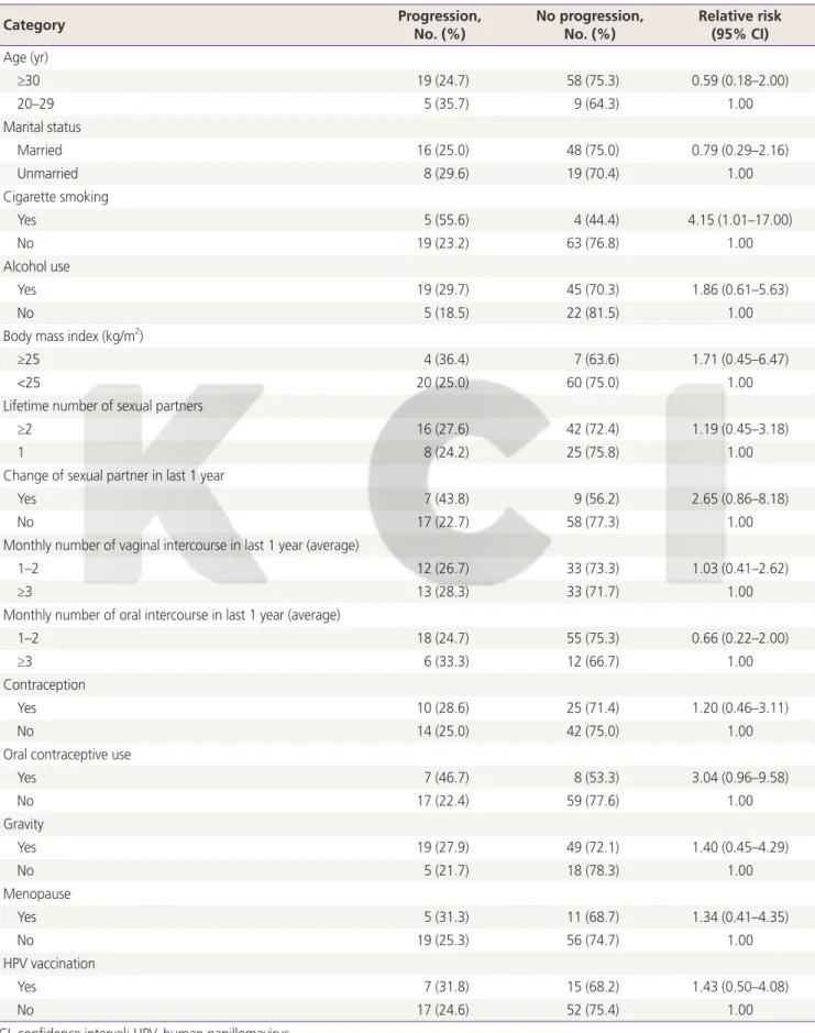
Table 3. Analysis of epidemiologic risk factors for cytological progression in human papillomavirus 16 infected women (n=91)
Category Progression,
No. (%) No progression,
No. (%) Relative risk (95% CI) Age (yr)
≥30 19 (24.7) 58 (75.3) 0.59 (0.18–2.00)
20–29 5 (35.7) 9 (64.3) 1.00
Marital status
Married 16 (25.0) 48 (75.0) 0.79 (0.29–2.16)
Unmarried 8 (29.6) 19 (70.4) 1.00
Cigarette smoking
Yes 5 (55.6) 4 (44.4) 4.15 (1.01–17.00)
No 19 (23.2) 63 (76.8) 1.00
Alcohol use
Yes 19 (29.7) 45 (70.3) 1.86 (0.61–5.63)
No 5 (18.5) 22 (81.5) 1.00
Body mass index (kg/m2)
≥25 4 (36.4) 7 (63.6) 1.71 (0.45–6.47)
<25 20 (25.0) 60 (75.0) 1.00
Lifetime number of sexual partners
≥2 16 (27.6) 42 (72.4) 1.19 (0.45–3.18)
1 8 (24.2) 25 (75.8) 1.00
Change of sexual partner in last 1 year
Yes 7 (43.8) 9 (56.2) 2.65 (0.86–8.18)
No 17 (22.7) 58 (77.3) 1.00
Monthly number of vaginal intercourse in last 1 year (average)
1–2 12 (26.7) 33 (73.3) 1.03 (0.41–2.62)
≥3 13 (28.3) 33 (71.7) 1.00
Monthly number of oral intercourse in last 1 year (average)
1–2 18 (24.7) 55 (75.3) 0.66 (0.22–2.00)
≥3 6 (33.3) 12 (66.7) 1.00
Contraception
Yes 10 (28.6) 25 (71.4) 1.20 (0.46–3.11)
No 14 (25.0) 42 (75.0) 1.00
Oral contraceptive use
Yes 7 (46.7) 8 (53.3) 3.04 (0.96–9.58)
No 17 (22.4) 59 (77.6) 1.00
Gravity
Yes 19 (27.9) 49 (72.1) 1.40 (0.45–4.29)
No 5 (21.7) 18 (78.3) 1.00
Menopause
Yes 5 (31.3) 11 (68.7) 1.34 (0.41–4.35)
No 19 (25.3) 56 (74.7) 1.00
HPV vaccination
Yes 7 (31.8) 15 (68.2) 1.43 (0.50–4.08)
No 17 (24.6) 52 (75.4) 1.00
CI, confidence interval; HPV, human papillomavirus.
HPV types at the time of enrollment were excluded. Of the remaining women, 345 (47.5%), 184 (25.3%), and 198 (27.2%) showed persistent, incidental, and cleared infection, respectively. The most common type was HPV 16 (n=143, 12.3%) presenting as a single infection (n=85, 59.4%) and multiple infections (n=58, 40.6%). Among those showing cytological progression at the six-month follow-up, 27.0%
were infected with HPV 16, which represented a significantly higher progression rate than that observed with other HPV types (RR, 1.75; 95% CI, 1.08–2.84; P=0.028) (Table 1).
HPV 16 infection status at follow-up testing was associ- ated with cervical cytology prognosis (Table 2). Persistent HPV 16 infection was significantly associated with cytologi- cal progression over one year (P<0.001). At the six-month follow-up, the rate of persistent infection was 85.2% in the cytological progression and only 32.0% in the cytological re- gression group. Only 3.7% of the women in the cytological progression group had a negative HPV test at follow-up, and HPV 16 infection was cleared in 38.0% of the women in the cytological regression group. The rate of cytological progres- sion was 42.6% (23 of 54) in the HPV 16 persistence group and only 4.8% (1 of 21) in the clearance group. The rate of cytological regression was only 29.6% (16 of 54) in the HPV 16 persistence group and as high as 90.5% (19 of 21) in the clearance group. The results were similar at the 12-month follow-up. The cytological progression rate in women with multiple HPV 16 infections (27.9%) was similar to that in women with HPV 16 infection alone (26.3%).
We performed logistic regression analysis to evaluate the epidemiological risk factors affecting cytological progression in HPV 16-positive women (Table 3). Of the 91 women who underwent six-month follow-up, 53 women had an HPV 16 single infection and 38 had HPV 16 multiple infections at the time of enrollment. The RR of progression in cigarette smok- ers (including former smokers) was higher than that in non- smokers (RR, 4.15; 95% CI, 1.01–17.0). We performed mul- tivariate logistic regression analysis to assess the compound effects of variables on the risk of progression. Smoking was the only significant independent predictor of cytological pro- gression (odds ratio, 4.15; 95% CI, 1.01–17.00; P=0.048).
Young age, alcohol use, obesity, oral contraceptive use, change in sexual partners within a year prior to enrollment in the study, gravidity, menopause, and HPV vaccination were not significantly associated with a high risk of cytological progression.
Discussion
The results of this study suggest that HPV 16 infection is a significant risk factor for the development of cervical cancer.
A meta-analysis including 14,595 patients reported that most invasive cervical cancers worldwide are associated with HPV 16 infection [16]. Current guidelines recommend additional evaluation as a component of cervical cancer screening in women with HPV 16 infection [17]. We evaluated the persis- tent HPV 16 infection rates and the epidemiological factors associated with cytological progression in HPV 16-positive women. This study showed that the rate of development of high-grade lesions in women with HPV 16 infection was high- er than that in women with other types of HPV infection (RR, 1.75; 95% CI, 1.08–2.84; P=0.028). Among HPV 16-positive women, persistent infection was observed in 54% at the six- month follow-up and in 39.1% at the 12-month follow-up.
Both rates were higher than the persistence rates of 47.5%
and 34.1%, respectively in the overall HPV cohort, suggesting that HPV 16 infection is more likely to persist and is associ- ated with progression to high-grade cervical lesions.
Risk factors for cervical cancer include age, cigarette smok- ing, multiple sexual partners, gravidity, an immunocompro- mised status, and long-term use of oral contraceptives [18].
Among the HPV 16-positive women evaluated in this study, cigarette smokers showed a significantly higher risk for pro- gression to abnormal cervical cytology. Smoking has been significantly associated with an increased risk of cervical can- cer [19-21]. Smoking interferes with HPV clearance by im- pairing the immune response that decreases the number of CD4 lymphocytes and Langerhans cells, as well as reducing the activity of natural killer cells [22-27]. A higher risk of cer- vical cancer was observed in women with HPV 16 infection with a history of smoking (not necessarily current smokers).
Moreover, a dose-dependent relationship with the number of cigarettes smoked has been reported [28]. Additionally, young age, alcohol use, obesity, oral contraceptive use, change of sexual partners within a year prior to enrollment in the study, gravidity, and menopause were associated with a higher risk of cytological progression. However, several other factors correlated with cytological progression were statisti- cally non-significant owing to the short-term follow-up and small sample size of this study.
The strengths of this study include the long-term prospec- tive study design, the fact that we included women from
five university medical centers across four metropolitan cities in Korea, and that we minimized interobserver variability in result interpretation by performing all cytological evaluations at a single center (Cheil General Hospital). Limitations of our study are as follows: 1) The Korean HPV cohort was designed such that follow-up biopsies were not mandatory, and only cytology and HPV testing were performed at each follow- up visit. Although cytology and HPV testing are effective screening tools for precancerous lesions, colposcopy guided biopsies remain the diagnostic standard for cervical lesions.
2) An incomplete understanding of other potential risk factors (previous HPV infection status, age at which HPV vaccination was administered) may limit the interpretation of results. 3) The 12-month follow-up used in this study is relatively short. A previous study has shown that cytological regression from ASC-US to normal and from LSIL to ASC- US or normal cytology occurred in 16.8 and 13.8 months, respectively in women with oncogenic HPV types [29]. In this study, among women with cytological progression at the six- month follow-up, the progression rate in HPV 16-positive women was significantly higher than that in women infected with other HPV types (27.0% vs. 17.5%; RR, 1.75; 95% CI, 1.08–2.84; P=0.028). However, the 12-month follow-up re- sults did not show a statistically significant difference. Thus, long-term follow-up studies are needed to draw more accu- rate inferences. Although this study included a relatively short follow-up period, the Korean HPV cohort is an ongoing study.
Owing to the relatively large sample size and the systematic study design, the results of that cohort study are expected to be more representative of Korean women as a whole.
In conclusion, the cytological progression rate in HPV 16-positive women with ASC-US or LSIL is higher than that in women infected with other HPV types. Furthermore, smoking among women with HPV 16 infection is a major risk factor for the progression of cervical lesions. This study would provide a better understanding of the epidemiological risk factors that determine the prognosis of low-grade cervi- cal abnormalities in Korean women.
Acknowledgements
This work was supported by the Research Program funded by the Korea Centers for Disease Control and Prevention (2013- E51005-02).
Conflict of interest
No potential conflict of interest relevant to this article was reported.
References
1. Torre LA, Bray F, Siegel RL, Ferlay J, Lortet-Tieulent J, Je- mal A. Global cancer statistics, 2012. CA Cancer J Clin 2015;65:87-108.
2. Jung KW, Won YJ, Oh CM, Kong HJ, Cho H, Lee DH, et al. Prediction of cancer incidence and mortality in Korea, 2015. Cancer Res Treat 2015;47:142-8.
3. Jung KW, Won YJ, Oh CM, Kong HJ, Lee DH, Lee KH;
et al. Cancer statistics in Korea: incidence, mortal- ity, survival, and prevalence in 2014. Cancer Res Treat 2017;49:292-305.
4. Walboomers JM, Jacobs MV, Manos MM, Bosch FX, Kummer JA, Shah KV, et al. Human papillomavirus is a necessary cause of invasive cervical cancer worldwide. J Pathol 1999;189:12-9.
5. Choi YJ, Park JS. Clinical significance of human papillo- mavirus genotyping. J Gynecol Oncol 2016;27:e21.
6. Cho HW, So KA, Lee JK, Hong JH. Type-specific persis- tence or regression of human papillomavirus genotypes in women with cervical intraepithelial neoplasia 1: a pro- spective cohort study. Obstet Gynecol Sci 2015;58:40-5.
7. Koshiol J, Lindsay L, Pimenta JM, Poole C, Jenkins D, Smith JS. Persistent human papillomavirus infection and cervical neoplasia: a systematic review and meta-analy- sis. Am J Epidemiol 2008;168:123-37.
8. Muñoz N, Bosch FX, de Sanjosé S, Herrero R, Castell- sagué X, Shah KV, et al. Epidemiologic classification of human papillomavirus types associated with cervical cancer. N Engl J Med 2003;348:518-27.
9. Castellsagué X. Natural history and epidemiology of HPV infection and cervical cancer. Gynecol Oncol 2008;110 Suppl 2:S4-7.
10. Khan MJ, Castle PE, Lorincz AT, Wacholder S, Sherman M, Scott DR, et al. The elevated 10-year risk of cervical precancer and cancer in women with human papilloma- virus (HPV) type 16 or 18 and the possible utility of type- specific HPV testing in clinical practice. J Natl Cancer Inst 2005;97:1072-9.
11. Cox JT, Schiffman M, Solomon D; ASCUS-LSIL Triage Study (ALTS) Group. Prospective follow-up suggests simi- lar risk of subsequent cervical intraepithelial neoplasia grade 2 or 3 among women with cervical intraepithelial neoplasia grade 1 or negative colposcopy and directed biopsy. Am J Obstet Gynecol 2003;188:1406-12.
12. Pretorius RG, Peterson P, Azizi F, Burchette RJ. Subse- quent risk and presentation of cervical intraepithelial neoplasia (CIN) 3 or cancer after a colposcopic diagnosis of CIN 1 or less. Am J Obstet Gynecol 2006;195:1260-5.
13. Chen EY, Tran A, Raho CJ, Birch CM, Crum CP, Hirsch MS. Histological ‘progression’ from low (LSIL) to high (HSIL) squamous intraepithelial lesion is an uncommon event and an indication for quality assurance review.
Mod Pathol 2010;23:1045-51.
14. Kim JY, Nam BH, Lee JA. Is human papillomavirus geno- type an influencing factor on radiotherapy outcome?
Ambiguity caused by an association of HPV 18 geno- type and adenocarcinoma histology. J Gynecol Oncol 2011;22:32-8.
15. Lee WC, Lee SY, Koo YJ, Kim TJ, Hur SY, Hong SR, et al.
Establishment of a Korea HPV cohort study. J Gynecol Oncol 2013;24:59-65.
16. Smith JS, Lindsay L, Hoots B, Keys J, Franceschi S, Winer R, et al. Human papillomavirus type distribution in in- vasive cervical cancer and high-grade cervical lesions: a meta-analysis update. Int J Cancer 2007;121:621-32.
17. Saslow D, Solomon D, Lawson HW, Killackey M, Kulas- ingam SL, Cain J, et al. American Cancer Society, Ameri- can Society for Colposcopy and Cervical Pathology, and American Society for Clinical Pathology screening guide- lines for the prevention and early detection of cervical cancer. Am J Clin Pathol 2012;137:516-42.
18. Wang SS, Zuna RE, Wentzensen N, Dunn ST, Sherman ME, Gold MA, et al. Human papillomavirus cofactors by disease progression and human papillomavirus types in the study to understand cervical cancer early endpoints and determinants. Cancer Epidemiol Biomarkers Prev 2009;18:113-20.
19. Castellsagué X, Muñoz N. Chapter 3: cofactors in hu- man papillomavirus carcinogenesis--role of parity, oral contraceptives, and tobacco smoking. J Natl Cancer Inst Monogr 2003;31:20-8.
20. IARC Working Group on the Evaluation of Carcinogenic
Risks to Humans. Tobacco smoke and involuntary smok- ing. IARC Monogr Eval Carcinog Risks Hum 2004;83:1- 1438.
21. Appleby P, Beral V, Berrington de González A, Colin D, Franceschi S, Goodill A, et al. Carcinoma of the cervix and tobacco smoking: collaborative reanalysis of indi- vidual data on 13,541 women with carcinoma of the cervix and 23,017 women without carcinoma of the cervix from 23 epidemiological studies. Int J Cancer 2006;118:1481-95.
22. Vaccarella S, Herrero R, Snijders PJ, Dai M, Thomas JO, Hieu NT, et al. Smoking and human papillomavirus in- fection: pooled analysis of the International Agency for Research on Cancer HPV Prevalence Surveys. Int J Epide- miol 2008;37:536-46.
23. Poppe WA, Ide PS, Drijkoningen MP, Lauweryns JM, Van Assche FA. Tobacco smoking impairs the local immuno- surveillance in the uterine cervix. An immunohistochemi- cal study. Gynecol Obstet Invest 1995;39:34-8.
24. Poppe WA, Peeters R, Drijkoningen M, Ide PS, Daenens P, Lauweryns JM, et al. Cervical cotinine and macrophage- Langerhans cell density in the normal human uterine cervix. Gynecol Obstet Invest 1996;41:253-9.
25. Barton SE, Maddox PH, Jenkins D, Edwards R, Cuzick J, Singer A. Effect of cigarette smoking on cervical epi- thelial immunity: a mechanism for neoplastic change?
Lancet 1988;2:652-4.
26. de Jong A, van Poelgeest MI, van der Hulst JM, Drijfhout JW, Fleuren GJ, Melief CJ, et al. Human papillomavirus type 16-positive cervical cancer is associated with im- paired CD4+ T-cell immunity against early antigens E2 and E6. Cancer Res 2004;64:5449-55.
27. Evans EM, Man S, Evans AS, Borysiewicz LK. Infiltration of cervical cancer tissue with human papillomavirus-spe- cific cytotoxic T-lymphocytes. Cancer Res 1997;57:2943- 50.
28. Herrero R, Brinton LA, Reeves WC, Brenes MM, Tenorio F, de Britton RC, et al. Invasive cervical cancer and smok- ing in Latin America. J Natl Cancer Inst 1989;81:205-11.
29. Schlecht NF, Platt RW, Duarte-Franco E, Costa MC, Sobrinho JP, Prado JC, et al. Human papillomavirus infection and time to progression and regression of cervical intraepithelial neoplasia. J Natl Cancer Inst 2003;95:1336-43.