Ⅰ. 서 론
현재까지 골 결손부를 재건하기 위한 골 이식 재료로 가장 널리 사용되고 있는 것은 자가골이며1-2)
,자가골은 다른 종류의
골 이식 재료와 달리 골 형성 기전에 의해 치유되며 면역학적 반응이 문제되지 않는 장점을 가지고 있다. 악골의 큰 골 결손 부는 주로 장골 이식에 의해 수복되며, 이 경우 골 채취를 위한부가적 수술이 요구되는 단점을 가지고 있다3)
. 작은 골 결손부
일 경우에는 부가적인 수술이 필요 없어 주변에서 손쉽게 자 가골을 채취하여 이식하게 되므로 수술 시간을 절약할 수 있 고 환자의 불편감을 감소시킬 수 있다. 악골과 같은 막성골 형 태(intramembranous bone type)의 경우 늑골, 경골, 장골 등과 같 은 구강외 공여부에 해당하는 연골내화성 골 형태(endochondri-al bone type)보다 치유 기간을 줄일 수 있으며, 이식 후 흡수율
이 적고 많은 양의 망상골(cancellous bone)을 이용할 수 있어 빠 른 골 형성과 재혈관화를 촉진할 수 있다는 장점을 지니고 있 다4-5).
지금까지 악골로 부터 자가골을 채취하기 위한 다양한 기구 가 소개되었는데, 그중 1998년 Tomaso Vercellotti에 의해 개발된
Piezosurgery가 새롭게 각광을 받고 있다
6-10). Piezosurgery는 일반
Abstract (J. Kor. Oral Maxillofac. Surg. 2008;34:428-434)권 대 근
700-721 대구광역시 중구 삼덕2가50 경북대학교 치과대학 구강악안면외과학교실 Tae-Geon Kwon
Dept. of OMFS, School of Dentistry, Kyungpook National University 50 Samduk 2-ga, Jung-gu, Daegu, 700-721, Korea
Tel: 82-53-420-5911,5912 Fax: 82-53-426-5365
가토 두개골에 이식한 다양한 형태의 자가골에 의한 골성 회복 양상 비교 평가
최소영∙이수연∙김진욱∙김진수∙이상한∙신홍인*∙권대근
경북대학교 치과대학 구강악안면외과학교실, *경조직-바이오치아 재생 연구소, 구강병리학교실
A COMPARATIVE STUDY OF VARIOUS TYPE OF AUTOGENEOUS BONE GRAFT ON THE RABBIT-SKULL DEFECT HEALING
So-Young Choi, Su-Youn Lee, Jin-Wook Kim, Chin-Soo Kim, Sang-Han Lee, Hong-In Shin*, Tae-Geon Kwon
Dept. of Oral & Maxillofacial Surgery, *IHBR, Dept. of Oral Pathology, School of Dentistry, Kyungpook National University, Kyungpook National University
Introduction: Piezosurgery device is one of the most commonly used instrument on the intraoral surgery such as maxillary sinus lift and autoge- neous bone graft. Piezosurgery instrument also contains the tips that are manufactured especially for the convenient bone graft, which now many sur- geons apply them for collecting bone graft materials in the curettage method for the restoration of skull defects. However, objective data has not been shown concerning the effects about bone graft with using Piezosurgery. Therefore we investigated the effects of Piezosurgery on the rabbit-skull defect healing.
Materials & Methods: To investigate the regeneration of the bony defect with various bone graft, 10 adult New Zealand white rabbits (average weight : 2.8 ± 0.3kg, about 12weeks) were used. The four circular bony defects measuring 6mm in diameter were made with Piezosurgery device on each rabbit cranial bone. The harvested bone tissues during defect formation were also used for autogeneous bone graft. They were grafted into the defects in a various type; block type (Group 1), particulated type by the bone mill (Group 2), chopped type by curette shaped Piezosurgery tip (Group 3), the defect without any graft was served as control (control group). The animals were sacrificed after 6 weeks and bone regeneration capacity was evaluated histomorphometrically.
Result & Conclusion: Autogeneous bone graft harvested using a Piezosurgery instrument showed satisfactory bone regeneration. There was no conspicuous difference bone prepared amomg by bone mill or Piezosurgery and block bone graft. Therefore, the bone harvested from the intraoral site near the operation field using the piezosurgery device can be a feasible and reliable graft for intraoral bony defects.
Key words: Autogeneous bone graft, Skull defects of the rabbits, Piezosursery device
서 30kHz의 주파수를 갖는 특별한 진동을 가짐으로써 선택적 인 골 절단이 가능하다. 사용 시 충분한 주수로 인해 열 발생을 감소시킬 수 있으며, 연조직에 손상을 주지 않기 때문에 다양 한 구강 내 술식에 유용하게 활용될 수 있다. Piezosurgery는 용 도에 맞게 다양한 tip을 제공하고 있다. 골 표면을 긁어내어 손 쉽게 골을 채취할 수 있는 tip도 포함되어 있어, 인접된 골부위 로부터 curettage하듯 골 이식편을 얻어 이식하는 방법이 임상 적으로 많이 적용되고 있다. 그러나 아직 객관적인 Piezosurgery 를 이용한 골 이식 효과에 대한 자료가 많지 않아 이번 연구를 통해 Piezosurgery를 이용한 골 이식의 효과에 대해 입증하고자 하였다.
Ⅱ. 연구 대상 및 연구 방법 1. 연구대상
본 실험은 12주령의 New Zealand산 백색 가토 10마리(평균 무 게 2.8 ± 0.3kg)를 사용하였다. 각 실험 동물의 두개골에
Piezosurgery(Mectron Piezosurgery
�, Medical Technology, Carasco, GE, Italy) tip(OT 5)을 이용하여 지름 6mm의 원형 골 결손부 4개
를 형성하였고 다음과 같이 골을 이식하지 않은 대조군과 서로 다른 형태의 자가골을 이식한 실험군으로 구분하여 관찰하 였다.
대조군 : 골 이식을 하지 않은 군
실험 1군 : 채취한 원형의 블록형 골조직을 그대로 이식한 실 험군
실험 2군 : 채취한 블록형 골을 bone mill(Bone milling Forceps,
USTOMED INSTRUMENTE Ulrich Storz GmbH &
Co. KG)로 분쇄하여 이식한 실험군
실험 3군 : 결손부를 형성하는 과정 중에 Piezosurgery tip(OT 7) 을 이용하여 골조직을 긁어서, 골수집기(Osseous
bone collector-OTA 100, Osseous Technologies of America)를 이용하여 골 입자(particulated bone)를 모
아 이식한 실험군2. 연구방법
(1) 실험
실험동물을 수술 30분 전 Ketamine hydrochloride(Ketamine�
, 유
한양행, 한국) 30mg/kg와 Xylazine hydrochloride(Rompun�,
바이 엘코리아, 한국) 10mg/kg을 근주하여 전신마취를 시행하였다.실험동물을 고정하고 수술 부위를 제모하고 10% Povidone-
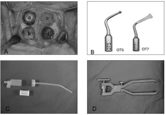
Fig. 1. (A) Defect preparation (a : Group 1, b : Group 2, c : Group 3, d : Control) (B) Piezosurgery tip OT 5 & OT 7
(C) Osseous bone collector (OTA 100, Osseous Technologies of America)
(D) Bone mill (Bone milling Forceps, USTOMED INSTRUMENTE Ulrich Storz GmbH & Co. KG)
Iodine 용액으로 수술 부위를 소독하였다. 수술 중 지혈을 목적
으로 1:100,000 에피네프린이 함유된 2% 염산리도카인으로 수 술 부위를 국소마취 하였다. 두개골 정중부를 따라 약 4 cm의 절개선을 형성하고, 양쪽 안와상연까지 골막 손상 없이 골막 을 박리하였다. 봉합선을 피하여 노출된 두개골에 Piezosurgerytip(OT 5)을 이용하여 지름 6mm의 원형 골 결손부 4개를 형성
하였다. 각각의 골 결손부를 앞에서 서술한 바와 같이 서로 다 른 방법으로 얻은 자가골을 이식하였다. 그리고 3-0 black silk를 이용하여 층별 봉합하였다. 감염 예방을 목적으로 술 후 이틀 간 Enrofloxacin(바이트릴�, 유한양행, 한국) 0.2 ml/kg을 근주하
였다. 그리고 술 후 일주일 째 발사하였다.(2) 조직학적 관찰
실험 6주경과 후 2% 염산리도카인 2ml, 생리식염수 10ml를 섞 어 심장에 주사하여 토끼를 희생시켰다. 그리고 골막이 부착된 상태로 두개골을 분리하여 10% formaldehyde로 고정하고 10%
EDTA 3일, 10% formic-acid로 3일간 탈회시켜 이식부 최대 지름
부위에서 절단한 후, 통법에 준하여 파라핀 포매 표본을 제작하 였다. 박절기를 이용하여 5 ㎛ 두께 절편을 얻어 Masson'sTrichrome 염색을 시행한 후 광학현미경으로 관찰하였다.
(3) 골양 평가 (Quantitative analysis)
골이식의 골성 회복 정도를 평가하기 위해 현미경에 부착된 디지털 카메라로 영상을 얻어 IMT(VT) image analysis
(iMTechnology Co, KOREA) 프로그램을 이용하였다. 골성 회복
정도 평가는 골 결손부에 새롭게 형성된 골의 면적으로 평가 하였으며, 결손부와 정상골의 경계를 우선 설정하고 결손부 전체면적을 100으로 하여 새롭게 형성된 골조직의 면적을 퍼 센트로 산출하였다. 각 이식부 간의 통계학적 평가는 ANOVATest를 이용하였다.
Ⅲ. 연구 결과 1. 광학현미경적 소견
대조군. 골 결손부 주변으로부터 중심으로 골화가 진행되면 서 지방을 함유한 골 조직을 이루었다. 골과 접하는 하방면은 미성숙된 골 조직이 판상을 이루고 있었으나 완전한 연속성을 이루지 못하였고 이 부위는 섬유성 골로 채워지고 있었다(Fig.
2).
Fig. 2.Non-grafted defects at 6th week. Central ingrowth of new bone from the marginal bone was relatively well formed but was not fused at center. Note the isolated ossicles surrounded by dense fibrous connective tissue. (Trichrome stain, A: ×10, B: ×100)
Fig. 3.Defect grafted with block type autogeous bone at 6th week. The grafted bone was markedly replaced
A B
실험 1군. 이식된 원형의 블록형 골조직은 자기 형태를 잘 유 지하고는 있었으나, 주변조직과 명확한 융합을 이루고 있지 못하였고, 그 사이에 결체조직이 게재되어 있었다. 내부는 새 로 형성된 골에 의해 부분적으로 흡수 대치되었으며 지방 성 분보다는 혈관을 함유한 소성 결체 조직 양상의 골수를 함유 하고 있었다(Fig. 3).
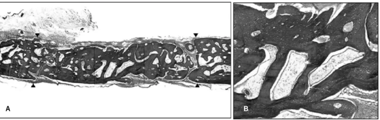
실험 2군. 이식된 골 기질을 중심으로 양호한 골 형성이 이루 어지면서 판상을 이루었고 많은 신생골은 미성숙된 골 형태의 섬유성 골 조직을 나타내었다. 내부에 이식된 다수의 골편에 는 골세포가 함유되어 있었고 신생골과 잘 유합되었으며 부분 적으로 신생골에 의해 대체되고 있었다(Fig. 4).
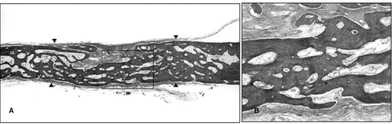
실험 3군. 실험 2군과 비슷한 양상으로 판상의 골성 회복을 이루고 있었으나, 실험 2군에 비해 다수의 혈관과 지방을 함유 한 골수를 형성하면서 그물상 골판을 이루고 있었으나 이식골 입자들은 새롭게 형성된 미성숙 골조직에 의해 양호하게 유합 되었다(Fig. 5).
2. 골양 평가 (Quantitative analysis)
이식 후 6주간 생성된 골양은 대조군에서 23.18±9.42%, 실험
1군이 42.63±8.87%, 실험 2군이 37.84±10.32% 그리고 실험 3
군이 36.95±11.00%을 나타내었다(Fig. 6).원형의 블록형 골조직을 이식한 부위에서 가장 많은 골양을 나타내고 있으며, bone mill과 Piezosurgery 기구를 이용한 파쇄 골편 이식부가 그 다음 순이었으며 골이식을 하지 않은 대조 군에서 가장 적은 양의 골 형성이 이루어졌음을 알 수 있었다.
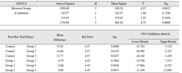
ANOVA test 결과 통계학적으로 유의한 차이(P=0.0012)를 나
타내었으며 대조군과 실험 1군(P=0.0008), 대조군과 실험 2군(P=0.0145), 대조군과 실험 3군(P=0.0237)은 통계학적으로 유의
할 만한 차이가 있음을 알 수 있었다. 그러나 실험 1군과 실험 2 군(P=0.7064), 실험 1군과 실험 3군(P=0.5836), 실험 2군과 실험 3 군(P=0.9971)간에는 통계학적으로 유의할 만한 차이를 보이지 않았다(Table 1).Fig. 4.Defect grafted with particulated autogenous bone particles prepared using a bone at 6th week. The newly formed bone around the grafted particulated bone was fused and formed a thin a thin reticular form bone plate filling the bone defect. Note the abundant fat marrow within reticular spaces. (Trichrome stain, A: ×10, B: ×100)
Fig. 5. Defect grafted with harvested autogenous bone particles using a piezosurgery at 6th week. The defect was compeletely covered with newly formed bone plate. Note the regularly arranged delicate fibrovascular marrow spaces. (Trichrome stain, A: ×10, B: ×100)
A B
A B
Ⅳ. 총괄 및 고찰
가토 10마리의 두개골 결손부에 이식한 자가골을 조직학적 으로 관찰한 결과 실험 2군과 실험 3군은 비슷한 조직 소견을 보이고 있었으며, 미성숙 신생골이 주위 정상골과 양호한 유 합을 이루고 있는 것을 볼 수 있었다. 실험 2군과 실험 3군에서 신생골이 대조군 및 실험 1군에 비해 많이 형성되었다. 실험 1 군의 원형의 블록형 이식골은 주위 골과 유합을 이루고 있지 못하였으나 이식골의 형태를 잘 유지하고 있었다. 그리고 부 분적으로 새로운 골로 대치되고 있었다. 대조군은 미성숙된 골조직이 얇은 판상을 이루고 있었으나 주변골과 완전히 연결 됨 없이 많은 부분이 섬유성 골로 채워졌다. 신생골양은 실험 1 군에서 가장 많은 양이 계측되었으며. 실험 2군, 실험 3군 그리
에서 기존의 골은 붉은 색을 나타내며, 새로 형성된 골은 좀 더 푸른색을 띄고 있고, 붉은색과 푸른색이 공존하는 부분은 기 존의 골이 신생골로 대체되고 있음을 시사한다. 신생골과 기 존 골이 공존하는 부분에서 신생골만을 명확히 경계 짓는 것 이 불가능하여 신생골의 양만을 따로 측정하지는 않았으며 계 측된 골의 양은 새로 형성된 신생골의 양만을 말하는 것이 아 니라 기존의 골도 모두 포함된 양이 된다. 따라서 본 연구에서 는 원형의 블록형 골조직을 이식한 경우 가장 많은 골이 계측 되었다. 이는 블록형 골조직인 경우 기존의 골 흡수가 서서히 진행되어 기존의 골과 신생골이 동시에 계측되었기 때문으로 다른 실험군보다 더 많은 신생골이 형성되었다고 하기에는 다 소 무리가 있다고 사료된다.
ANOVA test 결과에서 알 수 있듯 대조군과 실험 1군 (P=0.0008), 대조군과 실험 2군(P=0.0145), 대조군과 실험 3군 (P=0.0237)은 통계학적으로 유의할 만한 차이가 있었으나, 실
험 1군과 실험 2군(P=0.7064), 실험 1군과 실험 3군(P=0.5836), 실 험 2군과 실험 3군(P=0.9971)은 통계학적으로 유의할 만한 차 이를 보이지 않았다. 골을 이식하지 않은 대조군보다 골을 이 식한 곳에서 골 형성이 좀 더 양호하게 이루어졌으며, 골을 이 식하는 방법에 있어서는 서로 큰 차이를 보이지 않았다고 할 수 있었다.골이식하기에 적당한 자가골의 크기에 대한 의견은 다양하 다. Urist12)는 250�420㎛의 크기의 골 입자는 연골 형성과 골화 를 방해하기에 1000�2000㎛의 큰 입자들이 더 효과적이라고 보고하였으나, Rivault 등13)은 큰 입자들보다는 100㎛ 정도의 자 가골이 골모 세포의 활성을 돕는다고 하였다. Jonck14)의 최근 보고에 의하면 12�25㎛의 미세 골 입자들이 임플란트시에 골 화를 증가시킨다고 하였으며, Shapoff 등15)은 적절한 골 입자의
Fig. 6. Percentage of bone regeneration in surgical defect for each group.
Table 1.Statistical comparison with ANOVA
ANOVA Sum of Squares df Mean Square F Sig.
Between Groups 1950.60 3 650.20 6.57 0.0012
(Combined) 243.57 1 243.57 2.46 0.1258
219.63 1 219.63 2.22 0.1454
1730.98 2 865.49 8.74 0.0008
95% Confidence Interval
Post-Hoc Test(Tukey) Mean
Std. Error Sig.
Difference
Lower Bound Upper Bound
Control Group 1 -19.45 4.57 0.0008 -31.783 -7.120
Control Group 2 -14.66 4.57 0.0145 -26.996 -2.333
Control Group 3 -13.77 4.57 0.0237 -26.102 -1.439
Group 1 Group 2 -4.79 4.45 0.7064 -16.790 7.215
Group 1 Group 3 -5.68 4.45 0.5836 -17.684 6.322
Group 2 Group 3 0.89 4.45 0.9971 -11.109 12.896
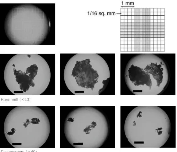
골이식 시에 골개조 속도가 빠르기 때문에 0.5�2㎣ 크기의 입 자가 10㎣보다 선호된다고 보고하고 있다. 본 실험에서 사용한 골 입자의 크기는 실험 2군에서처럼 bone mill을 사용한 경우에 는 약 3 × 3 mm, 실험 3군에서처럼 Piezosurgery를 사용한 경우 약 0.3 × 1.5 mm정도의 크기로 관찰되었으나 그 크기의 범주 가 매우 넓기 때문에(0.05 × 0.05 � 3.5 × 2.5) 평균적인 크기를 계산하는 것은 의미가 없는 것으로 사료되나, 최근의 Pallesen 등의 연구에서 골이식시 선호되는 입자 크기와는 유사하다고 할 수 있다(Fig. 7).
본 실험에서처럼 구강외에서는 문제되지 않지만 구강 내에 서 Piezosurgery를 이용하여 골조직을 긁은 후 골수집기(bone
collector)로 골입자를 모아서 이식할 때는 타액 내의 세균에 의
한 감염에 대해 유의하여야 한다. 이러한 세균에 의한 감염은 여러 방법으로 연구되어 왔으며, 수술방법, 골수집 기구(bonecollector device)의 형태, 환자의 치아 및 치주 건강 상태에 따라
서 달라 질 수 있다. Sivolella 등17)은 Piezosurgery 기구와 bone trap 을 이용한 실험을 통해 골 조각을 rifamycin SV로 처치하였을 때 세균의 감염이 감소하였다고 보고하고 있다. 골조직을 모 을 때 사용하는 suction tip과 타액 등을 모을 때 사용되는 suctiontip을 구분하여 사용하는 것도 타액 내의 세균의 감염을 감소시
킬 수 있는 한 방법이라 생각된다.
구강 내에서 블록형으로 골조직을 채취할 때나 bone mill을 이용해 골 입자를 형성해야 할 경우에는 이식부위 인접골 조 직에서 블록형 골조직을 채취하기 위해 좀 더 광범위한 수술 범위가 형성되어야 하므로, 술 후 부종이나 동통 등 환자의 불 편감이 증가될 수 있다. Piezosurgery를 이용하여 주위 골조직을 채취하는 경우에는 추가적으로 수술 범위를 형성할 필요가 없 고, Piezosurgery tip을 이용해 손쉽게 골조직을 긁어 채취할 수 있다. 미세 진동으로 정밀한 삭제가 가능하며, 선택적인 골 삭 제가 가능하므로 연조직의 손상을 최소화 할 수 있는 장점이 있다. 이번 실험 결과에서 Piezosurgery tip을 이용하여 골조직을 긁어 이식한 골 이식부는 골양은 블록상태의 골조직을 그대로 이식한 골 이식부나 bone mill을 이용하여 골조직을 분쇄하여 이식한 골 이식부보다 다소 적게 나타났지만 통계학적인 차이 를 보이지는 않았다. 조직학적으로는 미성숙된 신생골들이 골 입자와 골 입자 사이에 양호하게 형성되어 있었으며 주위골과 도 양호한 유합을 보였다. 따라서 구강 내 작은 골 결손부의 골 이식시 Piezosurgery를 이용하여 주위골을 긁어 이식하는 것은 임상적으로 유용한 방법 중의 하나라고 할 수 있다.
Fig. 7.Measurement of the particle sizes (The black bar indicates 1000㎛).
Bone mill (×40)
Piezosurgery (×40)
Ⅴ. 결 론
상악동 거상술과 자가골 채취 등의 구강내 술식에 있어 다양 하게 사용되고 있는 기구인 Piezosurgery는 손쉽게 골을 채취할 수 있는 tip이 포함되어 있어, 구강내 작은 크기의 골 결손부를 수복하기 위한 인근 골조직의 채취방법으로 새롭게 적용되고 있으나 이러한 Piezosurgery를 이용한 골 이식 효과에 대한 자료 가 많지 않아 이번 연구를 통해 Piezosurgery를 이용한 골 이식 의 유용성을 평가 하였다.
실험은 12주령의 New Zealand산 백색 가토 10마리를 대상으 로 하였으며 Piezosurgery 기구를 이용하여 두개골에 4개의 직 경 6mm 크기의 원형 결손부를 형성한 후 채취된 원형의 블록 형 골조직을 그대로 이식한 군(실험 1군), bone mill을 이용하여 자가골을 분쇄 후 이식한 군(실험 2군), Piezosurgery tip을 이용 하여 curettage 방식으로 채취하여 골수집기를 이용하여 골편 을 모아 이식한 군(실험 3군), 아무것도 이식하지 않은 군(대조 군)로 구분하여 관찰 하였다. 실험 6주경과 후 각 실험 동물을 희생하여 Masson`s Trichrome 염색을 시행하여 조직학적 소견 을 관찰하였으며 조직형태학적으로 골양을 계측하였다.
Piezosurgery
를 이용하여 형성한 골조직을 이식한 경우 조직학적으로 양호한 골 생성을 보이며 골양 역시 bone mill이나 블 럭형 골조직을 이용하여 이식한 경우와 비교하여 통계학적으 로 유의할만한 차이를 보이지 않아 Piezosurgery를 이용하여 골 을 긁어 채취하여 구강내 작은 골 결손부에 이식하는 방법은 유용하고 믿을만한 방법 중의 하나로 사료된다.
참고문헌
1. Hopp SG, Dahners LE, Gilbert JA: A study of the mechanical strength of long bone defects treated with various bone autograft substitutes : An experimental investigation in the rabbit. J Orthop Res 1989;7:579-584.
2. Goldberg VM, Stevenson S: Nature history of autograft and allo- graft. Clin Orthop 1987;7:225.
3. Arrington ED, Smith WJ, Chambers HG: Complications of iliac crest bone graft harvesting. Clin Orthop 1996;329:300-309.
4. Mulliken JB, Glowacki J, Kaban LB, Forkman J, Murray JE:
Application of demineralized allogenic bone implants for the cor- rection of maxillocranial facial deformities. Ann Surg 1981;3:633.
5. Kainulainen V, Oikarinen K: Comparison of four bone collectors designed for oral and maxillofacial surgery : An in vitro study.
Clin Oral Implants Res 1998;9:327-324.
6. Hoigne DJ, Stu¨binger S, Kaenel OV, Shamdasani S, Hase- nboehler P: Piezoelectric osteotomy in hand surgery: First experi- ences with a new technique. BMC Musculoskeletal Disorders 2006;7:36.
7. Stubinger S, Kuttenberger J, Filippi A, Sader R, Zeilhofer HF:
Intraoral Piezosurgery : Preliminary results of a new technique. J Oral Maxillofac Surg 2005;63:1283-1287.
8. Akita S, Fukui M, Nakagawa H, Fujii T, Akino K: Cranial bone defect healing is accelerated by mesenchymal stem cells induced by coadministration of bone morphogenetic protein-2 and basic fibroblast growth factor. Wound Repair Regen 2004;12:252-259.
9. Sohn DS, Ahn MR, Lee WH, Yeo DS, Lim SY: Piezoelectric os-
10. Siervo S, Ruggli-Milic S, Radici M, Siervo P, J a¨ger K:
Piezoelectric surgery. : An alternative method of minimally inva- sive surgery. Schweiz Monatsschr Zahnmed 2004;114:365-377.
11. Chiriac G, Herten M, Schwarz F, Rothamel D, Becker J:
Autogenous bone chips: Influence of a new piezoelectric device (Piezosurgery) on chip morphology, cell viability and differentia- tion. J Clin Periodontol 2005;32:994-999.
12. Urist MR, Silverman BF, Bu¨ring K, Dubuc FL, Rosenberg JM:
The bone induction principle. Clin Orthop Relat Res 1967;53:243 13. Rivault AF, Toto PD, Levy S, Gargiulo AW: Autogenous bone grafts : Osseous coagulum and osseous retrograde procedures in primates. J Periodontol 1971;42:787-796.
14. Jonk LM: bone induction effect of fine bone shavings in polyester fiber. An experimental study. S Afr Med J 1975;49:697-702.
15. Shapoff CA, Bowers GM, Levy B, Mellonig JT, Yukna RA: The effect of particle size on the osteogenic activity of composite grafts of allogeneic freeze-dried bone and autogenous marrow. J Periodontol 1980;51:625-630.
16. Pallesen L, Schou S, Aaboe M, Hjorting-Hansen E, Nattestad A, Melsen F: Influence of particle size of autogenous bone grafts on the early stages of bone regeneration: A histologic and stereolog- ic study in rabbit calvarium. Int J Oral Maxillofac Implants 2002;17:498-506.
17. Sivolella S, Berengo M, Scarin M, Mella F, Martinelli F:
Autogenous particulate bone collected with a Piezo-electric sur- gical device and bone trap: A microbiological and histomorpho- metric study. Arch Oral Biol 2006;51:883-891.
18. Schepers EJ, Ducheyne P: Bioactive glass particles of narrow size range for the treatment of oral bone defects : A 1-24 month experiment with several materials and particle sizes and size ranges. J Oral Rehabil 1997;24:171-181.
19. Xu H, Shimizu Y, Asai S, Ooya K: Experimental sinus grafting with the use of deproteinized bone particles of different sizes.
Clin Oral Impl Res 2003;14:548-555.
20. Shand JM, Heggie AA, Holmes AD, Holmes W: Allogeneic bone grafting of calvarial defects: An experimental study in the rabbit.
Int J Oral Maxillofac Surg. 2002;31:525-531.
21. Haddad AJ, Peel SA, Clokie CM, Sandor GK: Closure of rabbit calvarial critical-sized defects using protective composite allo- geneic and alloplastic bone substitutes. J Craniofac Surg 2006;17:926-934.
22. Robiony M, Polini F, Costa F, Vercellotti T, Politi M:
Piezoelectric bone cutting in multipiece maxillary osteotomies. J Oral Maxillofac Surg 2004;62:759-761.
23. Moghadam HG, Sandor GK, Holmes HH, Clokie CM:
Histomorphometric evaluation of bone regeneration using allo- geneic and alloplastic bone substitutes. J Oral Maxillofac Surg 2004;62:202-213.
24. Stal S, Tjelmeland K, Hicks J, Bhatia N, Eppley B, Hollier L:
Compartmentalized bone regeneration of cranial defects with biodegradable barriers : An animal model. J Craniofac Surg 2001;12:41-47.
25. Young MP, Worthington HV, Lloyd RE, Drucker DB, Sloan P, Carter DH: bone collected during dental implant surgery : A clinical and histological study. Clin Oral Implants Res 2002;13:298-303.
26. Oikarinen K, Kainulainen V, Kainulainen T: A method of har- vesting corticocancellous bone chips for reconstructive maxillo- facial surgery. Int J Oral Maxillofac Surg 1997;26:103-105.
27. Schlee M, Steigmann M, Bratu E, Garg AK: Piezosurgery basics and possibilities. Implant Dent. 2006;15:334-340.
28. Lambrecht JT: Intraoral piezo-surgery. Schweiz Monatsschr Zahnmed. 2004;114:28-36.
29. Vercellotti T, Pollack AS: A new bone surgery device: Sinus grafting and periodontal surgery. Compend Contin Educ Dent 2006;27:319-325.
30. Clokie CM, Moghadam H, Jackson MT, Sandor GK: Closure of critical sized defects with allogenic and alloplastic bone substi-