ABSTRACT
Purpose: Various crosslinking methods have been introduced to increase the longevity of collagen membranes. The aim of this study was to compare and evaluate the degradation and bone regeneration patterns of 3 collagen membranes.
Methods: Four 8-mm-diameter circular bone defects were created in the calvaria of 10 rabbits. In each rabbit, each defect was randomly allocated to 1) the sham control group, 2) the non-crosslinked collagen sponge (NS) group, 3) the chemically crosslinked collagen membrane (CCM) group, or 4) the biphasic calcium phosphate (BCP)-supplemented ultraviolet (UV)-crosslinked collagen membrane (UVM) group. Each defect was covered with the allocated membrane without any graft material. Rabbits were sacrificed at either 2 or 8 weeks post-surgery, and radiographic and histologic analyses were done.
Results: New bone formed underneath the membrane in defects in the CCM and UVM groups, with a distinctive new bone formation pattern, while new bone formed from the base of the defect in the NS and control groups. The CCM maintained its shape until 8 weeks, while the UVM and NS were fully degraded at 8 weeks; simultaneously, sustained inflammatory infiltration was found in the margin of the CCM, while it was absent in the UVM. In conclusion, the CCM showed longer longevity than the UVM, but was accompanied by higher levels of inflammation.
Conclusions: Both the CCM and UVM showed distinctive patterns of enhancement in new bone formation in the early phase. UV crosslinking can be a biocompatible alternative to chemical crosslinking.
Keywords: Bone regeneration; Collagen; Ultraviolet rays
INTRODUCTION
Guided bone regeneration (GBR) is a widely used surgical procedure to overcome bone deficiency for dental implant surgery [1,2]. Among the various materials utilized for GBR, a combination of xenograft bone material and resorbable collagen membrane has been the most frequently used for lateral augmentation of narrow alveolar ridges [3]. Numerous
Research Article
Received: Jul 5, 2020 Revised: Aug 25, 2020 Accepted: Nov 9, 2020
*Correspondence:
Seong-Ho Choi
Department of Periodontology, College of Dentistry, Yonsei University, 50 Yonsei-ro, Seodaemun-gu, Seoul 03722, Korea.
E-mail: [email protected] Tel: +82-2-2228-3189 Fax: +82-2-392-0398
†Inpyo Hong and Alharthi Waleed Khalid contributed equally to this study.
Copyright © 2021. Korean Academy of Periodontology
This is an Open Access article distributed under the terms of the Creative Commons Attribution Non-Commercial License (https://
creativecommons.org/licenses/by-nc/4.0/).
ORCID iDs Inpyo Hong
https://orcid.org/0000-0002-0486-9593 Alharthi Waleed Khalid
https://orcid.org/0000-0001-5668-5748 Hyung-Chul Pae
https://orcid.org/0000-0002-6365-3557 Young Woo Song
https://orcid.org/0000-0003-1835-5646 Jae-Kook Cha
https://orcid.org/0000-0002-6906-7209 Jung-Seok Lee
https://orcid.org/0000-0003-1276-5978 Jeong-Won Paik
https://orcid.org/0000-0002-5554-8503
Inpyo Hong †,Alharthi Waleed Khalid †, Hyung-Chul Pae , Young Woo Song , Jae-Kook Cha , Jung-Seok Lee , Jeong-Won Paik , Seong-Ho Choi *
Department of Periodontology, Research Institute of Periodontal Regeneration, Yonsei University College of Dentistry, Seoul, Korea
Diverse patterns of bone regeneration in rabbit calvarial defects depending on the type of collagen membrane
Periodontal Science
Seong-Ho Choi
https://orcid.org/0000-0001-6704-6124 Funding
This work was supported by a National Research Foundation of Korea (NRF) grant funded by the Korean government (Ministry of Science, ICT & Future Planning) (No. NRF- 2017R1A2B4002782).
Author Contributions
Conceptualization: Seong-Ho Choi; Formal analysis: Inpyo Hong; Investigation: Inpyo Hong, Alharthi Waleed Khalid, Hyung-Chul Pae; Methodology: Inpyo Hong, Hyung-Chul Pae; Project administration: Seong-Ho Choi;
Writing - original draft: Inpyo Hong, Alharthi Waleed Khalid; Writing - review & editing:
Young Woo Song, Jae-Kook Cha, Jung-Seok Lee, Jeong-Won Paik, Seong-Ho Choi.
Conflict of Interest
No potential conflict of interest relevant to this article was reported.
studies have proven the efficacy of GBR and the long-term stability of implants placed at GBR sites, implying that GBR is no longer an uncommon procedure for successful implant surgery [4].
However, the natural collagen matrix undergoes fast degradation, which causes limitations in terms of stable and long-lasting space maintenance [5]. Previous research has shown that the collagen membrane should be preserved for at least 4 weeks to accomplish its role in regeneration [6]. To overcome this limitation, crosslinking of collagen fibers has been applied to improve the mechanical characteristics of membranes and their resistance to enzymatic degradation [7,8]. Numerous studies have shown favorable outcomes of using crosslinked collagen membranes in GBR [9-12].
Multiple crosslinking methods have been developed for collagen membranes, of which chemically mediated crosslinking is one of the most commonly used due to its economic advantages and convenience in manufacturing. However, the chemicals used for crosslinking and the byproducts of chemical crosslinking reaction pose risks of cytotoxicity and foreign body reaction, which diminish the biocompatibility of the membrane. Furthermore, the consumption of the hydroxyl functional group by covalent bonding between collagen molecules impairs interactions with adjacent tissue and cells [13].
Ultraviolet (UV) crosslinking is considered to be more biocompatible than chemically mediated crosslinking, although it is weaker [13]. UV irradiation induces radical reactions that form crosslinks between collagen fibers, without consuming functional groups of collagen. However, the rigidity of the crosslinking is less favorable [14]. To supplement the mechanical stability of UV crosslinking, biphasic calcium phosphate (BCP) was added in a previous study [15].
The efficacy of various types of crosslinked membranes, including those discussed above, for GBR has been evaluated; however, still there is no gold standard in terms of membrane selection [10-12,16,17]. In the present study, we used 3 types of collagen membranes with different crosslinking methods for GBR in a rabbit calvarial model, and evaluated the outcomes in terms of bone regeneration and the tissue reaction.
MATERIALS AND METHODS
Experimental materials
Three different types of collagen membranes were prepared for the experiment:
1) Pure bovine, non-crosslinked type 1 collagen sponge (Collatape; Zimmer Biomet, Palm Beach, FL, USA). This material has a highly porous sponge structure that absorbs blood and wound exudate.
2) Chemically crosslinked porcine type 1 collagen membrane (Collagen Membrane P;
Genoss, Suwon, Korea). In this material, pure type 1 porcine collagen was crosslinked using carbodiimide and sterilized by gamma irradiation.
3) BCP-supplemented UV-crosslinked porcine type 1 collagen membrane (Ovis membrane;
Dentis, Daegu, Korea). To create this material, pure type 1 porcine collagen was prepared in a solution, and then BCP particles with a size of 7–10 μm were dispersed into the solution. The collagen-to-BCP ratio was 1:1 by weight. After mixture, the solution was poured into a mold, UV-irradiated, and freeze-dried.
Study design
Four round defects measuring 8 mm in diameter were produced in the calvaria of rabbits. In each rabbit, each of the 4 defects was randomly allocated to 1 of the following study groups, such that each rabbit had 4 defects, each of which was allocated to a different group (Figure 1):
1) Sham control group: defect filled with a blood clot but not covered.
2) NS group: defect filled with a blood clot and covered with a pure bovine non-crosslinked type 1 collagen sponge.
3) CCM group: defect filled with a blood clot and covered with a chemically crosslinked porcine type 1 collagen membrane.
4) UVM group: defect filled with a blood clot and covered with a BCP-supplemented UV- crosslinked porcine type 1 collagen membrane.
Five of the 10 rabbits were sacrificed at 2 weeks, and the remaining 5 were sacrificed at 8 weeks.
Animal experiment surgery procedure
The protocol of the animal experiment in this study followed the ARRIVE guideline and was approved by the Institutional Animal Care and Use Committee of Yonsei Medical Center, Seoul, Republic of Korea (approval number 2017-0117) [18]. Ten male New Zealand white rabbits with a body weight of 3 ± 0.2 kg were housed and cared under the Association for Assessment and Accreditation of Laboratory Animal Care International (AAALAC) guidelines.
The surgical procedure followed in this study was determined based on previous studies [9,10,15]. Isoflurane (2.5%) inhalation and an intravenous injection of alfaxan (5 mg/kg) were used for general anesthesia. Povidone-iodine was applied to disinfect the surgical site. After a subcutaneous injection of 2% lidocaine with 1:100,000 epinephrine for local anesthesia, an incision was made on the midline of the cranium and a full-thickness flap was elevated to expose the calvarium. Four circular bone defects with a diameter of 8 mm were created by an 8-mm-diameter trephine bur under saline irrigation to avoid heat-induced damage to the underlying dura mater and cerebrum. In each defect, except for the sham control group, the allocated membrane was trimmed into a 10×10 mm square and used to cover the defect without application of bone graft material. After placing the membrane on the defect, the elevated flap was replaced and sutured with absorbable 4-0 suture material (Vicryl; Ethicon, Somerville, NJ, USA). Enfloxacin (10 mg/day) was given for 5 days after the operation as
A B
Figure 1. Surgical preparation for the study. (A) Four round calvarial defects with 8-mm diameters were prepared using a trephine bur. (B) Each defect was randomly assigned to a study group. Clockwise from top left; NS, CCM, sham control and UVM.
NS: non-crosslinked collagen sponge group, CCM: chemically crosslinked collagen membrane group, UVM:
ultraviolet-crosslinked collagen membrane group.
a general antibiotic. With daily care, healing of the surgical site and the rabbits' general condition were clinically observed. Experimental animals were sacrificed at either 2 or 8 weeks post-surgery, according to their designated group, to acquire calvarial specimens.
Micro-computed tomography (CT) analysis
Specimens were immersed into a 10% formalin fixation solution. After 10 days, the specimens were scanned with micro-CT (SkyScan 1173; Bruker-CT, Kontich, Belgium) at a pixel size of 13.86 μm (130 kV, 60 μA). Digital Imaging and Communications in Medicine images were reconstructed with 3-dimensional reconstruction software (N Recon version 1.7.0.4; Bruker-CT).
For volume measurements, the region of interest (ROI) was selected according to a previous study [15]. Specifically, the bony margin of the 8-mm-diameter defect was selected as the lateral border of the ROI. The connective tissue layer covering the defect was selected as the superior border of the ROI, and the dura mater was selected as the inferior border of the ROI.
Then, 8-bit grayscale values were used to discriminate the radiopacity of defects. Areas with grayscale values of 55 to 255 were regarded as bone (of any type). Areas with grayscale values of 55 to 97 and lower than 55 were considered to be newly formed bone and connective tissue, respectively. Within the ROI, the volume of the newly formed bone volume was calculated as follows using 3-dimensional reconstruction software (N Recon version 1.7.0.4; Bruker-CT):
New bone volume (NBV; mm3): sum of newly formed bone within the defect.
Histologic and histomorphometric analyses
Specimens were decalcified after micro-CT by submerging them in 5% formic acid for 2 weeks. Each decalcified specimen was embedded in paraffin wax, and 5-μm-thick sections were made along the center of each 8-mm-diameter round defect. Hematoxylin and eosin (H&E) and Masson trichrome were used for staining, and the slides were then examined under a light microscope (DM LB; Leica Microsystems, Wetzlar, Germany) with an attached camera (DC300F; Leica Microsystems, Wetzlar, Germany). The entire slide images were converted into digital image files for histomorphometric analysis. With a computer-aided slide image analysis program (CaseViewer 2.2; 3DHISTECH Ltd., Budapest, Hungary), the ROI was selected according to a previous study [15]. Specifically, the lateral border of the ROI was selected as the bony margin of the defect made by the 8-mm trephine bur. If the collagen membrane had not been fully absorbed, the lower margin of the membrane remnant was used as the superior border, but otherwise the connective tissue layer covering the defects was selected as the superior border. The inferior border was the dura mater. Within the ROI, the following parameters were measured:
Total area (TA; mm2): the overall area within the ROI.
New bone area (NBA; mm2): sum of new bone area within the ROI.
Statistical analysis
Commercially available software (SPSS version 23, IBM Corp., Armonk, NY, USA) was used for the statistical analysis. The Shapiro-Wilk test was applied to confirm the distribution of the data. The histomorphometric results were summarized as median values and
interquartile ranges. To analyze the statistical significance (P<0.05) of differences in NBV, TA, and NBA between the groups at each time point and within the same study group between the 2 healing periods, the Kruskal-Wallis test and Mann-Whitney U test were used.
RESULTS
Clinical observations
No specific adverse reaction or noteworthy event was observed clinically. Neither specific inflammatory signs nor wound exposure was found at the surgical sites.
Micro-CT analysis
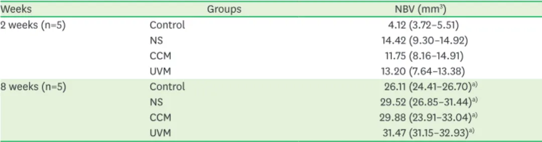
Micro-CT images are shown in Figure 2. In the UVM group, BCP microparticles, which were dispersed in the collagen membrane, were observed as scattered radiopaque points at 2 weeks. In the CCM and UVM groups, new bone was found near the upper area of the lateral defect margin at 2 weeks. At 8 weeks in both groups, most of the defects were filled with new bone. The volumetric measurements are summarized in Table 1. At both 2 weeks and 8 weeks, no statistically significant difference was found in NBV in among the groups at the same time point (P=0.427 at 2 weeks and P=0.782 at 8 weeks, respectively). In all groups, NBV was significantly larger at 8 weeks than at 2 weeks.
2 weeks Control
NS
CCM
UVM
8 weeks
Figure 2. Micro-computed tomography images. At 2 weeks, all groups showed new bone formation at the periphery of the defect, and dispersed BCP microparticles were observed in the UVM group. At 8 weeks, the defects of all groups were filled with regenerated new bone, and the CCM and UVM groups showed more continuous bone regeneration of the defect.
BCP: biphasic calcium phosphate, NS: non-crosslinked collagen sponge group, CCM: chemically crosslinked collagen membrane group, UVM: ultraviolet-crosslinked collagen membrane group.
Table 1. Median and interquartile range of NBV measured by micro-CT grayscale values
Weeks Groups NBV (mm3)
2 weeks (n=5) Control 4.12 (3.72–5.51)
NS 14.42 (9.30–14.92)
CCM 11.75 (8.16–14.91)
UVM 13.20 (7.64–13.38)
8 weeks (n=5) Control 26.11 (24.41–26.70)a)
NS 29.52 (26.85–31.44)a)
CCM 29.88 (23.91–33.04)a)
UVM 31.47 (31.15–32.93)a)
Values are presented as median (Q2–Q3) (mm3).
NBV: new bone volume, CT: computed tomography, NS: non-crosslinked collagen sponge group, CCM: chemically crosslinked collagen membrane group, UVM: ultraviolet-crosslinked collagen membrane group.
a)Statistically significant difference compared to the corresponding groups at 2 weeks (P<0.05).
Histologic findings
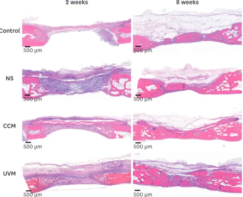
Representative histologic images at 2 and 8 weeks are depicted in Figure 3.
At 2 weeks, bone regeneration was found from the lateral periphery of defects in all groups (Figure 4). In the NS and control groups, new bone seemed to be growing from the lower peripheral border, adjacent to the inner periosteum of the rabbit calvaria. As a result, new bone formation was mainly observed in the lower portion of the defect, so that the overall shape of new bone formation formed a right triangle, the base of which corresponded to the inferior border of the defect. In the CCM and UVM groups, however, new bone formation was more abundant on the superior peripheral border, adjacent to the collagen membrane.
New bone seemed to be growing along the lower border of the membrane. Consequently, new bone formation was mainly observed in the superior portion of defect, so that the overall shape of new bone formation was a right triangle, the base of which was formed by the superior border of the defect.
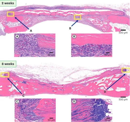
At 2 weeks, infiltration of inflammatory cells was observed around the UVM and CCM. Most of the inflammatory cells were found in the border of the membrane and tissue area. The distribution of inflammation was more intense in the CCM group (Figure 5). The membrane structure of the CCM remained intact, and cellular infiltration was not observed in the inner part of the membrane. The UVM also maintained its membrane structure, but cellular infiltration and tissue integration were observed around the boundary of the membrane (Figure 6). Blood vessels and scattered granulocytes invaded around the UVM. The distinction between the connective tissue area and the membrane was less clear in the UVM group than
2 weeks Control
NS
CCM
UVM
8 weeks
500 µm 500 µm
500 µm 500 µm
500 µm 500 µm
500 µm 500 µm
Figure 3. Histologic images under low magnification obtained at 2 weeks and 8 weeks (H&E staining, scale bar = 500 μm). At 2 weeks, in all defects, new bone formation was observed only in the periphery and the applied NS, CCM, and UVM were kept intact. At 8 weeks, the NS and UVM had been almost fully absorbed, while the CCM remained relatively intact.
NS: non-crosslinked collagen sponge group, CCM: chemically crosslinked collagen membrane group, UVM:
ultraviolet-crosslinked collagen membrane group, H&E: hematoxylin and eosin.
Control CCM
NS UVM
500 µm 500 µm
500 µm 500 µm
Figure 4. Masson trichrome-stained histologic images of new bone formation patterns at 2 weeks. In the NS and control groups, new bone formation was more abundant on the lower border of the periphery, adjacent to the inner periosteum of the rabbit calvaria. In the CCM and UVM groups, new bone formation was more abundant on the superior border of the periphery, adjacent to the collagen membrane.
NS: non-crosslinked collagen sponge group, CCM: chemically crosslinked collagen membrane group, UVM:
ultraviolet-crosslinked collagen membrane group.
2 weeks
A
C D
B
500 µm
500 µm
A B
C D
20 µm 20 µm
20 µm 20 µm
8 weeks
Figure 5. Histologic images of the CCM group (H&E staining). At 2 weeks, infiltration of inflammatory cells was observed around the CCM. Inflammatory cells were most invasive at the lateral border of the CCM (A), and were also observed below the membrane (B). Even at 8 weeks, inflammatory cells remained present along the lateral border of the membrane (C, D).
CCM: chemically crosslinked collagen membrane group, H&E: hematoxylin and eosin.
in the CCM group. In the NS group, cellular infiltration was found within the collagen sponge structure. A minimal level of inflammation was found in the NS and control groups.
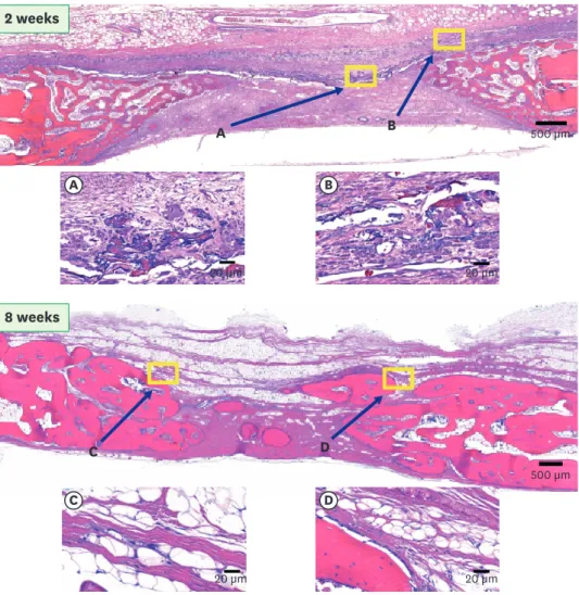
At 8 weeks, no remnants of the collagen sponge were found in the NS group. The UVM had almost fully degraded, with only a few remnants remaining in the form of a thin fibrous film (Figure 6). Due to resorption, infiltration of inflammatory cells was not observed in the NS, UVM, and control groups. However, notable degradation was only minimally seen in the CCM group, and a remarkable infiltration of inflammatory cells was still observed, especially around the lateral margin of the CCM (Figure 5). Regardless of inflammatory cell infiltration, all study groups and the control group at 8 weeks showed favorable healing and closed defects with mature bone formation.
Histomorphometric analysis
The histomorphometric measurements are summarized in Table 2. The TA of the control, NS, and UVM groups at 8 weeks were significantly larger than those of each respective group at 2 weeks. On the contrary, the CCM group, which showed the largest TA at 2 weeks, did not show a significant difference compared to 8 weeks (P=0.151). The NBA of each group
2 weeks
A
C D
B 500 µm
500 µm
A B
20 µm 20 µm
8 weeks
C D
20 µm 20 µm
Figure 6. Histologic images of the UVM group (H&E staining). At 2 weeks, mild infiltration of inflammatory cells was observed around the UVM (A, B). At 8 weeks, the UVM was almost fully degraded, with only a few thin fibrous films remaining (C). Even on the lateral side of the defect, there was no significant sign of inflammation (D).
UVM: ultraviolet-crosslinked collagen membrane group, H&E: hematoxylin and eosin.
at 8 weeks was significantly larger than that observed at 2 weeks. However, no statistically significant differences were found among the study groups at the same time points (P=0.175 at 2 weeks, P=0.851 at 8 weeks, respectively).
DISCUSSION
In this study, distinct new bone formation patterns were found depending on the
crosslinking of collagen membranes. Chemical crosslinking of the collagen membrane was advantageous in terms of longevity, but it also increased the inflammatory host response.
BCP-supplemented UV-crosslinking showed less inflammation and longevity than chemical crosslinking. Despite these differences in longevity and inflammation, both types of crosslinked collagen membranes exhibited prominent early bone formation patterns around the membrane. Although the new bone formation patterns were different, there was no significant difference in the amount of new bone formation.
Based on several previous studies, it is widely accepted that the membrane should be maintained for a considerable period of time to function as a barrier, which is a key
contributor to successful outcomes in GBR [2,6]. However, the time required for maintaining barrier function has not been clearly defined [19]. In this study, it was clear that the
degradation rate of the membrane depended on whether the membrane was crosslinked, whether mineralized particles were added, and how crosslinking was achieved. However, the different degradation rates did not have a significant effect on the amount of new bone formation when only the membrane was used.
The sole usage of a collagen membrane for regenerative treatment shows a clear limitation in terms of maintaining space against the pressure from adjacent soft tissue, which can cause the space to collapse. In this study, no improvement in bone regeneration was found in the defects covered by only collagen membranes compared to spontaneous healing, even for the CCM, which was barely resorbed. To support space maintenance, the use of bone graft material combined with a membrane as a scaffold is usually recommended, and this is the most widely used method for GBR [3,20]. Since the collagen membrane is naturally resorbed even after crosslinking, it is crucial to support space maintenance with bone graft material for successful outcomes in GBR.
Early bone regeneration of the defects with treated with the CCM and UVM showed different regeneration patterns compared to the other two groups; in particular, plentiful bone Table 2. Median and interquartile range of TA and NBA in the histometric analysis
Weeks Groups TA (mm2) NBA (mm2)
2 weeks (n=5) Control 4.37 (3.98–4.45) 1.17 (0.58–1.19)
NS 4.48 (4.20–5.25) 1.08 (1.00–1.21)
CCM 6.05 (4.93–8.18) 1.64 (1.25–1.99)
UVM 4.70 (4.69–5.03) 1.17 (0.93–1.26)
8 weeks (n=5) Control 5.60 (4.96–5.95)a) 2.48 (2.24–3.70)a)
NS 6.85 (6.24–6.90)a) 3.13 (3.06–3.80)a)
CCM 6.46 (6.23–7.50) 3.31 (1.93–3.43)a)
UVM 7.15 (6.89–7.71)a) 3.17 (2.87–3.80)a)
Values are presented as median (Q2–Q3) (mm2).
TA: total area, NBA: new bone area, NS: non-crosslinked collagen sponge group, CCM: chemically crosslinked collagen membrane group, UVM: ultraviolet-crosslinked collagen membrane group.
a)Statistically significant difference compared to the corresponding groups at 2 weeks (P<0.05).
regeneration was observed along the lower part of the collagen membranes. Although the CCM showed more inflammatory cell infiltration and a more clearly distinguishable margin of the membrane than was the case for the UVM, both types of membranes showed similar shapes in early bone regeneration, in that bone regeneration seemed to be enhanced close to the collagen membrane. This distinctive early bone regeneration pattern in the collagen membrane-applied groups may imply that collagen membranes are not only a physical barrier for soft tissue infiltration in GBR, but may also play a biologically active role in bone regeneration [19].
Previous immunohistochemical observations of GBR sites revealed high concentrations of bone-related proteins such as alkaline phosphatase, osteopontin, and osteocalcin under the membrane [21]. Furthermore, higher expression of bone regeneration-related growth factors such as bone morphogenic protein-2 was observed underneath the collagen membrane [22]. Based on these findings, a previous review suggested that a collagen membrane forms a biologically active local environment through abundant signals for cell recruitment, bone formation, and remodeling [19]. Similarly, our study presented different early bone regeneration patterns between defects with and without collagen membranes. This result might be related to the biologically active role of the collagen membrane.
Crosslinking methods delay the resorption of the natural collagen membrane. The CCM showed the strongest degree of crosslinking, which resulted in enhanced longevity and rigidity of the membrane. However, due to covalent bonding from the crosslinking mechanism, the functional groups of collagen molecules are consumed, resulting in a loss of tissue integrity [13]. Increased hardness and decreased tissue integration of the CCM may cause irritation around the membrane boundary and increase the risk of complications such as healing impairment and wound infection, which are associated with membrane exposure [16,23]. Furthermore, chemical residues from crosslinking agents can also cause inflammation around GBR sites [24]. In contrast, UV crosslinking is known to be more biocompatible; however, at the same time, the degree of crosslinking induced by UV radiation is relatively low and bonding strength is weak [14]. This could be a disadvantage in terms of resistance to degradation compared to chemical crosslinking.
Nonetheless, the necessity of crosslinking and the appropriate degree of crosslinking remain controversial. Schwarz et al. reported that there was no significant difference in angiogenesis and bone regeneration between crosslinked collagen membranes and non-crosslinked collagen membranes in GBR [25]. The increased stability of the barrier membrane resulting from crosslinking was expected to enhance bone regeneration in defects treated using GBR;
however, Becker et al. reported more complications (e.g., infection) when early exposure occurred in crosslinked collagen membranes than in non-crosslinked ones [23]. In the present study, complications could not be evaluated because they did not occur in all study groups. There were differences in collagen membrane degradation and the early bone regeneration pattern depending on the presence or the method of crosslinking, but these differences were not thought to be related to the late healing period, since bone regeneration at 8 weeks was similar among all groups.
In our study, microcrystalline BCP was immersed between the collagen fibers of the UVM. The addition of BCP microparticles to the collagen matrix has been shown to increase mechanical strength and resistance to enzymatic degradation [26]. However, a recent study reported that the addition of calcium phosphate nanoparticles to UV-crosslinked collagen membranes did
not result in any significant modification of the original system [27]. In our study, it was not possible to distinguish the effects of UV crosslinking from the effects of BCP supplementation, because none of the experimental groups used UVM without the addition of BCP particles.
Thus, further research is needed to evaluate the physicochemical changes that result from BCP addition to the collagen membrane and the effects thereof on bone regeneration.
There are some limitations of the present study. First, the defects in the study model were only covered with membranes, without applying bone graft materials to the defect. However, since collagen membranes are often used together with bone grafts as scaffolds in clinical situations, further research is necessary to evaluate the regeneration pattern when different membranes interact with bone substitutes. Second, only histologic and radiologic analyses were done in this study. An immunohistochemical analysis would be necessary to obtain more information on the biological activity of membranes related to cytokines and growth factors. Finally, due to limitations in the size of the rabbit calvaria, other types of membranes such as non-crosslinked collagen membrane or UVM without BCP microparticles could not be assigned to any study groups. Furthermore, the rabbit calvaria defect model did not involve movement or external pressure, limiting its usefulness for evaluating the space-maintaining ability of membranes under realistic conditions. Further investigation with a more
challenging model, such as a beagle canine model, might be more appropriate for evaluating the space-maintaining capacity of the membrane.
Within the limitations of this study, while the CCM showed greater resistance to degradation than the UVM, both the CCM and UVM showed distinctively enhanced new bone formation in the early phase of healing; therefore, it may be concluded that UV crosslinking might work as an alternative to chemical crosslinking due to its advantage in biocompatibility.
REFERENCES
1. Benic GI, Hämmerle CH. Horizontal bone augmentation by means of guided bone regeneration.
Periodontol 2000 2014;66:13-40.
PUBMED | CROSSREF
2. Retzepi M, Donos N. Guided bone regeneration: biological principle and therapeutic applications. Clin Oral Implants Res 2010;21:567-76.
PUBMED | CROSSREF
3. Sanz-Sánchez I, Ortiz-Vigón A, Sanz-Martín I, Figuero E, Sanz M. Effectiveness of lateral bone augmentation on the alveolar crest dimension: a systematic review and meta-analysis. J Dent Res 2015;94:128S-142S.
PUBMED | CROSSREF
4. Salvi GE, Monje A, Tomasi C. Long-term biological complications of dental implants placed either in pristine or in augmented sites: a systematic review and meta-analysis. Clin Oral Implants Res 2018;29 Suppl 16:294-310.
PUBMED | CROSSREF
5. Delgado LM, Bayon Y, Pandit A, Zeugolis DI. To cross-link or not to cross-link? Cross-linking associated foreign body response of collagen-based devices. Tissue Eng Part B Rev 2015;21:298-313.
PUBMED | CROSSREF
6. Owens KW, Yukna RA. Collagen membrane resorption in dogs: a comparative study. Implant Dent 2001;10:49-58.
PUBMED | CROSSREF
7. Jorge-Herrero E, Fernández P, Turnay J, Olmo N, Calero P, García R, et al. Influence of different chemical cross-linking treatments on the properties of bovine pericardium and collagen. Biomaterials 1999;20:539-45.
PUBMED | CROSSREF
8. Charulatha V, Rajaram A. Influence of different crosslinking treatments on the physical properties of collagen membranes. Biomaterials 2003;24:759-67.
PUBMED | CROSSREF
9. An YZ, Heo YK, Lee JS, Jung UW, Choi SH. Dehydrothermally cross-linked collagen membrane with a bone graft improves bone regeneration in a rat calvarial defect model. Materials (Basel) 2017;10:927.
PUBMED | CROSSREF
10. Park JY, Jung IH, Kim YK, Lim HC, Lee JS, Jung UW, et al. Guided bone regeneration using 1-ethyl-3-(3- dimethylaminopropyl) carbodiimide (EDC)-cross-linked type-I collagen membrane with biphasic calcium phosphate at rabbit calvarial defects. Biomater Res 2015;19:15.
PUBMED | CROSSREF
11. Elgali I, Omar O, Dahlin C, Thomsen P. Guided bone regeneration: materials and biological mechanisms revisited. Eur J Oral Sci 2017;125:315-37.
PUBMED | CROSSREF
12. Bunyaratavej P, Wang HL. Collagen membranes: a review. J Periodontol 2001;72:215-29.
PUBMED | CROSSREF
13. Sorushanova A, Delgado LM, Wu Z, Shologu N, Kshirsagar A, Raghunath R, et al. The collagen suprafamily: from biosynthesis to advanced biomaterial development. Adv Mater 2019;31:e1801651.
PUBMED | CROSSREF
14. Davidenko N, Bax DV, Schuster CF, Farndale RW, Hamaia SW, Best SM, et al. Optimisation of UV irradiation as a binding site conserving method for crosslinking collagen-based scaffolds. J Mater Sci Mater Med 2016;27:14.
PUBMED | CROSSREF
15. Hong I, Khalid AW, Pae HC, Cha JK, Lee JS, Paik JW, et al. Distinctive bone regeneration of calvarial defects using biphasic calcium phosphate supplemented ultraviolet-crosslinked collagen membrane. J Periodontal Implant Sci 2020;50:14-27.
PUBMED | CROSSREF
16. Jiménez Garcia J, Berghezan S, Caramês JMM, Dard MM, Marques DNS. Effect of cross-linked vs non- cross-linked collagen membranes on bone: a systematic review. J Periodontal Res 2017;52:955-64.
PUBMED | CROSSREF
17. Lee DW, Kim KT, Joo YS, Yoo MK, Yu JA, Ryu JJ. The role of two different collagen membranes for dehiscence defect around implants in humans. J Oral Implantol 2015;41:445-8.
PUBMED | CROSSREF
18. Kilkenny C, Browne WJ, Cuthill IC, Emerson M, Altman DG. Improving bioscience research reporting:
the ARRIVE guidelines for reporting animal research. PLoS Biol 2010;8:e1000412.
PUBMED | CROSSREF
19. Omar O, Elgali I, Dahlin C, Thomsen P. Barrier membranes: more than the barrier effect? J Clin Periodontol 2019;46 Suppl 21:103-23.
PUBMED | CROSSREF
20. Sanz M, Dahlin C, Apatzidou D, Artzi Z, Bozic D, Calciolari E, et al. Biomaterials and regenerative technologies used in bone regeneration in the craniomaxillofacial region: consensus report of group 2 of the 15th European Workshop on Periodontology on Bone Regeneration. J Clin Periodontol 2019;46 Suppl 21:82-91.
PUBMED | CROSSREF
21. Taguchi Y, Amizuka N, Nakadate M, Ohnishi H, Fujii N, Oda K, et al. A histological evaluation for guided bone regeneration induced by a collagenous membrane. Biomaterials 2005;26:6158-66.
PUBMED | CROSSREF
22. Turri A, Elgali I, Vazirisani F, Johansson A, Emanuelsson L, Dahlin C, et al. Guided bone regeneration is promoted by the molecular events in the membrane compartment. Biomaterials 2016;84:167-83.
PUBMED | CROSSREF
23. Becker J, Al-Nawas B, Klein MO, Schliephake H, Terheyden H, Schwarz F. Use of a new cross-linked collagen membrane for the treatment of dehiscence-type defects at titanium implants: a prospective, randomized-controlled double-blinded clinical multicenter study. Clin Oral Implants Res 2009;20:742-9.
PUBMED | CROSSREF
24. Rothamel D, Benner M, Fienitz T, Happe A, Kreppel M, Nickenig HJ, et al. Biodegradation pattern and tissue integration of native and cross-linked porcine collagen soft tissue augmentation matrices - an experimental study in the rat. Head Face Med 2014;10:10.
PUBMED | CROSSREF
25. Schwarz F, Rothamel D, Herten M, Wüstefeld M, Sager M, Ferrari D, et al. Immunohistochemical characterization of guided bone regeneration at a dehiscence-type defect using different barrier membranes: an experimental study in dogs. Clin Oral Implants Res 2008;19:402-15.
PUBMED | CROSSREF
26. Song JH, Kim HE, Kim HW. Collagen-apatite nanocomposite membranes for guided bone regeneration. J Biomed Mater Res B Appl Biomater 2007;83:248-57.
PUBMED | CROSSREF
27. Acevedo CA, Olguín Y, Briceño M, Forero JC, Osses N, Díaz-Calderón P, et al. Design of a biodegradable UV-irradiated gelatin-chitosan/nanocomposed membrane with osteogenic ability for application in bone regeneration. Mater Sci Eng C Mater Biol Appl 2019;99:875-86.
PUBMED | CROSSREF