서 론
골 형성 단백(bone morphogenetic protein, BMP)은 1965년에 Urist1)에 의해 처음으로 보고되었으며, Bostrom 등2)은 BMP-2,
BMP-4가 내연골성 및 막성 골절 치유에 관여한다는 것을 밝혔 다. 이후에 BMP-2를 이용한 골 재형성 실험 연구들이 보고되었 으며3-5) 현재 BMP-2는 골 결손이 있는 부위에 골 이식 대체물 로 상품화되어 임상적으로 이용되고 있다. BMP-2는 골 유도 작 용 이외에도 관절 연골의 당단백 합성을 자극하여 관절 연골의 복원을 유도하는 작용이 있으며, 이러한 BMP-2의 특성을 이용 하여 관절 연골의 복원을 시도하는 실험 연구 결과들이 보고되 고 있으나6,7) 염증성 관절의 관절 연골에서는 BMP-2 단독으로 관 절 연골의 복원을 유도하지 못하는 것으로 알려져 있다.8) 본 연구 는 BMP-2의 특성을 이용하여 관절 연골의 전층 결손 부위에 미 세골절(microfracture)을 한 후 BMP-2를 충전하여 BMP-2가 관절
Copyright © 2015 by The Korean Orthopaedic Association
“This is an Open Access article distributed under the terms of the Creative Commons Attribution Non-Commercial License (http://creativecommons.org/licenses/by-nc/4.0/) which permits unrestricted non-commercial use, distribution, and reproduction in any medium, provided the original work is properly cited.”
The Journal of the Korean Orthopaedic Association Volume 50 Number 3 2015 Received October 2, 2014 Revised February 6, 2015 Accepted March 7, 2015 Correspondence to: Seoung-Joon Lee, M.D., Ph.D.
Department of Orthopedic Surgery, Konkuk University Medical Center, Konkuk University School of Medicine, 120-1 Neungdong-ro, Gwangjin-gu, Seoul 143-729, Korea
TEL: +82-2-2030-7616 FAX: +82-2-2030-7369 E-mail: [email protected]
*This paper was supported by Konkuk University in 2009.
재조합 골 형성 단백-2가 가토의 관절 연골 결손의 연골하 골 재형성 및 연골 치유에 미치는 영향
이석하 • 이승준 • 이종수 • 안재성*
건국대학교 의학전문대학원 건국대학교병원 정형외과학교실, *충남대학교 의학전문대학원 충남대학교병원 정형외과학교실
Influence of Recombinant Human Bone Morphogenetic Protein-2 on the Remodeling of Subchondral Bone and Cartilage Healing
in the Articular Cartilage Defect of the Rabbit
Suk Ha Lee, M.D., Ph.D., Seoung-Joon Lee, M.D., Ph.D. , Jong Soo Lee, M.D., and Jaesung Ahn, M.D.*
Department of Orthopedic Surgery, Konkuk University Medical Center, Konkuk University School of Medicine, Seoul,
*Department of Orthopedic Surgery, Chungnam National University Hospital, Chungnam National University School of Medicine, Daejeon, Korea
Purpose: The purpose of this study is to evaluate the effects of recombinant human bone morphogenetic protein-2 (rhBMP-2) after
microfracture on the remodeling of subchondral bone and cartilage healing in a model of full-thickness articular cartilage injury in a rabbit.Materials and Methods: A full thickness articular cartilage defect of 6×3-mm-size was created in the trochlear groove of the right femur
in 24 rabbits. The defect was left empty in six rabbits, and microfracture was done in 18 rabbits. After microfracture, no treatment was done in six rabbits, defect was filled with fibrin glue in six rabbits, and with fibrin glue and rhBMP-2 in six rabbits. The effect of rhBMP-2 after microfracture was evaluated based on histological analysis and real-time polymerase chain reaction (PCR) for analysis of collagen type at 8 weeks after surgery.Results: The score of histological grade scale of six rabbits in which the defect was filled with fibrin glue and rhBMP-2 was better than
that of others and real-time PCR also showed a higher amount of collage type 1 and collage type 2 in these six rabbits.Conclusion: We consider that fibrin glue and rhBMP-2 after microfracture may accelerate cartilage healing in an articular cartilage defect
and maybe helpful in healing the articular cartilage defect into more closely native hyaline cartilage.Key words: articular cartilage defect, microfracture, bone morphogenetic protein-2
연골 치유에 미치는 영향을 알아보고자 한다.
대상 및 방법
1. 실험 재료 및 방법
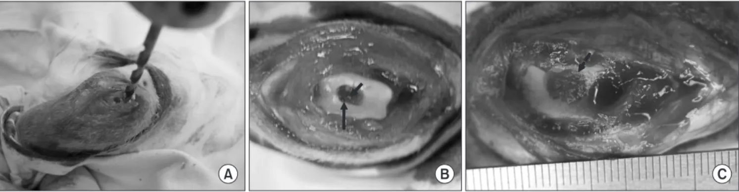
몸무게가 평균 3.5 kg (3.5±0.24 kg)인 생후 3-6개월 된 수컷 가 토(New Zealand white rabbit; Jung-Ang Lab Animal Inc., Seoul, Korea) 24마리의 우측 슬관절을 대상으로 하였으며 내측 관절 도 달법을 이용하여 가토의 대퇴골의 연골을 노출시킨 후에 대퇴골 의 슬개골 구에 3 mm 지름의 드릴을 이용하여 세로 6 mm, 가로 3 mm로 연골 결손을 만든 후 15번 수술칼을 이용하여 이 부위에 연골하 골의 손상이 없게 조금씩 연골을 제거하여 연골 전층 결 손을 만들었다. 총 24마리 중 18마리에서는 연골 전층 결손 부위 에 0.762 mm의 얇은 Kirschner 강선을 이용하여 미세골절을 하였 다(Fig. 1A, 1B).
2. 실험군 설정 및 처치
24마리의 가토의 슬관절을 6마리씩 무작위 배당하여 6마리는 슬 관절에 연골 전층 결손을 만든 후에 다른 처치를 하지 않고 대조 군으로 하였으며, 다른 18마리는 연골 전층 결손 부위에 미세골 절을 하였다. 연골 결손 부위에 미세골절을 만든 18마리 중 6마리 는 다른 처치를 하지 않았고 다른 6마리의 연골 결손 부위에는 섬 유성 아교(Green-plast; Korea Green Cross Co., Yongin, Korea) 0.1 ml를 충전하였고, 남은 6마리는 연골 전층 결손 부위에 0.1 ml 섬 유성 아교와 recombinant human bone morphogenetic protein-2 (rhBMP-2; R&D systems Inc., Minneapolis, MN, USA) 3 mg을 주
사기에 혼합하여 연골 결손 부위를 충전하였다(Fig. 1C). 실험 후 감염을 예방하기 위하여 겐타마이신(gentamycin; Kyung Dong Pharm Co., Seoul, Korea) 6 mg/kg을 실험 후 3일 동안 투여한 후 8 주간 동일한 조건으로 사육하였다.
실험 8주째에 각 가토를 희생시킨 후 내측 관절 도달법을 이용 하여 슬관절의 실험 부위를 노출한 후에 슬관절의 연골에 손상이 가지 않게 조심스럽게 슬관절 주위의 연부조직을 제거한 후에 실 험 부위를 포함한 대퇴골의 과상부를 채취하였다. 동일하게 처치 된 실험군 6마리 중 3마리는 현미경 분석을 위하여, 다른 3마리는 조직 성분 분석을 위하여 사용되었다.
3. 결과 분석 1) 현미경 관찰
각 표본을 10% 중성 포르말린에 3일간 고정하고 탈석회용액(de- calcifying agent, Calci-Clear Rapid; National Diagnostics, Atlanta, GA, USA)으로 탈석회화 과정을 거친 후 파라핀에 포매하여 6 mm 의 두께로 절제하여 조직 표본을 만들었다. H&E 염색을 하고, 글 리코스아미노글리칸 존재 여부를 보기 위해 alcian blue염색을 하 여 광학 현미경에서 관찰하였다. 조직학적 등급을 판정하기 위하 여 Pineda 등9)이 제안한 방법을 사용하였다.
2) 재생된 조직의 성분 분석
콜라겐 1형과 2형의 정량 분석을 위하여 real-time polymerase chain reaction (real-time PCR) (LightCycler 1.5 Instrument; Roche Diagnostics, Basel, Switzerland)을 이용하였는데, 15번 수술 칼을 이용하여 연골 결손 부위에 재생된 조직을 주변 조직과 경계 부
A B C
Figure 1. Experiment photographs. (A) A 3×6 mm defect is made at the intercondylar notch of rabbit’s femur with a 3 mm diameter drill hole (1–2 mm thickness) without subchondral bone damage. (B) Microfracture (arrows) is made with a fine K-wire. (C) The defect (dashed arrow) is filled with fibrin glue and recombinant human morphogenetic protein-2.
Table 1. Summary of Real-Time Polymerase Chain Reaction
Collagen type Forward primer Reverse primer Universal probe
1 agggagagcctggtgacaa gaagaccttgcaatccgttg #72, Cat. No. 04688953001 2 acagcaggttcacctataccg cccacttaccggtgtgtttc #46, Cat. No. 04688066001
위에서 잘라내어 real-time PCR에 사용할 조직을 준비하였고 각 콜라겐의 상대적 정량을 비교하기 위하여 가토의 실험 부위가 아 닌 반대측 슬관절의 슬개구 부위에서 정상 관절 연골을 채취하 였다. 1형 콜라겐의 정량 분석을 위하여 Universal ProbeLibrary probe (#72, cat.no. 04688953001; Roche Diagnostics)을 사용하였고 503에서 521 위치에 있는 agggagagcctggtgacaa 서열의 primer를 사 용하였다. 2형 콜라겐의 정량분석을 위하여 Universal ProbeLi- brary probe (#46, cat.no. 04688066001; Roche Diagnostics)과 578에 서 598 위치에 있는 acagcaggttcacctataccg 서열의 primer를 사용하 였고 real-time PCR은 45회 cycle을 시행하였다(Table 1).
3) 통계 분석
각 실험군의 Pineda 점수와 콜라겐 정량 결과를 PASW Statistics ver. 18 (IBM Co., Armonk, NY, USA) 프로그램을 이용하여 Krus- kal-Wallis test로 분석하였고 p<0.05의 결과를 통계적으로 유의 한 것으로 하였다.
결 과
1. 현미경 관찰 1) 조직 염색
H&E 염색 결과, 연골 결손 부위를 섬유성 아교와 rhBMP-2로 충 전한 실험군의 연골 결손 부위는 주변 경계 연골과 비슷한 두께 의 연골이 형성되었으며, 연골하 골 조직에서도 골 재형성이 되 었으며 주위 정상 연골하 골과 비슷한 두께로 재형성되었다(Fig.
2). Alcian blue 염색 결과, 섬유성 아교와 rhBMP-2로 충전한 실험 군의 연골 결손 부위가 뚜렷이 염색되었으며, 연골의 배열이 일 정하게 정렬되어 연골 조직과 연골하 골 조직이 명확하게 구분되 었다(Fig. 3).
2) Pineda의 조직학적 등급
미세골절 후에 섬유성 아교와 rhBMP-2를 충전한 실험군에서 4.00±0.00점으로 가장 낮은 점수를 보였다(p<0.05). 섬유성 아교
A
B
C
D
Figure 2. Photographs of H&E stain (×40). (A) Cartilage defect only. (B) Treatment with microfracture. (C) Treatment with microfracture and fibrin glue. (D) Treatment with microfracture, fibrin glue, and recombinant human bone morphogenetic protein-2 (rhBMP-2). The defect filled with fibrin glue and rhBMP-2 demonstrated more cartilagenous tissue and replacement of subchondral bone. The arrows indicate the boundary between the preexisting articular cartilage and the repair tissues. RC, repair cartilage; NC, normal cartilage.
A
B
C
D
Figure 3. Photographs of Alcian blue stain (×40). (A) Cartilage defect only. (B) Treatment with microfracture. (C) Treatment with microfracture and fibrin glue. (D) Treatment with microfracture, fibrin glue, and recombinant human bone morphogenetic protein-2 (rhBMP-2). These findings demonstrated the presence of greater amounts of hyaline cartilage in the group treated with microfracture, fibrin glue and rhBMP-2. The arrows indicate the boundary between the preexisting articular cartilage and the repair tissues. RC, repair cartilage; NC, normal cartilage.
만 충전한 실험군과 섬유성 아교와 rhBMP-2를 충전한 실험군의 조직 등급 각 항목을 비교하였을 때 Pineda의 조직 등급에 기재되 어 있는 여덟 가지 항목 중 integration at margin, staining of matrix, cellular morphology, bone repair, reformation of tidemark의 다섯 가지 항목에서 섬유성 아교와 rhBMP-2를 충전한 실험군이 더 낮 은 점수를 보였다(p<0.05; Table 2).
2. 재생된 조직의 성분 분석
Real-time PCR을 이용한 콜라겐의 정량 분석은 정상 연골의 콜 라겐 성분에 대한 상대적 정량 방법으로, 정상 연골의 콜라겐 정 량을 1이라는 절대값으로 하고 각각의 결과를 상대적 정량 값을 표시하였다. 연골 결손만 만든 실험군의 콜라겐 1형은 평균 0.5±
0.03, 콜라겐 2형은 평균 0.5±0.03였으며, 연골 결손 부위에 미세 골절을 한 실험군의 콜라겐 1형은 평균 3.98±0.2, 콜라겐 2형은 평균 0.95±0.36이었다. 연골 결손 부위에 미세골절 후 섬유성 아 교를 충전한 실험군의 콜라겐 1형은 평균 1.42±0.17, 콜라겐 2형 은 평균 0.84±0.05였고 연골 결손 부위에 미세골절 후 섬유성 아 교와 rhBMP-2를 충전한 실험군의 콜라겐 1형은 평균 3.92±0.12,
콜라겐 2형은 평균 8.82±0.41이었다(Fig. 4). 이들 값을 Kruskal- Wallis test를 이용하여 검정한 결과, 콜라겐 1형과 2형 모두에서 차이를 보였고, 이는 통계적으로 유의하였다(p<0.05).
고 찰
관절 연골은 불충분한 혈액 공급을 갖고 있는 조직으로 관절 연 골에 손상이 발생하였을 경우에 관절 연골의 재생 및 복원이 어 렵다고 알려져 있다.10) 관절 연골이 부분 손상되었을 경우에는 손상 부위에 연골 모세포의 접근이 불가능하기 때문에 자연 치 유가 되지 않으나 관절 연골의 전층 손상에서는 연골하 골 조직 의 노출로 골수 줄기 세포가 손상 부위에 접근할 수 있어서 연골 의 재형성이 가능하다고 알려져 있다.11,12) 이런 특성을 고려하여, Steadman 등13)이 인위적으로 연골 결손 부위에 연골하 골 조직을 노출시킨 후에 여러 개의 구멍을 만들어서 연골 결손 부위로 골 수 줄기 세포가 도달할 수 있게 하여 연골의 재형성을 촉진시키 는 미세골절 기법을 소개한 이후로 슬관절 및 족관절의 관절 연 골 병변에 대하여 임상적으로 많이 사용되고 있다.14-16) 그러나 장 기간의 추시 관찰에서 미세골절 후에 재형성된 연골 부위는 관 절 연골의 초자 연골 성분이 아닌 섬유 연골 조직으로 복원되며, 이 섬유연골은 초자 연골보다 물리적으로 약하기 때문에 퇴행 성 변화가 빨리 발생하는 문제점들이 보고되고 있다.17,18) 최근에 는 이러한 조직학적 문제를 극복하기 위하여 transforming growth factor (TGF)-P, fibroblast growth factor (FGF)-2, bFGF 같은 성장 인자 등을 이용한 조직 공학의 연구가 진행되고 있다.7,19,20) BMP 중에 BMP-7이 미세골절과 상호 작용하여 관절 연골이 초자 연 골로 치유되는 것을 촉진한다고 보고되었으며,21) BMP-2는 염증 성 관절의 관절 연골에서는 interleukin-1 (IL-1)의 반작용 때문에 BMP-2 단독으로는 관절 연골의 복원을 유도하지 못하는 것으로 알려져 있으나,8) IL-1의 영향이 없는 환경에서 연골과 골 표지자 의 표현을 유도하는 것으로 알려져 있으며, 생체 내에서도 연골 과 골형성을 유도하는 것으로 알려져 있다.22,23)
본 연구는 관절 연골 전층 결손 부위에 BMP-2의 투여가 관절
Collagen type 1 Collagen type 2
1 10
9 8 7 6 5 4 3 2 1 0
0.5 0.5
3.98
0.95 1.42
0.84
3.92 8.82
Figure 4. Results of real-time polymerase chain reaction in each group.
Group filled with fibrin glue and recombinant human morphogenetic protein-2 after microfracture demonstrated a higher amount of collagen type 1 and collagen type 2.
Table 2. Histological Scores of Articular Cartilage Defect
Variable
Category Filling of
defect
Integration at margin
Staining of matrix
Cellular morphology
Architecture within defect
Architecture
of surface Bone repair Reformation of tidemark
Total
score
CD 1.66±0.57 2.33±0.57 2.66±0.57 3.66±1.15 2.66±1.15 2.66±0.57 2.66±0.57 3.33±0.57 21.66±3.05 CD+MF 0.33±0.57 1.33±0.57 2.33±0.70 2.33±0.57 1.66±0.57 1.66±0.57 2.33±0.57 1.66±0.57 13.66±2.08 CD+MF+FG 0.02±0.04 1.08±0.60 1.24±0.86 1.12±0.98 1.24±1.16 1.72±0.90 1.68±0.94 1.63±1.27 7.68±1.45 CD+MF+FG+rhBMP 0.00±0.00 1.00±0.47 0.83±0.81 0.71±0.36 1.12±1.24 1.50±0.85 0.40±0.05 0.42±0.76 4.50±2.10 p-value 0.062 0.0374 0.0387 0.0258 0.0863 0.0603 0.0406 0.0285 0.0158 Values are presented as mean±standard deviation. The score for defects treated with rhBMP-2 was significantly different (p<0.05) from that for the other defects. CD, cartilage defect; MF, microfracture; FG, fibrin glue; rhBMP, recombinant human morphogenetic protein-2.연골의 재생과 연골하 골조직의 재형성을 유도할 수 있는지를 알 기 위하여 실험을 시행하였다. 본 연구에서는 관절 연골 결손 부 위에서 재생 정도 및 재생의 질을 평가하기 위하여 Pineda의 조직 등급9)을 사용하였으며, 이는 8개의 항목에 대하여 각각의 점수를 책정하여 연골 조직의 재생 정도를 판정하는 방법으로, 총점이 0 점(정상 연골)에서 31점(재생이 안된 연골)으로 총점이 높아질수 록 연골의 재생이 불완전함을 의미한다. Pineda 조직 등급의 총 점수를 각 실험군별로 비교하였을 때 미세골절 후 섬유성 아교와 rhBMP-2를 같이 충전한 실험군이 5.00±0.00으로 가장 낮은 점 수를 보였으며 이는 rhBMP-2가 연골의 재생을 정상 연골에 보다 가깝게 촉진하는 것으로 해석할 수 있다.
연골하 골 조직이 불완전하게 재형성이 되었을 때에는 연골하 골 조직이 본연의 강성보다 더 경직되어 충격 흡수의 역할이 소 실되며, 불충분한 혈관 공급을 형성하여 관절 연골에 충분한 혈 액 공급을 하지 못 하게 되고, 연골하 골 조직에서 연골의 재형성 에 영향을 미치는 insunlin-like growth factor-1, TGF-1 등이 유리 되어 관절 연골의 복원을 억제되거나 골관절염으로 진행된다.24-26) 따라서, 관절 연골 결손의 복원에서 연골하 골 조직의 재형성도 연골의 복원 및 골관절염으로 진행을 억제하는 데 중요한 역할 을 한다. 본 연구에서 rhBMP-2를 충전한 실험군의 Pineda 조직 등급의 각 항목을 분석한 결과, 연골하 골 조직의 재배치 항목과 tidemark의 재형성 항목에서 다른 항목에 비교하여 낮은 점수를 보였는데, 이는 rhBMP-2의 골 유도의 특성 때문인 것으로 판단 된다. 즉 rhBMP-2는 연골아세포의 분화를 촉진할 뿐 아니라 골 아세포의 분화를 동시에 촉진하여 미세골절만을 한 다른 군보다 빨리 골의 재형성을 유도한 것으로 판단된다. 본 연구에서 관찰 된 연골하 골의 재형성은 재생된 연골이 보다 확고히 연골하 골 조직과 유착을 이뤄서 연골 조직이 물리적 외력에 더 저항력을 가질 수 있게 도움을 줄 수 있을 것으로 생각되며 충분한 혈액 공 급 및 골관절염 유발 인자들의 유리 억제를 통하여 연골 복원에 도 도움을 줄 수 있을 것으로 생각된다.
Real-time PCR로 재생된 조직의 콜라겐의 정량 분석을 한 결 과, 연골 결손만 만든 실험군에서는 콜라겐 1형 및 2형이 모두 낮 게 나왔는데 이는 연골 결손 부위에 복원이 잘되지 않아서 콜라 겐 1형 및 2형이 모두 낮게 검출되는 것으로 판단되며, 섬유성 아 교와 rhBMP-2를 충전한 실험군에서는 콜라겐 1형이 3.92, 콜라 겐 2형이 8.82로 높게 나왔는데, 이는 1군과는 달리 왕성한 복원이 이루어지고 있음을 나타낸다. 특히 콜라겐 2형은 다른 군보다 상 대적으로 높게 검출되어 재생된 관절 연골이 초자 연골에 가깝게 재생되었다는 것으로 해석할 수 있다. 따라서 관절 연골의 결손 부위에 미세골절만을 한 경우보다는 rhBMP-2를 투여하는 것이 초자 연골 형성을 촉진할 수 있을 것으로 판단된다. 다만 제 섬유 성 아교와 rhBMP-2를 충전한 실험군에서 콜라겐 1형이 다른 군 에 비하여 많이 검출된 이유는 조직의 치유가 왕성하게 진행되어
콜라겐 1형이 많이 검출된 것으로 판단된다. Real-time PCR의 결 과에서 예상과는 달리, 섬유성 아교만을 충전한 실험군의 콜라겐 1형은 0.84, 콜라겐 2형은 1.42로 미세골절만을 만든 2군에 비하여 콜라겐 1형과 2형 모두 낮게 검출되어 섬유성 아교가 치유 과정 을 저해하는 결과를 보였다. 조직의 특성에 따라 섬유성 아교의 작용에 대한 논란이 있기 때문에27-30) 본 연구에서는 섬유성 아교 를 단독으로 사용하였을 경우와 rhBMP-2를 혼합하여 사용하였 을 경우에 발생할 수 있는 실험 결과를 비교하기 위하여 섬유성 아교를 사용하였다.
섬유성 아교는 일종의 매개체로 많이 사용되고 있는데 혈관 이 풍부한 조직에서는 섬유성 아교가 fibrin clot의 형성을 억제하 여 치유 과정을 저해하며, 인대 또는 연골같이 혈관이 약한 부위 에서는 섬유성 아교와 성장 인자를 같이 투여하면 손상된 조직 의 재생을 촉진시킬 수 있다고 알려져 있다.27-30) 본 연구에서 섬 유성 아교를 단독으로 사용하였을 때에 보인 낮은 콜라겐 형성은 섬유성 아교가 미세골절 부위에서 fibrin clot이 형성되는 것을 오 히려 억제하여 치유 과정을 저해하는 것으로 생각할 수도 있으나 Pineda 점수에서는 섬유성 아교를 사용한 경우에 더 좋은 점수를 보인 것을 감안할 때, 본 연구의 제한점인 실험 개체수 부족에 따 른 데이터 오류도 고려해야 할 것으로 생각된다. 그러나 섬유성 아교와 rhBMP-2를 충전한 실험군의 콜라겐의 정량 분석 결과를 봤을 때, 섬유성 아교와 성장 인자를 같이 투여할 경우에는 섬유 성 아교가 성장 인자를 전달하기 위한 매개체로 작용하여 조직의 치유과정을 촉진하는 것으로 판단된다.
본 연구는 실험 개체수가 충분하지 않다는 큰 제한점이 있다.
총 24마리로 각 군별로 배정된 개체수가 6마리이며, 이를 다시 조 직 분석에 3마리씩, 콜라겐 정량 분석에 3마리씩 할당하였기 때문 실험 결과를 분석하고 통계적 의의를 부여하는 것에 제한이 있을 수 있다. 또한 real-time PCR을 이용한 콜라겐의 정량 분석 시에 house keeping 유전자를 통한 유전자들의 mRNA level을 보정하 지 못한 실험적 약점으로 본 연구의 실험값에 대한 절대적 신뢰 성이 부족하다는 약점이 있다. 그리고 실험 기간이 8주로 비교적 짧아서 rhBMP-2의 효과 시점과 장기 효과에 대한 분석이 부족 하다는 제한점이 있다. 다만 본 연구는 연골 전층 결손 부위에서 rhBMP-2가 단기간에 결손 부위 재생에 미치는 영향을 분석하는 데 의의가 있을 수 있으며, 앞서 기술한 실험적 약점을 보정하여 장기 효과에 대한 분석을 위한 실험 등이 추가적으로 필요할 것 으로 생각된다.
결 론
본 연구에서는 BMP 중 상용화되어 쉽게 이용할 수 있는 rhBMP- 2를 연골 결손 부위에 충전하여 연골 결손 부위에서 연골의 재생 을 보다 효과적으로 촉진하는 결과를 얻을 수 있었으며, 미세골
절 후 섬유성 아교와 rhBMP-2는 관절 연골 결손 부위에서 연골 치유를 촉진시키고 초자 연골에 근접하게 연골이 치유되게 도움 을 줄 수 있을 것으로 생각된다.
CONFLICTS OF INTEREST
The authors have nothing to disclose.
REFERENCES
1. Urist MR. Bone: formation by autoinduction. Science. 1965;
150:893-9.
2. Bostrom M, Lane JM, Tomin E, et al. Use of bone morpho- genetic protein-2 in the rabbit ulnar nonunion model. Clin Orthop Relat Res. 1996;327:272-82.
3. Jones AL, Bucholz RW, Bosse MJ, et al; BMP-2 Evaluation in Surgery for Tibial Trauma-Allgraft (BESTT-ALL) Study Group. Recombinant human BMP-2 and allograft compared with autogenous bone graft for reconstruction of diaphyseal tibial fractures with cortical defects. A randomized, con- trolled trial. J Bone Joint Surg Am. 2006;88:1431-41.
4. Boyne PJ, Lilly LC, Marx RE, et al. De novo bone induction by recombinant human bone morphogenetic protein-2 (rh- BMP-2) in maxillary sinus floor augmentation. J Oral Maxil- lofac Surg. 2005;63:1693-707.
5. McKay WF, Peckham SM, Badura JM. A comprehensive clinical review of recombinant human bone morphogenetic protein-2 (INFUSE Bone Graft). Int Orthop. 2007;31:729-34.
6. Siebert CH, Schneider U, Sopka S, Wahner T, Miltner O, Niedhart C. Ingrowth of osteochondral grafts under the in- fluence of growth factors: 6-month results of an animal study.
Arch Orthop Trauma Surg. 2006;126:247-52.
7. Siebert CH, Miltner O, Weber M, Sopka S, Koch S, Niedhart C. Healing of osteochondral grafts in an ovine model under the influence of bFGF. Arthroscopy. 2003;19:182-7.
8. Wim B, Peter M, Scharstuhl A, Henk M. Tissue engineering, cells, scaffolds, and growth factors. Clin Orthop Relat Res.
2001;391:244-50.
9. Pineda S, Pollack A, Stevenson S, Goldberg V, Caplan A. A semiquantitative scale for histologic grading of articular car- tilage repair. Acta Anat (Basel). 1992;143:335-40.
10. Mankin HJ. The response of articular cartilage to mechanical injury. J Bone Joint Surg Am. 1982;64:460-6.
11. Buckwalter JA. Articular cartilage: injuries and potential for healing. J Orthop Sports Phys Ther. 1998;28:192-202.
12. Wei X, Gao J, Messner K. Maturation-dependent repair of untreated osteochondral defects in the rabbit knee joint. J Biomed Mater Res. 1997;34:63-72.
13. Steadman JR, Rodkey WG, Singleton SB, Britts KK. Micro- fracture technique for full-thickness chondral defects: tech- nique and clinical results. Oper Tech Orthop. 1997;7:300-4.
14. Steadman JR, Briggs KK, Rodrigo JJ, Kocher MS, Gill TJ, Rodkey WG. Outcomes of microfracture for traumatic chon- dral defects of the knee: average 11-year follow-up. Arthros- copy. 2003;19:477-84.
15. Blevins FT, Steadman JR, Rodrigo JJ, Silliman J. Treatment of articular cartilage defects in athletes: an analysis of functional outcome and lesion appearance. Orthopedics. 1998;21:761-7.
16. Steadman JR, Rodkey WG, Rodrigo JJ. Microfracture: surgi- cal technique and rehabilitation to treat chondral defects.
Clin Orthop Relat Res. 2001;391 Suppl:S362-9.
17. Frisbie DD, Trotter GW, Powers BE, et al. Arthroscopic sub- chondral bone plate microfracture technique augments heal- ing of large chondral defects in the radial carpal bone and medial femoral condyle of horses. Vet Surg. 1999;28:242-55.
18. Steadman RJ, Rodkey WG, Singleton SB, et al. Microfracture procedure for treatment of full-thickness chondral defects:
technique, clinical results, and current basic science status.
In: Harner CD, Vince KG, Fu FH, ed. Techniques in knee sur gery. Philadelphia: Lippincott Williams & Wilkins; 2001.
23-31.
19. Kang SW, Bada LP, Kang CS, et al. Articular cartilage regen- eration with microfracture and hyaluronic acid. Biotechnol Lett. 2008;30:435-9.
20. Ishii I, Mizuta H, Sei A, Hirose J, Kudo S, Hiraki Y. Healing of full-thickness defects of the articular cartilage in rabbits using fibroblast growth factor-2 and a fibrin sealant. J Bone Joint Surg Br. 2007;89:693-700.
21. Kuo AC, Rodrigo JJ, Reddi AH, Curtiss S, Grotkopp E, Chiu M. Microfracture and bone morphogenetic protein 7 (BMP-7) synergistically stimulate articular cartilage repair. Osteoar- thritis Cartilage. 2006;14:1126-35.
22. Katagiri T, Yamaguchi A, Ikeda T, et al. The non-osteogenic mouse pluripotent cell line, C3H10T1/2, is induced to dif- ferentiate into osteoblastic cells by recombinant human bone morphogenetic protein-2. Biochem Biophys Res Commun.
1990;172:295-9.
23. Thies RS, Bauduy M, Ashton BA, Kurtzberg L, Wozney JM, Rosen V. Recombinant human bone morphogenetic pro- tein-2 induces osteoblastic differentiation in W-20-17 stro- mal cells. Endocrinology. 1992;130:1318-24.
24. Li B, Aspden RM. Mechanical and material properties of the subchondral bone plate from the femoral head of pa- tients with osteoarthritis or osteoporosis. Ann Rheum Dis.
1997;56:247-54.
25. Conaghan PG, Vanharanta H, Dieppe PA. Is progressive os- teoarthritis an atheromatous vascular disease? Ann Rheum Dis. 2005;64:1539-41.
26. Shibakawa A, Yudoh K, Masuko-Hongo K, Kato T, Nishioka K, Nakamura H. The role of subchondral bone resorption
pits in osteoarthritis: MMP production by cells derived from bone marrow. Osteoarthritis Cartilage. 2005;13:679-87.
27. Thomopoulos S, Soslowsky LJ, Flanagan CL, et al. The effect of fibrin clot on healing rat supraspinatus tendon defects. J Shoulder Elbow Surg. 2002;11:239-47.
28. Lusardi DA, Cain JE Jr. The effect of fibrin sealant on the strength of tendon repair of full thickness tendon lacerations in the rabbit Achilles tendon. J Foot Ankle Surg. 1994;33:443-7.
29. Stemberger A, Blümel G. Fibrinogen-fibrin conversion and inhibition of fibrinolysis. Thorac Cardiovasc Surg. 1982;
30:209-14.
30. Schlag G, Redl H. Fibrin sealant in orthopedic surgery. Clin Orthop Relat Res. 1988;227:269-85.
재조합 골 형성 단백-2가 가토의 관절 연골 결손의 연골하 골 재형성 및 연골 치유에 미치는 영향
이석하 • 이승준 • 이종수 • 안재성*
건국대학교 의학전문대학원 건국대학교병원 정형외과학교실, *충남대학교 의학전문대학원 충남대학교병원 정형외과학교실
목적: 관절 연골 전층 결손이 있는 가토 모델에서 재조합 골 형성 단백-2가 가토의 관절 연골 결손의 연골하 골 재형성 및 연골 치유 에 미치는 영향을 분석하고자 한다.
대상 및 방법: 24마리의 수컷 가토의 우측 대퇴골 활차 구에 6×3 mm의 전층 연골 결손을 만든 후에 24마리 중 6마리는 더 이상 처 치를 하지 않았으며, 18마리는 결손 부위에 미세 골절을 하였다. 미세골절한 18마리 중 6마리는 다른 처치를 하지 않았고 다른 6마리 는 결손 부위에 섬유성 아교를 충전하였으며 남은 6마리에는 결손 부위에 섬유성 아교와 재조합 골 형성 단백-2를 충전하였다. 수술 8주째에 조직학 등급 점수와 real-time polymerase chain reaction을 이용한 콜라겐의 성분 분석으로 미세골절 부위에 충전된 재조 합 골 형성 단백-2가 관절 연골의 치유에 미치는 효과를 분석하였다.
결과: 연골 결손 부위를 섬유성 아교와 재조합 골 형성 단백-2로 충전한 6마리에서 조직학적 등급 점수가 더 좋았으며, 콜라겐 1형 과 2형도 더 많이 검출되었다.
결론: 미세골절 후 섬유성 아교와 재조합 골 형성 단백-2는 관절 연골 결손 부위에서 연골 치유를 촉진시키고 초자 연골에 근접하게 연골이 치유되게 도움을 줄 수 있을 것으로 생각된다.
색인단어: 관절 연골 결손, 미세골절, 골 형성 단백-2
접수일 2014년 10월 2일 수정일 2015년 2월 6일 게재확정일 2015년 3월 7일 책임저자 이승준
서울시 광진구 능동로 120-1, 건국대학교 의학전문대학원 건국대학교병원 정형외과학교실 TEL 02-2030-7616, FAX 02-2030-7369, E-mail [email protected]
*이 논문은 2009학년도 건국대학교의 지원에 의하여 연구되었음.
Copyright © 2015 by The Korean Orthopaedic Association
“This is an Open Access article distributed under the terms of the Creative Commons Attribution Non-Commercial License (http://creativecommons.org/licenses/by-nc/4.0/) which permits unrestricted non-commercial use, distribution, and reproduction in any medium, provided the original work is properly cited.”