저작자표시-비영리-변경금지 2.0 대한민국 이용자는 아래의 조건을 따르는 경우에 한하여 자유롭게 l 이 저작물을 복제, 배포, 전송, 전시, 공연 및 방송할 수 있습니다. 다음과 같은 조건을 따라야 합니다: l 귀하는, 이 저작물의 재이용이나 배포의 경우, 이 저작물에 적용된 이용허락조건 을 명확하게 나타내어야 합니다. l 저작권자로부터 별도의 허가를 받으면 이러한 조건들은 적용되지 않습니다. 저작권법에 따른 이용자의 권리는 위의 내용에 의하여 영향을 받지 않습니다. 이것은 이용허락규약(Legal Code)을 이해하기 쉽게 요약한 것입니다. Disclaimer 저작자표시. 귀하는 원저작자를 표시하여야 합니다. 비영리. 귀하는 이 저작물을 영리 목적으로 이용할 수 없습니다. 변경금지. 귀하는 이 저작물을 개작, 변형 또는 가공할 수 없습니다.
Layered approach with autogenous bone
and bone substitute for ridge augmentation
on implant dehiscence defects in dogs
In-Kyeong Lee
Department of Dentistry
Layered approach with autogenous bone
and bone substitute for ridge augmentation
on implant dehiscence defects in dogs
Directed by Professor Ui-Won Jung
The Doctoral Dissertation
submitted to the Department of Dentistry
the Graduate School of Yonsei University
in partial fulfillment of the requirements for the degree of
Ph.D. in Dental Science
In-Kyeong Lee
December 2015
This certifies that the Doctoral Dissertation
of In-Kyeong Lee is approved.
The Graduate School
Yonsei University
감사의 글
이 논문이 완성되기까지 부족한 저를 이끌어 주시고 연구에
매진할 수 있도록 끊임없는 지도와 아낌없는 격려를 해주신 정의원
지도교수님께
진심으로
감사드립니다. 연구자로서
가져야
하는
마음가짐을 잊지 않게 해주시고 학문에 대한 열정을 알려주신
김종관 교수님, 언제나 따뜻한 관심과 격려를 아끼지 않으신 채중규
교수님, 냉철한 비판적 자세와 안목을 가질 수 있게 이끌어 주신
조규성 교수님, 작은 부분까지 조언해 주시고 힘이 되어주신 최성호
교수님, 연구에 있어 창의성의 중요함을 깨닫게 해주신 김창성
교수님, 세심한 부분까지 성심껏 지도해주신 이중석 교수님께 깊이
감사드립니다. 바쁘신 와중에도 심사를 맡아주시고 조언을 아끼지
않으신 김성태 교수님께도 감사드립니다. 연구가 무사히 완료될 수
있도록
도움을
준
치주과의
모든
의국원들과
연구원들에게도
고마움을 전합니다
항상 넘치는 사랑과 무한한 신뢰로 저를 지켜봐 주시는 부모님,
언제나 저에게 귀감과 의지가 되어주는 든든한 오빠에게 사랑과
고마움을 전합니다.
2015년 12월
저자 씀
Table of Contents
List of figures
··· ii
List of tables
··· iii
Abstract (English)
··· iv
I. Introduction
··· 1
II. Materials & Methods
··· 4
1. Experimental Animals ··· 4
2. Experimental Materials ··· 4
3. Experimental Design ··· 5
4. Surgical Procedures ··· 5
5. Radiographic Analysis
··· 8
6. Histologic and Histomorphometric Analyses ··· 9
7. Statistical Analysis ··· 10
III. Results
··· 11
1. Clinical Findings ··· 11
2. Radiographic Analysis ··· 11
3. Histologic and Histomorphometric Analyses
··· 12
IV. Discussion
··· 13
References
··· 17
Figure Legends
··· 21
Tables
··· 23
Figures
··· 25
Abstract (Korean)
··· 32
List of figures
Figure 1.
Schematic drawing of the surgical procedure.Figure 2.
Schematic illustration of histomorphometric measurements.Figure 3.
Three-dimensional reconstruction and two-dimensional cross-sectional views of micro-computed tomography scans in the three experimental groups.Figure 4.
Histologic views of the SBC group.Figure 5.
Histologic views of the IAB group.Figure 6.
Histologic views of the outer autogenous bone group.Figure 7.
Graph representing the composition in the area of interest in the three experimental group.List of tables
Table 1.
Volumetric radiographic measurements (n = 4).Abstract
Layered approach with autogenous bone and bone substitute
for ridge augmentation on implant dehiscence defects in dogs
In-Kyeong Lee, D.D.S., M.S.D
Department of Dentistry
The Graduate School, Yonsei University
(Directed by Professor Ui-Won Jung, D.D.S., M.S.D., PhD.)
Objective: This study compared the efficacies of different layered approaches using autogenous bone and synthetic bone substitute for ridge augmentation on implant dehiscence defects in dogs.
Materials and methods: Right mandibular second, third, and fourth premolars and the first molar were extracted, followed by standardized one-wall defect preparation in five dogs. After a healing period of 12 weeks, three implants (Implantium®) were
installed. Each of the three implant dehiscence defects was grafted with a different material as follows: (i) synthetic bone substitute combined with collagen (SBC; SBC group), (ii) inner autogenous bone layer and outer SBC layer (IAB group), and (iii) inner SBC layer and outer autogenous bone layer (OAB group). The grafted sites
were covered with a resorbable collagen membrane. Quantitative and qualitative analyses of the subsequent bone regeneration were performed at 12 weeks postoperatively.
Results: The dome-like augmented shape was relatively well maintained in the IAB and OAB groups, while the graft particles in the SBC group were dispersed. The bone-to-implant contact values tended to be significantly higher in the OAB group (49.51%) than in the SBC (36.58%) group. The amounts of newly formed bone within an area designated as 1 x 3 mm (width x height) from the implant platform in the IAB, OAB, and SBC groups were 35.59%, 28.10%, and 16.71%, respectively. Conclusion: Application of the layered approach using autogenous bone and synthetic biomaterial resulted in substantial new bone formation and volume maintenance on implant dehiscence defects, irrespective of the position of the autogenous bone layer.
Keywords: animal study, autogenous bone graft, bone substitutes, dental implants, guided bone regeneration
Layered approach with autogenous bone and bone substitute
for ridge augmentation on implant dehiscence defects in dogs
In-Kyeong Lee, D.D.S., M.S.D
Department of Dentistry
The Graduate School, Yonsei University
(Directed by Professor Ui-Won Jung, D.D.S., M.S.D., PhD.)
I. Introduction
The presence of sufficiently thick coronal bone around an implant is a prerequisite for long-term stability and to prevent biomechanical complications such as peri-implantitis and implant failure1-4. The tendency toward buccal bone resorption
has been accepted as a natural healing pattern of extraction sockets and might be the cause of the high prevalence of buccal dehiscence defects around the implants5,6.
Guided bone regeneration (GBR) has been introduced to compensate for such bone remodeling and to reestablish stable hard tissue over the implant surface7-10. Among
the various bone graft materials used for GBR, autogenous bone has been widely regarded as an ideal material due to its osteogenic properties and immunogenic
safety11. Jensen et al.12 compared the bone healing achieved using autografting,
anorganic bovine bone-derived mineral (ABBM), and synthetic b-tricalcium phosphate (b-TCP) in intraosseous defects in the mandible of minipigs. Autografting resulted in faster bone regeneration than grafting with either ABBM or synthetic b-TCP in the early healing phase. Although newly formed bone ultimately regenerated in all defects after 8 weeks regardless of the graft material used, autografting resulted in increased osseous maturity during all healing periods. Other studies have also shown that autogenous bone can induce faster and more mature bone regeneration in the initial healing period of lateral ridge augmentation13,14. In spite of these
advantages, the unpredictable resorption rate, limited availability, and patient morbidity limit its common use. Bone substitutes may overcome these potential shortcomings, and a good solution when autogenous bone is harvested may be combining that harvested bone with an osteoconductive bone substitute15.
Wang et al.16 introduced the concept of the sandwich bone augmentation
technique, which employs two layers of bone graft materials and a resorbable collagen membrane, whereby an inner autogenous bone layer is grafted close to the implant surface to provide an osteogenic environment around it, while the outer hydroxyapatite (HA) layer is placed over the inner autogenous bone layer. This bone substitute has slow-resorbing properties and is thus utilized to resist external compression. However, as the bone substitute is mostly osteoconductive, its bone-forming ability is inferior. Thus, bone substitute particles located distant from the alveolar bone as the main osteogenic source may be less involved in bone
regeneration and be vulnerable to fibrosis formation. A clinical study reported that horizontal ridge augmentation using ABBM particles layered onto autogenous bone blocks with a collagen membrane showed fibrous encapsulation and no evidence of osseous integration17. Furthermore, the long-lasting remnant materials located at the
outermost layer may lead to unwanted clinical events such as the protrusion or discharge of bone substitute particles, especially in thin biotype.
With the layered approach using autogenous bone and bone substitute for GBR, the order of the layers could be reconsidered. Positioning the bone substitute within the vicinity of the osteogenic source would be critical for predictable bone regeneration. Implants which are placed with a gap around them could have osseointegration, which could be explained by contact and distance osteogenesis18, 19.
Distance osteogenesis depends on the “jumping” of osteogenic potential. When harvested autogenous bone is grafted in the outer layer, it could be expected that viable osteoblasts and osteoprogenitor cells in the grafted bone transfer osteogenic potential toward the inside. Therefore, layering of the osteogenic autogenous bone over the bone substitute layer, which is an inverted protocol to the original sandwich bone augmentation technique, may provide an environment that is more favorable for the delivery of osteogenic cells from multiple sources to the osteoconductive bone substitute.
This study compared the efficacies of different layered approaches using autogenous bone and synthetic bone substitute for ridge augmentation on implant dehiscence defects.
II. Materials and methods
Experimental Animals
Five male mongrel dogs (mean age of 12–15 months, body weight of approximately 30 kg) with no systemic disease, intact dentition, and a healthy periodontium were used in this study. Animal selection, management, and the surgical protocol followed routine protocols approved by the Animal Care and Use Committee, Yonsei Medical Center, Seoul, Korea (Permission no. 2011-0188).
Experimental Materials
Sandblasted, acid-etched, and tapered titanium implants (3.8 mm in diameter and 8 mm in length; Implantium®
, Dentium, Seoul, Korea) were used. The synthetic bone substitute used in this study was the product combined with type I collagen (SBC; Osteon Collagen, Genoss, Suwon, Korea) and was cylindrical in shape (6 mm in diameter, 10 mm in length). SBC comprised a mixture of 70% HA and 30% b-TCP (particle size 0.5-1.0 mm, porosity 77%) and collagen; 93wt% graft and 7wt% collagen. Autogenous bone was obtained (~1 g) from the buccal wall of a previously extracted first molar site using a rotary instrument (ACM bone drill, Neobiotech, Seoul, Korea). The resorbable collagen membranes (HA Collagen membrane, Genoss,
Suwon, Korea) that were used to cover the grafted materials composed of type I collagen (50wt%) and HA particles (50wt%).
Experimental Design
Each of the three implant dehiscence defects in each dog was grafted with one of the following materials, with the material being allocated randomly (Fig. 1):
1. SBC alone (SBC group);
2. Inner autogenous bone layer and outer SBC layer (IAB group); 3. Inner SBC layer and outer autogenous bone layer (OAB group).
Surgical Procedures
Anesthesia
All surgical procedures were performed under sterile conditions and general anesthesia induced by intravenous injection of atropine (0.05 mg/kg; Kwangmyung Pharmaceutical, Seoul, Korea) and intramuscular injection of a combination of xylazine (2 mg/kg; Rompun, Bayer Korea, Seoul, Korea) and ketamine (10 mg/kg; Ketalar, Yuhan, Seoul, Korea), followed by inhalation anesthesia (Gerolan, Choongwae Pharmaceutical, Seoul, Korea). Local infiltration anesthesia (2% lidocaine HCL with epinephrine 1 : 80,000; Kwangmyung Pharm., Seoul, Korea) was
used at the surgical sites. Scaling and plaque control for all dentition was performed before the surgical procedure.
Preparation of the chronic narrow edentulous ridge
A crevicular incision was made from the right mandibular second premolar (P2) to the first molar (M1), and two buccal vertical incisions were made at the mesial side of P2 and the distal side of M1. The mucoperiosteal flap was reflected, and P2, the third and fourth premolars (P3 and P4, respectively), and M1 were carefully extracted after performing hemisection. A standardized one wall defect including P2– P4 mesiodistally, 3 mm apicocoronally, and 5 mm buccolingually was prepared using a high-speed carbide bur. Primary closure was obtained by interrupted suture with resorbable suture material (Monosyn® 4.0 Glyconate Monofilament, B. Braun, Tuttlingen, Germany). The sutures were removed 10 days after the surgical procedure.
Implantation and GBR procedure
A healing period of 12 weeks was allowed after preparation of a narrow edentulous ridge. A midcrestal incision was made from the mesial side of the second molar to the distal side of the first premolar, followed by two vertical incisions at both ends of the midcrestal incision. The mucoperiosteal flap was then reflected. The
defect bottom that had been created in a previous surgical procedure was flattened slightly using a ridge-contouring bur under copious irrigation with sterile saline. Following serial drilling according to a standard protocol, three implants were placed in the P2, P3, and P4 sites such that the implant platform was located at the level of lingual bone crest, with an insertion torque of approximately 20 N.cm. After installation of the implant, a buccal dehiscence of approximately 3 mm was made. After perforating the buccal cortical plate using a Lindemann drill, the P2, P3, and P4 sites were allocated randomly to one of the three groups (SBC, IAB, and OAB) and grafted accordingly.
In the SBC group, the total amount of a single SBC unit (i.e., a 6-mm-diameter x 10-mm-long cylinder) was grafted, while in the IAB and OAB groups, half of an SBC unit (i.e., 6-mm-diameter x 5-mm-long cylinder) was grafted together with ~0.5 g of harvested autogenous bone. The bone materials were covered with a resorbable collagen membrane. After a periosteal releasing incision was made to relieve the tension, the flap was advanced coronally and primary wound closure was achieved using interrupted sutures with resorbable suture material (Monosyn® 4.0 Glyconate Monofilament, B. Braun, Tuttlingen, Germany). The sutures were removed after 10 days. After a healing period of 12 weeks, the dogs were sacrificed by injecting an overdose of sodium pentobarbital. The mandibles were dissected, and the experimental sites including soft tissues were harvested for analysis.
Histologic preparation
Block sections including implants and grafted sites with surrounding hard and soft tissues were prepared. After fixation in 10% neutral buffered formalin for 10 days, the sections were scanned using micro-computed tomography (micro-CT; SkyScan 1076, SkyScan, Kontich, Belgium) at a resolution of 35 lm (100 kV, 100 μA). The specimens were trimmed and dehydrated in ethanol and then embedded in methyl methacrylate without being decalcified. They were then sectioned in the middle of the implants in a buccolingual direction. After being reduced to about 20 lm, they were stained with hematoxylin– eosin and Masson’s trichrome stains.
Radiographic Analysis
Two-dimensional cross-sectioned views and three-dimensionally reconstructed views were obtained using micro-CT. The total augmented volume was analyzed within the region of 5 mm mesial and distal from the center of the implant. Any augmented volume on either side of the boundary in its mesiodistal aspect was excluded. The overall augmented material was colored artificially in the three-dimensionally reconstructed views for easy recognition, using OnDemand 3D software (Cybermed, Seoul, Korea). After sectioning at mesial and distal boundaries, the cross-sectioned images were colored subsequently from mesial end to distal end.
Histologic and Histomorphometric Analyses
The specimens were examined histologically using a light microscope (BX-50, Olympus Optical, Tokyo, Japan) and analyzed histomorphometrically using image-analysis software programs (Image-Pro Plus, Media Cybernetics, Silver Spring, MD, USA; Adobe Photoshop CS6, Adobe Systems, San Jose, CA, USA). Linear and areal measurements were performed by two examiners. For linear analysis, the following parameters were recorded (Fig. 2):
1. Bone-to-implant contact (BIC, %): length of the implant boundary in contact with the newly formed bone;
2. P-B (mm): distance between the implant platform to the most-coronal point of BIC parallel to the implant axis;
3. P-C (mm): distance between the implant platform to the most-coronal bone level parallel to the implant axis.
In addition, the following parameters were measured within an area of interest [AOI; designated as 1 x 3 mm (width x height), from the implant platform]:
1. New bone (NB, mm2): area of newly formed bone.
2. Residual bone material (RBM, mm2): area of residual synthetic bone
substitute materials.
3. Mineralized tissue area (mm2): sum of NB and RBM.
4. Fibrovascular tissue (FVT, mm2): area left after subtracting NB and RBM
Statistical Analysis
The statistical analyses were performed using a software R, version 3.1.1 (http://www.r-pro ject.org). The nonparametric mixed model was used for comparing radiographic and histomorphometric parameters among the three experimental groups20. Statistical significance level was 5%. The Bonferroni correction was used
III. Results
Clinical Findings
Wound dehiscence was observed in three dogs at the point of suture removal (in the IAB group in one dog, in the SBC and IAB groups in another dog, and in all three groups in a third dog). The wound dehiscence in the first two cases closed completely during the healing period; however, in the third dog, the cover screws remained exposed until sacrifice. As there was barely any grafted material left around the implants in the last animal, it was excluded from the analyses. Except for that single exception, all other implants and grafted materials remained submerged and no complications were observed throughout the experimental period. Therefore, the data from four dogs were included in the analyses.
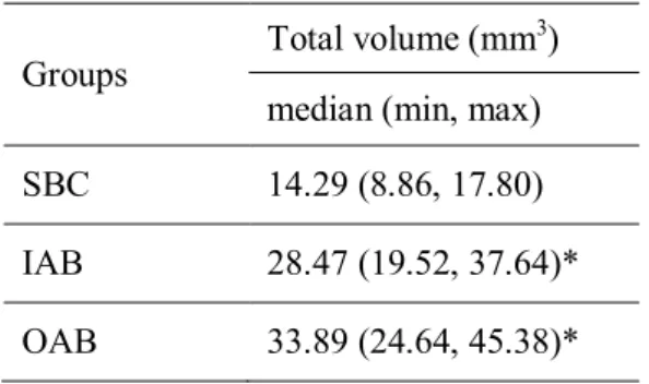
Radiographic Analysis
The dome-like augmented shape was relatively well maintained in both the IAB and OAB groups, while the grafted biomaterial particles tended to be dispersed in the SBC group (Fig. 3). The total augmented volume did not differ significantly between the IAB (28.47 mm3) and OAB (33.89 mm3) groups, but was significantly
Histologic and Histomorphometric Analyses
In all three experimental groups (i.e., IAB, OAB, and SBC), the synthetic biomaterial particles were linked to one another by a bridge of newly formed bone. In the SBC group, the newly formed bone tended to project from the bottom of defect in the coronal direction (Fig. 4). In some samples, the collagen membrane was collapsed, and a small amount of grafted material was observed around the coronal part of the implants.
RBM and NB were greater in the layered GBR groups (IAB and OAB) than in the SBC group. The regions of newly formed bone were connected to one another throughout the whole grafted area, and osseointegration was clearly visible (Figs 5 and 6). In the OAB group, new vital bone that was separated from the inner grafted bone materials was also observed in the outer area of the graft (Fig. 6). The aspect of the separated new bone could barely be seen, and almost all of the regions of new bone were connected to each other in the SBC and IAB groups.
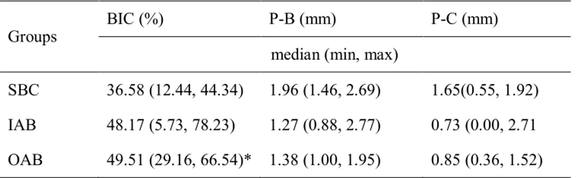
The median BIC appeared to be significantly higher in the OAB group (49.51%) than in the SBC group (36.58%). The median P-B and P-C values appeared to be lower in both the IAB (1.27 and 0.73 mm, respectively) and OAB (1.38 and 0.85 mm, respectively) groups compared with the SBC group (1.96 and 1.65 mm, respectively). However, these parameters did not differ significantly between the three groups (Table 2). With respect to the areal analyses, the median mineralized tissue area (the sum of NB and RBM) did not differ significantly in the SBC, IAB, and OAB groups (20.11%, 41.14%, and 38.56%, respectively; Fig. 7).
IV. Discussion
The scientific validity of the GBR approach using a layered composite has been verified in both animal and clinical studies, allowing rapid substitution of new bone and volume maintenance16,21-23. A combination of autogenous bone and synthetic
HA was found to induce a similar degree of bone regeneration compared to autogenous bone alone in the rat calvarium21. In a clinical study to resolve implant
dehiscence defect, there was no significant difference in the remaining depth of defect between ABBM and synthetic bone substitute used as an outer bone layer22. In all of
these studies, the autogenous bone was layered in the inner layer, which is in direct contact with the exposed implant surface. The biggest reason for this positioning is to achieve autogenous bone contact with the implant surface in order to promote osseointegration and support implant stability. However, most of the implant stability can be obtained from the high secondary stability of the other original bony envelope after a certain period of healing. Moreover, it has been shown that ABBM or synthetic bone used for GBR exhibits BIC values comparable to those obtained with autogenous bone24,25. Therefore, there is no clear rationale to support placement of the
autogenous layer on the inside, in apposition with the implant.
A clinical trial of lateral ridge augmentation with a mixture of ABBM and autogenous bone produced graft healing patterns that differed with the position of the graft26. The graft closest to the residual bone exhibited more particles surrounded by
bone, while the graft closest to the periosteum demonstrated more particles surrounded by soft tissue. In other words, the outer layer that was far from the regenerative source of native bone might be in a relatively unfavorable condition for bone regeneration. Furthermore, esthetic problems such as bulging of bone particles on the gingiva are possible, especially in the thin biotype. Therefore, it was hypothesized that the outer layer of autogenous bone is advantageous for the formation of a viable bone shell and in facilitating bone formation in the inner synthetic biomaterial layer.
The aim of the present study was to clarify how the position of the autogenous bone layer within the grafted mass influences bone regeneration. The layered approach with autogenous bone and synthetic biomaterial showed no statistically significant difference in dehiscence reduction between the two layered groups, regardless of the incremental sequence. In the OAB group, although new vital bone separated from the inner bone was observed in the outer area, a finding that was not clearly observed in the SBC and IAB groups, a mature autogenous bone shell was not found in the outer layer. However, significantly higher BIC was observed when autogenous bone was positioned at the outer layer than grafting synthetic biomaterial alone. This suggests that autogenous bone layered over the bone substitute could be advantageous for bone regeneration. Moreover, it was not possible to confirm the rationale for placing the autogenous bone on the inner layer. Due to the small sample size of the present study, further investigations with larger sample sizes are necessary to clarify this issue.
The chronic peri-implant dehiscence model used in this study allowed for a 12-week postextraction healing period. The healed narrow ridge was lined with a cortical layer and sloped naturally, which was close to clinical situation. However, soft tissue over the atrophied alveolar bone might be vulnerable to exposure following submersion of the graft materials. In addition, bone harvesting from adjacent region, it could increase postoperative swelling and tension of flap. These factors could be contributed to a quite high frequency of wound dehiscence.
The size of harvested autogenous bone may affect the resorption rate. In the study comparing the different harvesting methods, the size of the bone chip affected cell viability27. Particles with a size of 1–2 mm were associated with less damage on
the surface and higher cell viability than those sized 0.1–0.3 mm. In several studies that used particulated autogenous bone for bone augmentation, the bone was obtained from block bone first and then particulated using a bone mill or a trephine bur26,28.
The rotary instrument (ACM bone drill, Neobiotech, Seoul, Korea) used in the present study made it possible to harvest autogenous particulated bone directly. This was much easier and faster than the manual milling procedure. The particles harvested were larger than 1 mm; however, the wood shaving-like appearance might have accelerated the resorption of the autogenous bone.
The two layered GBR approaches implemented herein resulted in no significant difference in new bone formation and a well-maintained augmented contour. Therefore, in clinical situations with limited availability of autogenous bone, the layered GBR approach can be a useful alternative for reinforcing the lack of graft
volume, while maintaining the bone-regenerative capacity. Further studies are required to evaluate the effect of the outer autogenous bone layer using larger bone particles in a larger sample. In addition, an experimental protocol that does not involve a barrier membrane would clarify the exoskeleton effect of the outer autogenous bone layer.
Within the limitations of this study, it can be concluded that the layered approach using autogenous bone and synthetic biomaterial resulted in substantial bone formation and volume maintenance in peri-implant dehiscence defects, irrespective of the position of the autogenous bone layer.
References
1. Buser, D., Martin, W. & Belser, U.C. (2004) Optimizing esthetics for implant restorations in the anterior maxilla: anatomic and surgical considerations. The International Journal
of Oral and Maxillofacial Implants 19(Suppl): 43–61.
2. Bashutski, J.D. & Wang, H.L. (2007) Common implant esthetic complications. Implant
Dentistry 16: 340–348.
3. Fu, J.H., Hsu, Y.T. & Wang, H.L. (2012) Identifying occlusal overload and how to deal with it to avoid marginal bone loss around implants. European Journal of Oral
Implantology 5(Suppl.): S91–S103.
4. Chae, S.W., Kim, Y.S., Lee, Y.M., Kim, W.K., Lee, Y.K. & Kim, S.H. (2015) Complication incidence of two implant systems up to six years: a comparison between internal and external connection implants. Journal of Periodontal & Implant Science 45: 23–29.
5. Araujo, M.G. & Lindhe, J. (2005) Dimensional ridge alterations following tooth extraction. An experimental study in the dog. Journal of Clinical Periodontology 32: 212–218.
6. Araujo, M.G., Sukekava, F., Wennstrom, J.L. & Lindhe, J. (2005) Ridge alterations following implant placement in fresh extraction sockets: an experimental study in the dog. Journal of Clinical Periodontology 32: 645–652.
7. Buser, D., Dula, K., Belser, U.C., Hirt, H.P. & Berthold, H. (1995) Localized ridge augmentation using guided bone regeneration. II. Surgical procedure in the mandible.
8. Fugazzotto, P.A., Shanaman, R., Manos, T. & Shectman, R. (1997) Guided bone regeneration around titanium implants: report of the treatment of 1,503 sites with clinical reentries. International Journal of Periodontics and Restorative Dentistry 17: 293–299. 9. Hammerle, C.H., Jung, R.E. & Feloutzis, A. (2002) A systematic review of the survival
of implants in bone sites augmented with barrier membranes (guided bone regeneration) in partially edentulous patients. Journal of Clinical Periodontology 29(Suppl. 3): 226– 231; discussion 232–223.
10. Donos, N., Mardas, N. & Chadha, V. (2008) Clinical outcomes of implants following lateral bone augmentation: systematic assessment of available options (barrier membranes, bone grafts, split osteotomy). Journal of Clinical Periodontology 35: 173– 202.
11. Jang, H.Y., Kim, H.C., Lee, S.C. & Lee, J.Y. (2010) Choice of graft material in relation to maxillary sinus width in internal sinus floor augmentation. Journal of Oral and
Maxillofacial Surgery 68: 1859–1868.
12. Jensen, S.S., Broggini, N., Hjorting-Hansen, E., Schenk, R. & Buser, D. (2006) Bone healing and graft resorption of autograft, anorganic bovine bone and beta-tricalcium phosphate. A histologic and histomorphometric study in the mandibles of minipigs.
Clinical Oral Implants Research 17: 237–243
13. Buser, D., Dula, K., Hirt, H.P. & Schenk, R.K. (1996) Lateral ridge augmentation using autografts and barrier membranes: a clinical study with 40 partially edentulous patients.
Journal of Oral and Maxillofacial Surgery 54: 420–432; discussion 432–423.
14. Buser, D., Hoffmann, B., Bernard, J.P., Lussi, A., Mettler, D. & Schenk, R.K. (1998) Evaluation of filling materials in membrane–protected bone defects. A comparative histomorphometric study in the mandible of miniature pigs. Clinical Oral Implants
Research 9: 137–150.
15. Zerbo, I.R., de Lange, G.L., Joldersma, M., Bronckers, A.L. & Burger, E.H. (2003) Fate of monocortical bone blocks grafted in the human maxilla: a histological and histomorphometric study. Clinical Oral Implants Research 14: 759–766.
16. Wang, H.L., Misch, C. & Neiva, R.F. (2004) “Sandwich" bone augmentation technique: rationale and report of pilot cases. International Journal of Periodontics and Restorative
Dentistry 24: 232–245.
17. von Arx, T. & Buser, D. (2006) Horizontal ridge augmentation using autogenous block grafts and the guided bone regeneration technique with collagen membranes: a clinical study with 42 patients. Clinical Oral Implants Research 17: 359–366.
18. Jung, U.W., Kim, C.S., Choi, S.H., Cho, K.S., Inoue, T. & Kim, C.K. (2007) Healing of surgically created circumferential gap around non-submergedtype implants in dogs: a histomorphometric study. Clinical Oral Implants Research 18: 171–178.
19. Davies, J.E. (2003) Understanding peri-implant endosseous healing. Journal of Dental
Education 67: 932–949.
20. Brunner, E. & Langer, F. (2000) Nonparametric analysis of ordered categorical data in designs with longitudinal observations and small sample sizes. Biometrical Journal 42: 663–675.
21. Oginuma, T., Sato, S., Udagawa, A., Saito, Y., Arai, Y. & Ito, K. (2012) Autogenous bone with or without hydroxyapatite bone substitute augmentation in rat calvarium within a plastic cap. Oral Surgery, Oral Medicine, Oral Pathology and Oral Radiology 114: S107–S113.
22. Van Assche, N., Michels, S., Naert, I. & Quirynen, M. (2013) Randomized controlled trial to compare two bone substitutes in the treatment of bony dehiscences. Clinical
Implant Dentistry & Related Research 15: 558–568.
23. Fu, J.H., Oh, T.J., Benavides, E., Rudek, I. & Wang, H.L. (2014) A randomized clinical trial evaluating the efficacy of the sandwich bone augmentation technique in increasing buccal bone thickness during implant placement surgery: I. Clinical and radiographic parameters. Clinical Oral Implants Research 25: 458–467.
24. Choi, J.Y., Jung, U.W., Lee, I.S., Kim, C.S., Lee, Y.K. & Choi, S.H. (2011) Resolution of surgically created three-wall intrabony defects in implants using three different biomaterials: an in vivo study. Clinical Oral Implants Research 22: 343– 348.
25. De Santis, E., Lang, N.P., Cesaretti, G., Mainetti, T., Beolchini, M. & Botticelli, D. (2013) Healing outcomes at implants installed in sites augmented with particulate autologous bone and xenografts. An experimental study in dogs. Clinical Oral Implants
Research 24: 77–86.
26. Mordenfeld, A., Johansson, C.B., Albrektsson, T. & Hallman, M. (2014) A randomized and controlled clinical trial of two different compositions of deproteinized bovine bone and autogenous bone used for lateral ridge augmentation. Clinical Oral Implants
Research 25: 310–320.
27. von See, C., Rucker, M., Kampmann, A., Kokemuller, H., Bormann, K.H. & Gellrich, N.C. (2010) Comparison of different harvesting methods from the flat and long bones of rats. The British Journal of Oral & Maxillofacial Surgery 48: 607– 612.
28. Dasmah, A., Thor, A., Ekestubbe, A., Sennerby, L. & Rasmusson, L. (2012) Particulate vs. block bone grafts: three-dimensional changes in graft volume after reconstruction of the atrophic maxilla, a 2-year radiographic follow-up. Journal of Cranio-Maxillo-Facial
Figure Legends
Figure 1.
Schematic drawings of the surgical procedure: (a) SBC group, (b) IAB group, and (c) OAB group. Red circles = autogenous bone; blue circles = synthetic bone; dark blue line = collagen membrane; SBC = synthetic bone material coated with collagen and covered with a collagen membrane; IAB = inner autogenous bone and outer synthetic bone material covered with a collagen membrane; OAB = inner synthetic bone material and outer autogenous bone covered with a collagen membrane.Figure 2.
Schematic illustration of histomorphometric measurements. P = implant platform; C = the mostcoronal bone crest level; B = the first bone-to-implant contact; red box = area of interest (hematoxylin–eosin stain).Figure 3.
Three-dimensional reconstruction and two-dimensional cross-sectional views of micro-computed tomography scans in the three experimental groups: (a) oblique views, (b) occlusal views, and (c) buccolingual cross-sectional views.Figure 4.
Histologic views of the SBC group: (a) low-magnification view and (b) high-magnification view (hematoxylin–eosin stain). I = implant; NB = newly formed bone; RBM = remaining bone material.Figure 5.
Histologic views of the IAB group: (a) low-magnification view and (b) high-magnification view (hematoxylin–eosin stain). I = implant; NB = newly formed bone; RBM = remaining bone material.Figure 6.
Histologic views of the outer autogenous bone group: (a) low-magnification view and (b) high-low-magnification view (hematoxylin–eosin stain). I = implant; NB = newly formed bone; RBM = remaining bone material.Figure 7.
Graph representing the composition in the area of interest in the three experimental groups. FVT = fibrovascular tissue, RBM = remaining bone material, NB = newly formed bone, SBC = synthetic bone material combined with collagen and covered with a collagen membrane, IAB = inner autogenous bone and outer synthetic bone material covered with a collagen membrane, OAB = inner synthetic bone material and outer autogenous bone covered with a collagen membrane.Tables
Table 1.
Volumetric radiographic measurements (n = 4).Groups Total volume (mm
3)
median (min, max)
SBC 14.29 (8.86, 17.80)
IAB 28.47 (19.52, 37.64)*
OAB 33.89 (24.64, 45.38)*
SBC, synthetic bone material coated with collagen and covered with a collagen membrane; IAB, inner autogenous bone and outer synthetic bone material covered with a collagen membrane; OAB, inner synthetic bone material and outer autogenous bone covered with a collagen membrane; min, minimum; max, maximum.
Table 2.
Linear histomorphometric measurements (n = 4).Groups BIC (%) P-B (mm) P-C (mm)
median (min, max)
SBC 36.58 (12.44, 44.34) 1.96 (1.46, 2.69) 1.65(0.55, 1.92)
IAB 48.17 (5.73, 78.23) 1.27 (0.88, 2.77) 0.73 (0.00, 2.71
OAB 49.51 (29.16, 66.54)* 1.38 (1.00, 1.95) 0.85 (0.36, 1.52)
BIC, bone-to-implant contact; P-B, distance between the implant platform and the first BIC; P-C, distance between the implant platform and the most-coronal bone crest level; SBC, synthetic bone material coated with collagen and covered with a collagen membrane; IAB, inner autogenous bone and outer synthetic bone material covered with a collagen membrane; OAB, inner synthetic bone material and outer autogenous bone covered with a collagen membrane; min, minimum; max, maximum.
Figures
국문요약
성견에서 임플란트 주위 열개형 골결손부에 자가골과
골대체제의 적층 순서를 달리 적용한 수평적 치조제증대술
<지도교수 정 의 원> 연세대학교 대학원 치의학과이 인 경
발치 후 치유 과정 중에 일어나는 협측골의 흡수는 종종 임플란트 주위에 열개형 결손부를 만든다. 자가골은 골재생능과 면역학적인 안정성 측면에서 골유도재생술 시행시 이상적인 재료로 받아들여지고 있지만, 자가골 채득양은 제한적이다. 이처럼 자가골이 충분하지 않은 경우, 자가골을 임플란트 표면에 먼저 적용하고 그 상방에 골대체제를 적용하는 적층법이 제시된 바 있다. 본 연구는 골유도재생술 시행시 자가골이 반드시 임플란트에 접촉해야 골재생능이 증가하는가에 대한 의문과 자가골의 위치에 대해 재고해보기 위하여 계획되었으며, 임플란트 주위 열개형 결손부에 골유도재생술을 시행시 자가골과 골대체제의 적층 순서를 다르게 적용하고 그 효과를 비교하는 것을 목적으로 한다.총 5 마리의 성견을 대상으로 하악 우측 제 2, 3, 4 소구치 및 제 1 대구치를 발거하고, 임상적으로 무치악 부위의 좁은 치조제를 재현하기 위하여 근원심으로는 제 2 소구치에서 제 4 소구치까지, 치관치근상으로는 3mm, 협설측으로는 5mm 의 표준화된 일벽성 결손부를 형성하였다. 12 주의 치유기간을 거친 후 하악 우측 제 2, 3, 4 소구치 부위에 각각 하나씩 총 3 개의 임플란트(Implantium® )를 약 20N 의 초기고정으로 식립하였다. 임플란트 협측 3mm 의 열개형 결손부에 1) 콜라겐이 혼합된 골대체제만을 적용한 군 (SBC 군), 2) 자가골을 임플란트 표면에 적용하고 골대체제를 그 상방에 적용한 군 (IAB 군), 3) 골대체제를 임플란트 표면에 적용하고 자가골을 그 상방에 적용한 군(OAB 군) 중 하나를 이식하고 흡수성 차단막으로 피개하였다. 12 주 후, 방사선학적으로 2 차원적인 단면영상과 3 차원적인 재건영상을 분석하고, 조직계측학적으로는 선형분석으로 골-임플란트 접촉, 임플란트 플랫폼으로부터 치조골 최상방까지의 거리, 임플란트 플랫폼으로부터 첫번째 골-임플란트 접촉점까지의 거리를 측정하였으며, 면적분석으로 신생골양, 잔존 골대체제양, 광화조직양, 섬유혈관조직양을 분석하였다. IAB, OAB 군은 이식 당시의 돔 형태를 비교적 잘 유지하고 있는 반면에, SBC 군은 이식재가 주변으로 흩어진 형태를 보였다. 골-임플란트 접촉은 OAB 군 (49.51%)에서 SBC 군 (36.58%) 보다 통계학적으로 유의성
있게 높은 수치를 나타냈다. 신생골 형성은 IAB, OAB, SBC 에서 각각 35.59%, 28.10%, 16.71%를 보였다. 이식된 총부피의 경우, 적층법을 시행한 IAB 군과 OAB 군 사이에는 통계학적으로 유의한 차이가 없었으나, SBC 군은 통계학적으로 적은 수치를 나타냈다. 임플란트 플랫폼에서부터 치조골 최상방까지의 거리와 첫번째 골-임플란트 접촉점까지의 거리는 SBC 군과 비교하여 IAB 군과 OAB 군에서 더 큰 값을 나타냈지만 통계학적인 유의차는 존재하지 않았다. 이상의 연구를 통해, 자가골과 골대체제를 이용하여 서로 다른 순서의 적층법으로 치조제증대술을 시행하는 것은, 자가골층의 위치와 무관하게, 임플란트 주위 열개형 결손부에 상당한 양의 신생골 형성과 부피 유지를 가능하게 함을 확인할 수 있었다. _____________________________________________________________________ 핵심되는 말 : 동물실험, 자가골이식, 골대체제, 치과 임플란트, 골유도재생술