INTRODUCTION
It is generally accepted that autogenous bone is the gold standard among the many different types of bone graft materials for hard tissue defect restoration in maxillofacial area, although there are several shortcomings such as sec- ondary defect of donor site and limited amount of avail- able bone. Therefore, in order to overcome such shortcom- ings of autogenous bone, homogeneic bone, xenogeneic bone and alloplastic materials were developed and widely used in clinics. However, there are still unsolved problems related with graft materials such as unpredictable bony healing, immune response, risk of inflammation and high cost.
A prospective study on the effectiveness of newly developed autogenous tooth bone graft material for sinus bone graft procedure
Sang-Ho Jun1, Jin-Soo Ahn2,3, Jae-Il Lee3,4, Kyo-Jin Ahn5, Pil-Young Yun5, Young-Kyun Kim3,5*
1Department of Oral and Maxillofacial Surgery, Section of Dentistry, Korea University Anam Hospital, Seoul, Republic of Korea
2Department of Dental Biomaterials Science and Dental Research Institute, School of Dentistry, Seoul National University, Seoul, Republic of Korea
3Department of Dentistry & Dental Research Institute, School of Dentistry, Seoul National University, Seoul, Republic of Korea
4Department of Oral Pathology and Dental Research Institute, School of Dentistry, Seoul National University, Seoul, Republic of Korea
5Department of Oral and Maxillofacial Surgery, Section of Dentistry, Seoul National University Bundang Hospital, Seongnam, Republic of Korea
PURPOSE. The purpose of this prospective study was to evaluate the effectiveness of newly developed autogenous tooth bone graft material (AutoBT)application for sinus bone graft procedure. MATERIALS AND METHODS. The patients with less than 5.0 mm of residual bone height in maxillary posterior area were enrolled.
For the sinus bone graft procedure, Bio-Oss was grafted in control group and AutoBT powder was grafted in experimental group. Clinical and radiographic examination were done for the comparison of grafted materials in sinus cavity between groups. At 4 months after sinus bone graft procedure, biopsy specimens were analyzed by microcomputed tomography and histomorphometric examination for the evaluation of healing state of bone graft site. RESULTS. In CT evaluation, there was no difference in bone density, bone height and sinus membrane thickness between groups. In microCT analysis, there was no difference in total bone volume, new bone volume, bone mineral density of new bone between groups. There was significant difference trabecular thickness (0.07 μm in Bio-Oss group Vs. 0.08 μm in AutoBT group) (P=.006). In histomorphometric analysis, there was no difference in new bone formation, residual graft material, bone marrow space between groups. There was significant difference osteoid thickness (8.35 μm in Bio-Oss group Vs. 13.12 μm in AutoBT group) (P=.025).
CONCLUSION. AutoBT could be considered a viable alternative to the autogenous bone or other bone graft materials in sinus bone graft procedure. [J Adv Prosthodont 2014;6:528-38]
KEY WORDS: Tooth; Bio-Oss; Sinus
Corresponding author:
Young-Kyun Kim
Department of Oral and Maxillofacial Surgery, Section of Dentistry, Seoul National University Bundang Hospital, 300, Gumi-dong, Bundang-gu, Seongnam 463-707, Republic of Korea
Tel. 82 31 787 7541: e-mail, [email protected]
Received 7 May, 2014 / Last Revision 8 August, 2014 / Accepted 18 August, 2014
© 2014 The Korean Academy of Prosthodontics
This is an Open Access article distributed under the terms of the Creative Commons Attribution Non-Commercial License (http://creativecommons.
org/licenses/by-nc/3.0) which permits unrestricted non-commercial use, distribution, and reproduction in any medium, provided the original work is properly cited.
This study was supported by a grant of the Korea Healthcare technology R&D Project, Ministry of Health and Welfare, Republic of Korea (A102065).
Anorganic bovine bone was used for a long time and confirmed as osteoconductive material. Bio-Oss (Geistlich Pharma AG, Wolhusen, Switzerland) is the representative commercial product. Anorganic bovine bone supplies scaf- fold for de novo bone formation. It was reported that anor- ganic bovine bone showed better bone healing properties in comparison to other hydroxyapatite materials.1-4
In 2008, autogenous tooth bone graft material which was made from the extracted patient’s own teeth was devel- oped in Korea, and the commercial product was named as AutoBT (Korea Tooth Bank Co., Seoul, Korea). In previous experimental study, AutoBT was proved as biocompatible material showing both osteoinductive and osteoconductive healing process.5-7 Since 2009, successful clinical outcomes of AutoBT were reported when used in cases of sinus bone graft, ridge augmentation, guided bone regeneration, tooth transplantation, and extraction socket graft.8-14 Also, from the retrospective clinical study, it was confirmed that there was no immune reaction and low risk of infection in AutoBT graft site.15
However, until now, there was no systematic clinical study for the evaluation of clinical efficacy and safety of AutoBT. The purpose of this study was to evaluate effec- tiveness of AutoBT application in comparison with Bio- Oss for sinus bone graft procedure.
MATERIALS AND METHODS
This study and the consent forms were approved by Seoul National University Bundang Hospital Institutional Review Board (IRB Number: E-1110-067-003), and the guidelines for Good Clinical Practice were respected. Clinical and radiographic examination of the participants was done for the initial screening procedure to confirm inclusion and exclusion criteria. Inclusion criteria were 1) minimum age requirement of 18 years, 2) generally healthy or with con- trolled systemic disease, 3) less than 5.0 mm of residual bone height (RBH), 4) existing occluding dentition, 5) nor temporomandibular joint symptom neither occlusion prob- lems. Exclusion criteria were 1) any pathologic condition of maxilla including acute maxillary sinusitis, 2) previous radia- tion therapy history on maxilla, 3) any psychological prob- lem, 4) accidental occurrence of large perforation during sinus lift procedure.
According to the sample size calculation on the focus of new bone formation, total 43 participants (21 in control and 22 in experimental group) were enrolled. The allocation of the participants was done by random sequence genera- tor. The participants were allocated either in the control group (Bio-Oss group, group 1) or experimental group (AutoBT group, group 2). The first computed tomography (CT) was taken pre-operatively to all the participants for the determination of baseline characteristics. After the alloca- tion, two participants in Bio-Oss group and three partici- pants in AutoBT group were declined to participate. Finally, data from 38 participants (19 in Bio-Oss group and 19 in AutoBT group) were collected and analyzed.
Considering the preparation time of autogenous tooth for the participants allocated in experimental group, the teeth needed to be extracted (such as hopeless teeth involved in advanced periodontal disease, non-restorable decayed teeth or non-functional third molars) were extract- ed 2 weeks before sinus bone graft procedure. Extracted teeth were put into a storage container and kept refrigerat- ed or frozen. The material was produced in the form of powder (0.5-1.0 mm) through the process of removing the soft tissue attached to the collected teeth. Tooth was sec- tioned as crown and root portion and pulp tissue was removed. The ground teeth powder was washed to remove adhering contaminants and remaining soft tissues. The washed AutoBT is then dehydrated, defatted, and freeze- dried. Next, it was sterilized using ethylene oxide gas, packed, and sent back to the hospital.
We are investigating the written documents of screening test every quarter for the quality control such as the particle test on facility to keep Class 10,000 as well as measure- ments of remained reagent after processing, i.e. demineral- ization, defatting, sterilization and remained water content after freeze drying on the processed tooth. In addition, documents of the microbiologic test are conducting on random samples of tooth materials after the processing.
For non-contamination, each tooth should be enrolled into the process separately and independently. Microbiologic culture test on tooth materials after processing are also per- formed and documented as a tool for quality control and assurance according to Technical Manual of Korea Tooth Bank. The safety of the AutoBT is guaranteed through proper quality assurance procedures.16
All patients who underwent surgery took antibiotics (Augmentin, Ilsung Pharmaceuticals Co., Seoul, Korea) and a non-steroidal anti-inflammatory drug (Somalgen, Kunwha Pharmaceutical Co., Seoul, Korea) starting from 1 day before surgery to 5-7 days after surgery. They were made to gargle with 0.1% chlorhexidine solution (Hexamedine, Bukwang Pharm. Co., Ltd., Seoul, Korea) right before sur- gery and were instructed to gargle 3 times a day for 5 days after surgery. Surgery was performed under local anesthe- sia. Crestal incision with anterior releasing and mucoperios- teal flap elevation was done on edentulous area. A small oval-shaped window was prepared in the sinus wall. After the meticulous elevation of sinus membrane, Bio-Oss 1.5- 2.0 cc in control group and AutoBT powder 1.5-2.0 cc was grafted in experimental group. The suture was removed 10 days after surgery.
At 4 months after sinus bone graft procedure, implant surgery was planned.17,18 Under the additional consent of participants, biopsy was preceded using 3.0 mm diameter trephine bur (Dentium Co., Suwon, Korea) at the site of implant placement to evaluate the grafted site for the microcomputed tomography (microCT) and histomorpho- metric analysis. All implants were placed according to the manual that was recommended by the manufacturer.
For the baseline, pre-operative CT (Brilliance 64, Philips Medical, Cleveland, OH, USA) was taken in all the patients.
At the time of patient screening process, panoramic radiog- raphy (Orthoceph OC 100 CR, Instrumentarium Imaging, Tuusula, Finland) was taken. At 4 months after sinus bone graft procedure, post-operative CT follow-up was done for the evaluation of grafted bone density, grafted bone height, and sinus membrane thickness.
Pre-operative and post-operative bone density and bone height were measured using SimplantTM (Columbia Scientific Inc., Columbia, MD, USA) software. Sinus membrane thick- ness was evaluated using INFINITT PACS (INFINITT Healthcare Co., Seoul, Korea) software.
For the bone density evaluation, mean Hounsfield unit (HU) of residual bone was measured at the sinus lift and implant placement planned site from pre-operative CT.
Then mean HU of grafted bone site was measured from post-operative CT and compared (Fig. 1). Based on the guideline of bone quality suggested in the software (HU ≥ 1250 = D1, 850-1249 = D2, 350-849 = D3, 150-349 = D4), bone density was calculated. This measurement was performed by one oral and maxillofacial surgeon who does not know the control/experimental group.
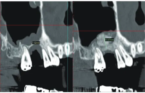
For the evaluation of bone height, RBH was measured at the sinus bone graft and implant placement planned site from pre-operative CT. Then grafted bone height (the dis- tance between alveolar crest and the most upper part of bone graft) was measured from post-operative CT and compared with pre-operative height at the same location (Fig. 2).
Fig. 1. Measurement of bone density by Simplant software.
Fig. 2. Measurement of bone height by Simplant software.
For the evaluation of sinus membrane thickness, the thickest area of sinus membrane of pre-operative bone graft planned site was measured in sagittal view.19,20 Then the thickest area of sinus membrane of post-operative bone graft site was measured and compared (Fig. 3).
As described, at 4 months after sinus bone graft proce- dure, biopsy specimen were taken at the site of implant placement using 3.0 mm trephine bur. Five participants in Bio-Oss group and one participant in AutoBT group were declined to take out trephine biopsy before implant place- ment. So, 14 specimens from Bio-Oss group and 18 speci- mens from AutoBT group were taken. After fixation of tre- phine bur in 10% buffered formalin solution, bony speci- mens (including trephine bur) were sent to Department of Dental Biomaterials Science and Dental Research Institute, School of Dentistry, Seoul National University for microCT analysis. Bone tissue contained in trephine bur was embed- ded in methyl-methacrylate resin. Trephine bur and outer resin embedding was removed using diamond disk without tissue damage. Cylindrical specimen was fabricated.
Cylindrical samples embedded in resin were imaged with a microCT system (SKYscan 1172, Bruker-Microct, Kontich, Belgium) in high resolution scanning mode (pixel matrix:
683×2000×1048, pixel size: 10.89 μm). X-ray source was set at 70 kV and 141 μA with the aid of 0.5 mm thick alu- minum filter to optimize the contrast, a 360° rotation step of 0.3° and 590 ms exposure time. Before microCT taking of specimens, two phantoms (0.25 and 0.75 BMD) were taken for the calibration. NRECON reconstruction soft- ware (NRecon v.1.4.4, SkyScan) was used to create two- dimensional, 2000×1048 pixel images. After image recon- struction, the region of interest (ROI) was selected within the reconstructed images of water to calibrate the standard
unit of X-ray CT density (HU) using CTAn Ver. 1.6.0 (SkyScan, Bruker-Microct, Kontich, Belgium) analysis soft- ware. Firstly, total core height composed of residual bone and new bone portion was measured, and then image analy- sis was performed after setting up graft material and new bone area as ROI. To distinguish new bone from graft material, graft material was taken and range of HU was analyzed. Minimum HU value of new bone was set as 350 considering Tajima et al.21’s suggestion and HU of D3 bone.
Maximum HU value of new bone was calculated by sub- traction of HU of graft material. The area higher than low ranked 5% of HU value distribution of binary image was considered as HU of graft material. HU value of Bio-Oss was more than 1900. In the case that the range of HU val- ue was 350-1900 in Bio-Oss specimen, it was considered as new bone and analyzed. As the same concept, the HU of AutoBT was more than 2100. Applying HU guideline as described, total bone amount and new bone amount of Bio-Oss and AutoBT specimen were calculated. Also, mean HU value, BMD and mean trabecular thickness of new bone were calculated.
After microCT analysis, in the process of specimen preparation for histologic evaluation, 2 specimens in Bio- Oss group and 2 specimens in AutoBT group were exclud- ed due to bad quality. These specimens were not homoge- nous enough for proper analysis, resulting in analysis of a total of 28 biopsy specimens (12 in Bio-Oss group, 16 in AutoBT group). After microCT taking, secondary embed- ding of core sample was done using Technovit 7200 (Heraeus Kulzer, Wehrheim, Germany). Block type speci- men were trimmed and then attached on acrylic slide using EXAKT 4230 attach machine. After middle cutting and grinding of attached block, acrylic slide was attached. Slide Fig. 3. Measurement of sinus membrane thickness by INFINITT PACS software.
cutting and grinding was done on the slide attached side and 20-30 µm thickness histologic specimens were fabri- cated. Slide review for histologic evaluation was done by two authors (Lee & Jun) with two different staining tech- niques. Masson’s trichrome (MT) and toluidine blue (TB) stains were used. For the histomorphometric analysis, the tissue stained with MT was examined at high magnification (×100). Representative area of graft material and new bone were selected, and then using KAPPA mage base program, area of newly-formed bone, area of graft material, area of bone marrow space was calculated and expressed as a per- centage to total area. The tissue stained with TB was exam- ined at high magnification (×200). Five sites of osteoid forming site surrounding graft material were selected and measured and mean osteoid thickness surrounding graft material was calculated.
Statistical analysis was performed using SPSS 18.0 soft- ware (SPSS Inc., Chicago, IL, USA). Continuous variables were expressed using mean ± SD. Differences in propor- tions were tested with the chi square test or Fisher’s exact test. Independent t-test or Mann-Whitney U test were used for the comparison between groups of clinical, radiological and histological evaluation on bone volume-related, bone density-related or bone architecture-related parameters.
Paired t-test was used for the evaluation of time dependent changes such as sinus membrane thickness. The univariate normality assumptions were verified with the Shapiro-Wilk test and the Brown and Forsythe’s variation of Levene’s test statistics were used to verify the homogeneity of variances.
Mann Whitney U test was performed when these assump- tions were not fulfilled. P<.05 was considered statistically significant.
RESULTS
There were 24 male participants (11 in Bio-Oss group and 13 in AutoBT group) and 14 female participants (8 in Bio- Oss group and 6 in AutoBT group). There was no group
difference of gender distribution in this study (P=.501).
The mean age of participants was 58.21 years in Bio- Oss group (n=19) and 53.15 years in AutoBT group (n=19).
There was no age difference between groups (P=.158).
Implant primary stability measured using Osstell Mentor was in the range of 41-88 ISQ (mean=70.59 ISQ) (n=28 implants) in Bio-Oss group and 39-85 ISQ (mean=64.92 ISQ) (n=29 implants) in AutoBT group.
There was no significant difference between groups (P=
.166).
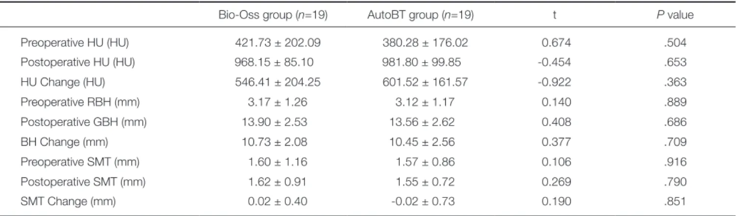
Pre-operative mean bone density of residual alveolar bone area was 421.73 HU in Bio-Oss group and 380.28 HU in AutoBT group. Mean pre-operative bone density of residual alveolar bone area was included in the category of D3 in both groups. Post-operative mean bone density of grafted bone area was 968.15 HU in Bio-Oss group and 981.80 HU in AutoBT group. Mean post-operative bone density of grafted bone area was included in the category of D2 in both groups.
Pre-operative mean residual bone height was 3.17 mm in Bio-Oss group and 3.12 mm in AutoBT group. Also, post-operative mean grafted bone height was 13.90 mm in Bio-Oss group and 13.56 mm in AutoBT group. Pre- operative mean sinus membrane thickness was 1.60 mm in Bio-Oss group and 1.57 mm in AutoBT group. Post- operative mean sinus membrane thickness was 1.62 mm in Bio-Oss group and 1.55 mm in AutoBT group. There was no significant differences between groups (Table 1).
In volume-related parameters, mean total bone volume (new bone + graft material) was 77.06% in Bio-Oss group and 64.98% in AutoBT group. There was no significant dif- ference between groups (P=.051). Mean new bone volume was 41.93% in Bio-Oss group and 37.91% in AutoBT group. There was no significant difference between groups (P=.192). The proportion of new bone volume to total bone volume (new bone/total bone) was 55.58% in Bio- Oss group and 60.34% in AutoBT group. There was no sig- nificant difference between groups (P=.377).
Table 1. CT evaluation on bone density, bone height, and sinus membrane thickness
Bio-Oss group (n=19) AutoBT group (n=19) t P value
Preoperative HU (HU) 421.73 ± 202.09 380.28 ± 176.02 0.674 .504
Postoperative HU (HU) 968.15 ± 85.10 981.80 ± 99.85 -0.454 .653
HU Change (HU) 546.41 ± 204.25 601.52 ± 161.57 -0.922 .363
Preoperative RBH (mm) 3.17 ± 1.26 3.12 ± 1.17 0.140 .889
Postoperative GBH (mm) 13.90 ± 2.53 13.56 ± 2.62 0.408 .686
BH Change (mm) 10.73 ± 2.08 10.45 ± 2.56 0.377 .709
Preoperative SMT (mm) 1.60 ± 1.16 1.57 ± 0.86 0.106 .916
Postoperative SMT (mm) 1.62 ± 0.91 1.55 ± 0.72 0.269 .790
SMT Change (mm) 0.02 ± 0.40 -0.02 ± 0.73 0.190 .851
RBH: residual bone height, GBH: grafted bone height, BH: bone height, SMT: sinus membrane thickness.
In density-related parameters, mean total core density was 1486.27 HU in Bio-Oss group and 1232.02 in AutoBT group. There was no significant difference between groups (P=.245). Also, mean bone mineral density (BMD: volumet- ric density of calcium hydroxyapatite) of total core was 0.34 g/cm2 in Bio-Oss group and 0.28 g/cm2 in AutoBT
group. There was no significant difference between groups (P=.245). Mean new bone density was 1223.08 HU in Bio- Oss group and 1274.31 in AutoBT group. There was no significant difference between groups (P=.217). Also, mean BMD of new bone was 0.27 g/cm2 in Bio-Oss group and 0.29 g/cm2 in AutoBT group. There was no significant dif- ference between groups (P=.245).
In architecture related parameter, trabecular thickness was 0.07 μm in Bio-Oss group and 0.08 μm in AutoBT group. There was significant difference between groups (P=.006) (Table 2).
In low magnification (×35) microscopic findings of AutoBT sample, the border between residual bone and new bone of sinus graft site was identified. Matured lamellar bone showing bony integration between residual bone and maxillary sinus was detected. Bone formation was found surrounding both enamel and dentin portion of graft mate- rial. New bone bridges between graft materials were also detected (Fig. 5). In high magnification (×400) microscopic findings of AutoBT sample, newly-formed and matured lamellar bone was detected surrounding graft material, osteoblasts covering newly-formed bone accumulated oste- oid. Active woven bone formation was also detected.
Medullary space formation composed of well vascularized connective tissue was also detected within new bone (Fig.
6). In histomorphometric analysis, the portion of new bone formation was 26.49% in Bio-Oss group and 31.07%
in AutoBT group. There was no significant difference between groups (P=.556). The portion of residual graft material was 31.12% in Bio-Oss group and 29.00% in AutoBT group. There was no significant difference between groups (P=.896). The portion of bone marrow space was 42.38%
in Bio-Oss group and 39.93% in AutoBT group. There was no significant difference between groups (P=.471). The mean osteoid thickness was 8.35 μm in Bio-Oss group and 13.12 μm in AutoBT group. There was significant differ- ence between groups (P=.025) (Table 3).
Fig. 4. MicroCT image.
Table 2. MicroCT evaluation on volume-related, density-related and architecture-related parameters
Bio-Oss group (n=14) AutoBT group (n=18) P value
Total Bone (%) 77.06 ± 14.21 64.98 ± 18.54 .051
New Bone (%) 41.93 ± 7.06 37.91 ± 8.49 .192
New Bone/Total Bone (%) 55.58 ± 10.49 60.34 ± 11.51 .377
Total Core HU (HU) 1486.27 ± 405.58 1232.02 ± 529.37 .245
Total Core BMD (g/cm2) 0.34 ± 0.09 0.28 ± 0.12 .245
New Bone HU (HU) 1223.08 ± 107.44 1274.31 ± 107.92 .217
New Bone BMD (g/cm2) 0.27 ± 0.02 0.29 ± 0.02 .245
Trabecular thickness (µm) 0.07 ± 0.02 0.08 ± 0.01 .006**
P value: ** <.01 by Mann-Whitney U test.
BMD: Bone mineral density.
Fig. 5. Matured lamellar bone showing bony integration between residual bone and maxillary sinus was detected. Bone formation was found surrounding both enamel and dentin portion of AutoBT material. New bone bridges between graft materials were also detected (Masson’s trichrome staining, ×35).
Fig. 6. Newly-formed and matured lamellar bone was detected surrounding AutoBT material, osteoblasts covering newly-formed bone accumulated osteoid. Active woven bone formation was also detected. Medullary space formation composed of well vascularized connective tissue was also detected within new bone (Masson’s trichrome staining, ×400).
Table 3. Histomorphometric analysis
Bio-Oss group (n=12) AutoBT group (n=16) P value
New bone formation (%) 26.49 ± 7.13 31.07 ± 14.52 .556
Residual graft material (%) 31.12 ± 14.51 29.00 ± 10.27 .896
Marrow space (%) 42.38 ± 16.37 39.93 ± 18.92 .471
Osteoid thickness (µm) 8.35 ± 3.94 13.12 ± 5.16 .025*
P value: * <.05 by Mann-Whitney U test.
DISCUSSION
Maxillary sinus cavity is a contained defect. However, max- illary sinus is not natural bone-forming area. These charac- teristics are why maxillary sinus is often selected for the evaluation of healing process of bone graft materials.22 So, in this study, as a model for the evaluation of healing pro- cess of two different bone graft material, maxillary sinus cavity was selected. In this study, there was no significant difference between groups in pre-operative RBH and bone density, which was the evidence that there was no selection bias. Also, implant primary stability measured at 4 months after graft was at favorable state, suggesting that the strength of bone graft site was excellent.
Transient swelling of the Schneiderian membrane occurred just after the sinus graft procedure post-operative- ly due to the inflammatory reaction. However, with the lapse of time, the change was recovered to normal thick- ness. In case that the biocompatibility of bone graft materi- al was not good, there is a risk of post-operative infection and sinus membrane remained thickened state. In this study, although there was considerable increase of bone height, the thickness of sinus membrane maintained nor- mally in both AutoBT and Bio-Oss groups post-operatively.
The findings that sinus mucosa thickness was maintained stably over time could be an evidence of biocompatibility of graft materials. So, it was proposed that both AutoBT and Bio-Oss were biocompatible in application in the sinus graft procedure.
Kühl et al.23 reported that increased bone density after sinus bone graft procedure with autogenous bone or mixed with other bone graft material was detected in microCT.
Huang et al.24 reported that BMD and bone volume of native bone (0.22 and 24.4%) were higher than BMD and bone volume of grafted bone (0.12 and 15.43%) at the time point of 4-5 months after iliac bone graft. Lundgren et al.25 evaluated bone volume using microCT analysis with core biopsy specimen at 6 months (40 ± 12%) and 12 months (48 ± 10%) respectively after sinus graft procedure with particulated mandibular symphysis bone. Chackartchi et al.26 performed microCT analysis with tissue specimen at 6-9 months after sinus graft procedure with bovine bone and reported that the volume of bone (7.99-14.64%) and bovine bone material (22.89-23.12%) seemed underestimat- ed in comparison with histomorphometric evaluation.
Chackartchi et al.26 suggested that partly due to the difficul- ty of distinguishment of exact borders showing direct con- nection between new bone and graft materials in microCT image, microCT technique had a chance to underestimate the volumes of the histological bone and graft materials.
In this study, all the same condition for CT taking was kept in detail. Under the guideline of HU value of graft material and previous study, the range of new bone was set up scientifically, bone graft material and new bone was ana- lyzed separately. For the determination of lower limit of HU of new bone, bone quality guideline of previous study of Tajima et al.21 was referred. For the determination of
upper limit of HU, using statistical method, microCT image of graft material were analyzed and the range of 95% HU was regarded as graft material. And equivalent to the lowest 5% HU was regarded as new bone.
In the microCT analysis of this study, though it was not statistically significant, total bone volume in the grafted area was higher in Bio-Oss group (77.06 ± 14.21) than in AutoBT group (64.98 ± 18.54). However, new bone por- tion in the total bone was higher in AutoBT group (60.34 ± 11.51%) than in Bio-Oss group (55.58 ± 10.49%).
Furthermore, trabecular thickness in AutoBT group was significantly higher than Bio-Oss group, suggesting that the ability of new bone formation of AutoBT graft material was not inferior to Bio-Oss. Also, at 4 months, bone vol- ume and bone density were relatively higher in comparison with other studies. HU and BMD of new bone at 4 months were corresponding with clinical findings showing D2 bone quality.
Histomorphometric researches related with bone graft evaluated and reported various parameters including total bone volume, new bone formation, woven/lamellar bone ratio, residual graft material amount, bone marrow includ- ing connective tissue. In this study, new bone formation, residual graft material ratio and bone marrow and osteoid thickness were measured. There have been many reports on histomorphometric analysis using tissue specimen harvest- ed after sinus bone graft with various bone graft material.
Many studies on allograft such as demineralized bone matrix with cancellous bone chips, human mineralized bone, cancellous block, and demineralized freeze-dried bone (DFDB) graft were performed. In the analysis of bone tissue specimens harvested at 6-11 months, wide range of new bone (24-75%) and residual bone graft mate- rial (9-25%) were measured and D3 bone density was detected with relatively high marrow portion.27-30 In the studies of autogenous bone graft for sinus bone graft pro- cedure, it was reported that newly formed bone portion was 38-40% and residual graft material was about 18%.31,32
Histomorphometric study with bone tissue samples har- vested after healing period was performed using various alloplastic materials for the sinus bone graft. In the studies of HA and TCP graft mixed at 6:4 ratio, it was reported that new bone formation was 26-40% and residual bone graft material was 27% at 4-9 months healing period.33,34 On the other hand, in studies of TCP graft at 6-8 months of healing period, 21-55% of new bone and 13-34% of residual graft material was detected.31,35,36 Whereas, in the studies of HA graft at 4-6 months, 20-35% of new bone formation and 30-45% of residual graft material were reported.31,37
From the studies on anorganic bovine bones graft, 12-50% of new bone formation and 20-30% of residual graft material were reported after 4-10 months of healing period.31,37-39 Orsini et al.40 performed the histologic and ultrastructural analysis of regenerated bone in maxillary sinus augmentation using a porcine bone-derived biomate- rial and reported that the portion of newly formed bone
was 36.0 ± 2.8% and residual grafted material was 31.0 ± 1.6%. The result of Orsini et al.’s study was relatively similar with the result of this study.
After mixing and grafting 2 different bone graft materi- als, many researches were reported on histomorphometric analysis. At 6-10 months after sinus bone graft with autoge- nous bone and xenograft, the proportion of new bone for- mation was reported as 27-46%, whereas residual bone graft material was 16-37%.41-43 Hanisch et al.44 measured new bone formation ratio after the graft with DFDBA and bovine HA at a one to one ratio, and reported 8.1 ± 3.0%
at 6 months, 9.0 ± 3.8% at 8 months, 20.7 ± 8.3% at 12 months respectively.
Gonshor et al.45 performed histologic and histomorpho- metric evaluation of allograft stem cell-based matrix sinus augmentation procedure. After mean 3.7 months of healing period, allograft cellular bone matrix (ACBM), containing native mesenchymal stem cells and osteoprogenitors graft group showed that vital bone content was 32.5 ± 6.8% and residual graft content was 4.9 ± 2.4%. On the other hand, conventional allograft group showed that vital bone con- tent was 18.3 ± 10.6% and residual graft content was 25.8
± 13.4%.
Some researchers proposed that histologic features of grafted site were influenced by harvesting technique of biopsy specimen. In comparison with crestal approach, the less bone formation tendency was detected in lateral win- dow biopsy.46,47 In this study, crestal approach with 3.0 mm trephine and surgical stent was selected for harvesting biop- sy specimen.
A slowly resorbable biomaterial might be suitable in sub-sinusal bone augmentation for preventing the re-expan- sion process and for augmenting the density of the regen- erated tissues. Bovine HA seems to be the most efficient fillers for 3 dimensional stability of sub-sinusal bone aug- mentation.48 In this study, AutoBT and Bio-Oss showed similar healing pattern, excellent graft volume maintenance and active new bone formation, in microCT and histomor- phometric evaluation of biopsy specimen 4 months after graft procedure. So, if it was applied for sinus bone graft, it would be useful material struggling repneumatization against intra-sinus pressure. Bone formation was found sur- rounding both enamel and dentin portion of AutoBT material. New bone bridges between graft materials were also detected in AutoBT group. Also newly-formed and matured lamellar bone was detected surrounding AutoBT material, osteoblasts covering newly-formed bone accumu- lated osteoid.
Bone healing of human sinus cavity showed different features according to the types of graft materials and heal- ing time. Also, vital bone formation ratio increased gradual- ly over time.49,50 In histomorphometric evaluation, the mean osteoid thickness was higher in AutoBT group (13.12 μm) than in Bio-Oss group (8.35 μm). Also, though it was not statistically significant, portion of new bone formation was higher in AutoBT group (31.07%) than in Bio-Oss group (26.49%).
CONCLUSION
In this prospective clinical study, sinus membrane thickness maintained stable in both graft materials. Also, there was no significant difference between groups on CT, microCT or histomorphometric analysis, except thicker trabecular and osteoid thickness in AutoBT group. Conclusively, AutoBT was not inferior to Bio-Oss as bone graft material for sinus bone graft procedures. So, AutoBT could be considered a viable alternative to the autogenous bone or other bone graft materials in sinus bone graft procedure. In terms of recycling autogenous tissue that would be usually discarded as medical waste, AutoBT is the new concept of graft material. It is marvelous that there is no donor site morbid- ity in spite of using autogenous tissue, and that there is no risk of immune response or spread of disease. In the future, more studies will be succeeded for the evaluation of AutoBT application for other bone graft procedures.
REFERENCES
1. Berglundh T, Lindhe J. Healing around implants placed in bone defects treated with Bio-Oss. An experimental study in the dog. Clin Oral Implants Res 1997;8:117-24.
2. Piattelli M, Favero GA, Scarano A, Orsini G, Piattelli A.
Bone reactions to anorganic bovine bone (Bio-Oss) used in sinus augmentation procedures: a histologic long-term report of 20 cases in humans. Int J Oral Maxillofac Implants 1999;
14:835-40.
3. Carmagnola D, Adriaens P, Berglundh T. Healing of human extraction sockets filled with Bio-Oss. Clin Oral Implants Res 2003;14:137-43.
4. Caubet J, Petzold C, Sáez-Torres C, Morey M, Iriarte JI, Sánchez J, Torres JJ, Ramis JM, Monjo M. Sinus graft with safescraper: 5-year results. J Oral Maxillofac Surg 2011;69:
482-90.
5. Kim YK, Kim SG, Byeon JH, Lee HJ, Um IU, Lim SC, Kim SY. Development of a novel bone grafting material using au- togenous teeth. Oral Surg Oral Med Oral Pathol Oral Radiol Endod 2010;109:496-503.
6. Kim YK, Kim SG, Oh JS, Jin SC, Son JS, Kim SY, Lim SY.
Analysis of the inorganic component of autogenous tooth bone graft material. J Nanosci Nanotechnol 2011;11:7442-5.
7. Kim YK, Lee JK, Kim KW, Um IW, Murata M. Healing Mechanism and Clinical Application of Autogenous Tooth Bone Graft Material. In: Pignatello R, ed. Advances in Biomaterials Science and Biomedical Applications [Internet].
Rijeka, Croatia; InTech; 2013;405-35.
8. Kim YK, Yi YJ. Horizontal ridge augmentation using ridge expansion and autogenous tooth bone graft: a case report. J Dent Rehabil Appl Sci 2011;27:109-15.
9. Kim YK, Kim SG, Um IW. Vertical and horizontal ridge aug- mentation using autogenous tooth bone graft materials: case report. J Korean Assoc Maxillofac Plast Reconstr Surg 2011;
33:166-70.
10. Kim YK, Kim SG, Kim KW, Um IW. Extraction socket pres- ervation and reconstruction using autogenous tooth bone
graft: Case report. J Korean Assoc Maxillofac Plast Reconstr Surg 2011;33:264-9.
11. Kim YK, Choi YH. Tooth autotransplantation with autoge- nous tooth- bone graft: a case report. J Korean Dent Sci 2011;4:79-84.
12. Lee JY, Kim YK, Kim SG, Lim SC. Histomorphometric study of sinus bone graft using various graft material. J Dent Rehab App Sci 2011;27:141-7.
13. Kim YK, Lee HJ, Kim KW, Kim SG, Um IW. Guide bone re- generation using autogenous teeth: case reports. J Korean Assoc Oral Maxillofac Surg 2011;37:142-7.
14. Jeong KI, Kim SG, Kim YK, Oh JS, Jeong MA, Park JJ.
Clinical study of graft materials using autogenous teeth in maxillary sinus augmentation. Implant Dent 2011;20:471-5.
15. Lee JY, Kim YK. Retrospective cohort study of autogenous tooth bone graft. Oral Biol Res 2012;36:39-43.
16. Kim YK, Um IW, Murata M. Tooth bank system for bone re- generation-Safety report- J Hard Tissue Biol 2014;23:371-6.
17. Jensen OT. The sinus bone graft. 2nd ed. Chicago: Quintessence Publ; 2006. p. 103-25.
18. Kim YK, Kim SG, Park JY, Yi YJ, Bae JH. Comparison of clinical outcomes of sinus bone graft with simultaneous im- plant placement: 4-month and 6-month final prosthetic loading.
Oral Surg Oral Med Oral Pathol Oral Radiol Endod 2011;
111:164-9.
19. Janner SF, Caversaccio MD, Dubach P, Sendi P, Buser D, Bornstein MM. Characteristics and dimensions of the Schneiderian membrane: a radiographic analysis using cone beam computed tomography in patients referred for dental implant surgery in the posterior maxilla. Clin Oral Implants Res 2011;22:1446-53.
20. Phothikhun S, Suphanantachat S, Chuenchompoonut V, Nisapakultorn K. Cone-beam computed tomographic evi- dence of the association between periodontal bone loss and mucosal thickening of the maxillary sinus. J Periodontol 2012;83:557-64.
21. Tajima N, Ohba S, Sawase T, Asahina I. Evaluation of sinus floor augmentation with simultaneous implant placement us- ing platelet-rich fibrin as sole grafting material. Int J Oral Maxillofac Implants 2013;28:77-83.
22. Nevins M, Kirker-Head C, Nevins M, Wozney JA, Palmer R, Graham D. Bone formation in the goat maxillary sinus in- duced by absorbable collagen sponge implants impregnated with recombinant human bone morphogenetic protein-2. Int J Periodontics Restorative Dent 1996;16:8-19.
23. Kühl S, Götz H, Brochhausen C, Jakse N, Filippi A, d'Hoedt B, Kreisler M. The influence of substitute materials on bone density after maxillary sinus augmentation: a microcomputed tomography study. Int J Oral Maxillofac Implants 2012;27:
1541-6.
24. Huang HL, Chen MY, Hsu JT, Li YF, Chang CH, Chen KT.
Three-dimensional bone structure and bone mineral density evaluations of autogenous bone graft after sinus augmenta- tion: a microcomputed tomography analysis. Clin Oral Implants Res 2012;23:1098-103.
25. Lundgren S, Moy P, Johansson C, Nilsson H. Augmentation of the maxillary sinus floor with particulated mandible: a his-
tologic and histomorphometric study. Int J Oral Maxillofac Implants 1996;11:760-6.
26. Chackartchi T, Iezzi G, Goldstein M, Klinger A, Soskolne A, Piattelli A, Shapira L. Sinus floor augmentation using large (1-2 mm) or small (0.25-1 mm) bovine bone mineral parti- cles: a prospective, intra-individual controlled clinical, micro- computerized tomography and histomorphometric study.
Clin Oral Implants Res 2011;22:473-80.
27. Kim DM, Nevins ML, Camelo M, Camelo JM, Schupbach P, Hanratty JJ, Uzel NG, Nevins M. The efficacy of demineral- ized bone matrix and cancellous bone chips for maxillary si- nus augmentation. Int J Periodontics Restorative Dent 2009;
29:415-23.
28. Gapski R, Neiva R, Oh TJ, Wang HL. Histologic analyses of human mineralized bone grafting material in sinus elevation procedures: a case series. Int J Periodontics Restorative Dent 2006;26:59-69.
29. Cammack GV, Nevins M, Clem DS, Hatch JP, Mellonig JT.
Histologic evaluation of mineralized and demineralized freeze-dried bone allograft for ridge and sinus augmenta- tions. Int J Periodontics Restorative Dent 2005;25:231-7.
30. Chaushu G, Vered M, Mardinger O, Nissan J. Histomorphometric analysis after maxillary sinus floor augmentation using can- cellous bone-block allograft. J Periodontol 2010;81:1147-52.
31. Scarano A, Degidi M, Iezzi G, Pecora G, Piattelli M, Orsini G, Caputi S, Perrotti V, Mangano C, Piattelli A. Maxillary si- nus augmentation with different biomaterials: a comparative histologic and histomorphometric study in man. Implant Dent 2006;15:197-207.
32. Szabó G, Huys L, Coulthard P, Maiorana C, Garagiola U, Barabás J, Németh Z, Hrabák K, Suba Z. A prospective mul- ticenter randomized clinical trial of autogenous bone versus beta-tricalcium phosphate graft alone for bilateral sinus ele- vation: histologic and histomorphometric evaluation. Int J Oral Maxillofac Implants 2005;20:371-81.
33. Kim YK, Yun PY, Lim SC, Kim SG, Lee HJ, Ong JL. Clinical evaluations of OSTEON as a new alloplastic material in si- nus bone grafting and its effect on bone healing. J Biomed Mater Res B Appl Biomater 2008;86:270-7.
34. Kolerman R, Goshen G, Joseph N, Kozlovsky A, Shetty S, Tal H. Histomorphometric analysis of maxillary sinus aug- mentation using an alloplast bone substitute. J Oral Maxillofac Surg 2012;70:1835-43.
35. Kurkcu M, Benlidayi ME, Cam B, Sertdemir Y. Anorganic bovine-derived hydroxyapatite vs β-tricalcium phosphate in sinus augmentation: a comparative histomorphometric study.
J Oral Implantol 2012;38:519-26.
36. Ozyuvaci H, Bilgiç B, Firatli E. Radiologic and histomorpho- metric evaluation of maxillary sinus grafting with alloplastic graft materials. J Periodontol 2003;74:909-15.
37. Karabuda C, Ozdemir O, Tosun T, Anil A, Olgaç V. Histological and clinical evaluation of 3 different grafting materials for si- nus lifting procedure based on 8 cases. J Periodontol 2001;72:
1436-42.
38. Wallace SS, Froum SJ, Cho SC, Elian N, Monteiro D, Kim BS, Tarnow DP. Sinus augmentation utilizing anorganic bo- vine bone (Bio-Oss) with absorbable and nonabsorbable
membranes placed over the lateral window: histomorphomet- ric and clinical analyses. Int J Periodontics Restorative Dent 2005;25:551-9.
39. Zhang Y, Tangl S, Huber CD, Lin Y, Qiu L, Rausch-Fan X.
Effects of Choukroun's platelet-rich fibrin on bone regenera- tion in combination with deproteinized bovine bone mineral in maxillary sinus augmentation: a histological and histomor- phometric study. J Craniomaxillofac Surg 2012;40:321-8.
40. Orsini G, Scarano A, Piattelli M, Piccirilli M, Caputi S, Piattelli A. Histologic and ultrastructural analysis of regenerated bone in maxillary sinus augmentation using a porcine bone- derived biomaterial. J Periodontol 2006;77:1984-90.
41. Galindo-Moreno P, Avila G, Fernández-Barbero JE, Aguilar M, Sánchez-Fernández E, Cutando A, Wang HL. Evaluation of sinus floor elevation using a composite bone graft mix- ture. Clin Oral Implants Res 2007;18:376-82.
42. Peleg M, Garg AK, Misch CM, Mazor Z. Maxillary sinus and ridge augmentations using a surface-derived autogenous bone graft. J Oral Maxillofac Surg 2004;62:1535-44.
43. Artzi Z, Kozlovsky A, Nemcovsky CE, Weinreb M. The amount of newly formed bone in sinus grafting procedures depends on tissue depth as well as the type and residual amount of the grafted material. J Clin Periodontol 2005;32:
193-9.
44. Hanisch O, Lozada JL, Holmes RE, Calhoun CJ, Kan JY, Spiekermann H. Maxillary sinus augmentation prior to place- ment of endosseous implants: A histomorphometric analysis.
Int J Oral Maxillofac Implants 1999;14:329-36.
45. Gonshor A, McAllister BS, Wallace SS, Prasad H. Histologic and histomorphometric evaluation of an allograft stem cell- based matrix sinus augmentation procedure. Int J Oral Maxillofac Implants 2011;26:123-31.
46. Tarnow DP, Wallace SS, Froum SJ, Rohrer MD, Cho SC.
Histologic and clinical comparison of bilateral sinus floor el- evations with and without barrier membrane placement in 12 patients: Part 3 of an ongoing prospective study. Int J Periodontics Restorative Dent 2000;20:117-25.
47. Froum SJ, Tarnow DP, Wallace SS, Rohrer MD, Cho SC.
Sinus floor elevation using anorganic bovine bone matrix (OsteoGraf/N) with and without autogenous bone: a clinical, histologic, radiographic, and histomorphometric analysis- -Part 2 of an ongoing prospective study. Int J Periodontics Restorative Dent 1998;18:528-43.
48. Lambert F, Léonard A, Drion P, Sourice S, Layrolle P, Rompen E. Influence of space-filling materials in subantral bone augmentation: blood clot vs. autogenous bone chips vs.
bovine hydroxyapatite. Clin Oral Implants Res 2011;22:538- 45.
49. Lee YM, Shin SY, Kim JY, Kye SB, Ku Y, Rhyu IC. Bone re- action to bovine hydroxyapatite for maxillary sinus floor aug- mentation: histologic results in humans. Int J Periodontics Restorative Dent 2006;26:471-81.
50. De Leonardis D, Pecora GE. Prospective study on the aug- mentation of the maxillary sinus with calcium sulfate: histo- logical results. J Periodontol 2000;71:940-7.