Purpose: The details of breast reconstruction and radiation therapy (RT) vary between institutions; therefore, we sought to investigate the practice patterns of radiation oncologists who specialize in breast cancer.
Materials and Methods: We identified the practice patterns and inter-hospital variations from a multi-center co- hort of women with breast cancer who underwent post-mastectomy RT (PMRT) to the reconstructed breast at 16 institutions between 2015 and 2016. The institutions were requested to contour the target volume and produce RT plans for one representative case with five different clinical scenarios and answer questionnaires which elicited infrastructural information. We assessed the inter-institutional variations in RT in terms of the target, normal or- gan delineation, and dose-volume histograms.
Results: Three hundred fourteen patients were included; 99% of them underwent immediate reconstruction. The most irradiated material was tissue expander (36.9%) followed by transverse rectus abdominis musculocutaneous flap (23.9%) and silicone implant (12.1%). In prosthetic-based reconstruction with tissue expander, most patients received PMRT following partial deflation. Conventional fractionation and hypofractionation RT were used in 66.6% and 33.4% patients, respectively (commonest: 40.05 Gy in 15 fractions [17.5%]). Furthermore, 15.6% of the patients received boost RT and 53.5% were treated with bolus. Overall, 15 physicians responded to the ques- tionnaires and six submitted their contours and RT plans. There was a significant variability in target delineations and RT plans between physicians, and between clinical scenarios.
Conclusion: Adjuvant RT following post-mastectomy reconstruction has become a common practice in Korea. The details vary significantly between institutions, which highlights an urgent need for standard protocol in this clini- cal setting.
Keywords: Radiotherapy, Mammaplasty, Breast neoplasm
Post-mastectomy radiation therapy in breast reconstruction: a patterns of care study of the Korean Radiation Oncology Group
Gowoon Yang
1, Jee Suk Chang
1, Kyung Hwan Shin
2, Jin Ho Kim
2, Won Park
3, Haeyoung Kim
3, Kyubo Kim
4, Ik Jae Lee
5, Won Sup Yoon
6, Jihye Cha
7, Kyu-Chan Lee
8, Jin Hee Kim
9, Jin Hwa Choi
10, Sung-Ja Ahn
11, Boram Ha
12, Sun Young Lee
13, Dong Soo Lee
14, Jeongshim Lee
15, Sei One Shin
16, Yong Bae Kim
11
Department of Radiation Oncology, Yonsei Cancer Center, Yonsei University College of Medicine, Seoul, Korea
2
Department of Radiation Oncology, Seoul National University College of Medicine, Seoul, Korea
3
Department of Radiation Oncology, Samsung Medical Center, Sungkyunkwan University School of Medicine, Seoul, Korea
4
Department of Radiation Oncology, Ewha Womans University College of Medicine, Seoul, Korea
5
Department of Radiation Oncology, Gangnam Severance Hospital, Yonsei University College of Medicine, Seoul, Korea
6
Department of Radiation Oncology, Korea University Ansan Hospital, Korea University College of Medicine, Seoul, Korea
7
Department of Radiation Oncology, Wonju Severance Christian Hospital, Yonsei University Wonju College of Medicine, Wonju, Korea
8
Department of Radiation Oncology, Gachon University Gil Medical Center, Incheon, Korea
9
Department of Radiation Oncology, Keimyung University Dongsan Medical Center, Keimyung University School of Medicine, Daegu, Korea
10
Department of Radiation Oncology, Chung-Ang University Hospital, Seoul, Korea
11
Department of Radiation Oncology, Chonnam National University Hwasun Hospital, Chonnam National University Medical School, Hwasun, Korea
12
Department of Radiation Oncology, Hallym University Dongtan Sacred Heart Hospital, Hwasung, Korea
13
Department of Radiation Oncology, Chonbuk National University Hospital, Jeonju, Korea
14
Department of Radiation Oncology, Uijeongbu St. Mary’s Hospital, College of Medicine, The Catholic University of Korea, Uijeongbu, Korea
15
Department of Radiation Oncology, Inha University Hospital, Inha University School of Medicine, Incheon, Korea
16
Department of Radiation Oncology, Andong Medical Group Andong Hospital, Andong, Korea
Radiat Oncol J 2020;38(4):236-243 https://doi.org/10.3857/roj.2020.00738
Received: September 27, 2020 Revised: November 15, 2020 Accepted: November 25, 2020 Correspondence:
Jee Suk Chang
Department of Radiation Oncology, Yonsei Cancer Center, Yonsei University College of Medicine, 50-1 Yonsei-ro, Seodaemun-gu, Seoul 03722, Korea
Tel: +82-2-2228-8117 Fax: +82-2-2227-7823 E-mail: [email protected] ORCID:
https://orcid.org/0000-0001-7685-3382
Copyright © 2020 The Korean Society for Radiation Oncology
This is an Open Access article distributed under the terms of the Creative Commons Attribution Non-Commercial License (http://creativecommons.org/licenses/by-nc/4.0/)
which permits unrestricted non-commercial use, distribution, and reproduction in any medium, provided the original work is properly cited.
Introduction
Breast reconstruction has become very common over the last two decades [1,2]. It significantly improves the quality of life and reduces adverse psychosocial effects following mastectomy [3]. However, the decision regarding breast reconstruction becomes complicated in pa- tients who anticipate to receive post-mastectomy radiation therapy (PMRT) [4]. The majority of radiation oncologists believe that their ability to deliver radiotherapy to the chest wall is challenged by im- mediate breast reconstruction [5]. Simultaneously, the majority of reconstructive surgeons believe that the cosmetic outcomes of breast reconstruction are negatively affected by PMRT [6,7].
Reconstructions can either be immediate or delayed or a com- bined approach called delayed-immediate reconstruction [8]. An implant, autologous tissue or a combination of an implant and a flap, can be used for the restoration of the breast mound [9]. All reconstruction approaches have their own pros and cons [10,11].
The techniques available for radiotherapy delivery in patients with breast cancer who have undergone reconstruction have im- proved over the past decade, which has reduced the challenges in radiotherapy planning that once seemed difficult to overcome.
Despite the improvements in both reconstruction and radiothera- py techniques, several issues still require to be answered, such as the type of reconstruction that is the most feasible with PMRT, the timing of reconstruction relative to PMRT, and optimization of radiotherapy to minimize treatment-related complications without compromising the oncological and cosmetic outcomes.
The practice patterns may vary between institutions regarding the reconstruction type and radiotherapy planning and delivery.
However, there are no standard protocols or guidelines in clinical settings. Therefore, the aim of this study was to investigate the practice patterns of PMRT in breast reconstruction in Korea.
Materials and Methods
1. Ethical statement
After approval from the review board of the Korean Radiation On- cology Group (KROG 18-04) and Severance Hospital Institutional Review Board (No. 4-2017-0931), a retrospective multi-center ob- servational study and a dummy run study were conducted in pa- tients with breast cancer who received RT and underwent breast reconstruction to identify the practice patterns.
2. Observational study
The inclusion criteria were as the following: histologically proven breast cancer, non-metastatic disease, female sex, and breast re- construction following mastectomy and subsequent adjuvant RT
between 2015 and 2016. The reconstruction rate has increased since 2015 after reimbursement by the Korean National Insurance Service and we surmised that it was actively performed during this period. Patients who underwent bilateral mastectomy for bilateral breast cancer or were male sex, or lost to follow-up were exclud- ed. The medical charts of the patients were collected and retro- spectively reviewed after approval from each participating center.
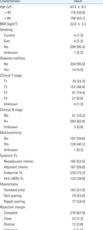
The following parameters were analyzed: age, body mass index, smoking history, history of diabetes, clinical T and N stages, mul- ticentricity, systemic therapy, mastectomy (standard, skin-sparing, and nipple-sparing), resection margin (clear, close, and positive), reconstruction sequence (one-stage vs. two-stage), reconstruc- tion timing (immediate vs. delayed), reconstruction options (au- tologous-based vs. prosthetic-based), reconstruction material at the time of RT (tissue expander, implant, transverse rectus ab- dominis musculocutaneous flap, deep inferior epigastric perfora- tor flap, latissimus dorsi flap, and others), bilateral reconstruction, operation time, RT technique (three-dimensional conformal, field- in-field, step-and-shot intensity-modulated radiotherapy, and volumetric arc therapy), RT dose/fraction, estimated maximum dose in chest wall on RT planning system, chest wall boost RT, bolus material used, and use of regional RT (Table 1).
3. Dummy run study
The institutions participating in the Division of Breast Cancer, KROG were invited to participate in the present dummy run. They were requested to answer questionnaires (Supplementary A) re- garding the infrastructural information, which included computed tomography (CT) simulation, radiation treatment planning (RTP) system, RT treatment details, and quality assurance (QA) activity for the RT machine. CT images from one anonymized patient was provided as a representative case with five different clinical sce- narios (cases 1–5) (Supplementary Fig. S1). The physicians from the participating institutions downloaded these CT images from a web- site and registered them in their own RTP systems; subsequently, they designed RT treatment plans for each clinical scenario. The target range, dose prescription, treatment technique, and beam in- formation were obtained through the questionnaire. RT files in the Digital Imaging and Communications in Medicine (DICOM) format for the structure sets and radiation doses were submitted along with the questionnaires.
4. Data analysis
All analyses were performed using IBM SPSS Statistics version 25.0
for Windows (IBM Corp., Armonk, NY, USA) using two-sided statis-
tical tests. Descriptive analyses were performed using Microsoft
Excel 2010 (Microsoft Corporation, Redmond, WA, USA).
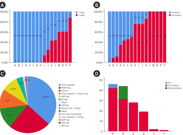
the same study period, approximately 1,105 patients were referred for PMRT. Table 2 and Fig. 1 present the details of the reconstruction treatments. Most of the patients (99%) underwent immediate recon- struction. One-stage or autologous reconstruction was more fre- quent than two-stage or prosthetic reconstruction when PMRT was administered. A variety of materials were used for the reconstruction.
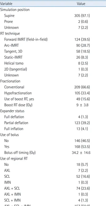
The most commonly used material was tissue expander (36.9%) fol- lowed by TRAM flap (23.9%), silicone implant (12.1%), and a combi- nation of tissue expander and silicone implant (11.2%). When tissue expander was used in prosthetic-based reconstruction, most patients (39.2%) received PMRT after partial deflation. Table 3 presents the details of radiotherapy. Conventional fractionation RT was used in 66.6% of the patients, while hypofractionation was used in 33.4% of patients (commonest: 40.05 Gy in 15 fractions [17.5%]). Additionally, 15.6% of the patients received boost RT and 53.5% of the patients were treated with a bolus.
2. Dummy run and questionnaire
Overall, 15 physicians responded to the questionnaires and six phy- sicians submitted their contours and RT plans (Fig. 1). According to the questionnaires, the prescribed dosage varied between physi- cians’. Generally, conventional fractionation with 50–50.4 Gy and Table 1. Baseline characteristics (n = 314)
Characteristic Value
Age (yr) 43.5 ± 8.3
< 40 116 (36.9)
≥ 40 198 (63.1)
BMI (kg/m
2) 22.6 ± 3.3
Smoking
Current 4 (1.3)
Ever 4 (1.3)
No 299 (95.2)
Unknown 7 (2.2)
Diabetes mellitus
No 300 (95.5)
Yes 14 (4.5)
Clinical T stage
T1 70 (22.3)
T2 152 (48.4)
T3 61 (19.4)
T4 27 (8.6)
Unknown 4 (1.3)
Clinical N stage
N0 51 (16.2)
N+ 260 (82.8)
Unknown 3 (0.6)
Multicentricity
No 187 (59.6)
Yes 126 (40.1)
Unknown 1 (0.3)
Systemic Tx
Neoadjuvant chemo 165 (52.5)
Adjuvant chemo 187 (59.6)
Endocrine Tx 230 (73.2)
Anti-HER2 Tx 125 (39.8)
Mastectomy
Standard total 163 (51.9)
Skin sparing 74 (23.6)
Nipple sparing 77 (24.5)
Resection margin
Complete 276 (87.9)
Close 23 (7.3)
Positive 12 (3.8)
Unknown 3 (1.0)
Values are presented as median ± standard deviation or number (%).
BMI, body mass index; Tx, treatment; HER2, human epidermal growth factor receptor 2.
Results
1. Observational study
Overall, 314 patients treated with PMRT for reconstructed breasts between 2015 and 2016 at the 16 institutions were included. During
Table 2. Reconstruction treatments (n = 314)
Variable Value
Reconstruction stage
One-stage 160 (51.0)
Two-stage 154 (49.0)
Reconstruction timing
Immediate 312 (99.4)
Delayed 2 (0.6)
Reconstruction type
Prosthetic-based 190 (60.5)
ADM use 171 (54.5)
Autologous-based 122 (23.0)
Both 2 (0.6)
Reconstruction status at the time of RT
Tissue expander 116 (36.9)
TRAM 75 (23.9)
Silicone implant 39 (12.0)
DIEP 29 (9.2)
LD 12 (3.8)
Others 43 (14.0)
Bilateral reconstruction, yes 34 (10.8)
Operation time (hr) 4.8 ± 3.1
Values are presented as number (%) or median ± standard deviation.
ADM, acellular dermal matrix; RT, radiotherapy; TRAM, transverse rectus
abdominis myocutaneous; DIEP, deep inferior epigastric perforator; LD,
latissimus dorsi.
over 25–28 fractions was the commonest practice. In a case where a patient was diagnosed with initial clinical internal mammary node metastasis (case 3), one physician responded with prescrip- tion of boost dose of 10 Gy in 5 fractions to the internal mammary node after 50 Gy in 25 fractions. Another physician responded with a total dose of 52.8 Gy in 16 fractions with simultaneous integrat- ed boost technique to the internal mammary node. In a case where a patient presented with positive surgical resection margin (case 5), one physician responded with prescription of 54 Gy (Fig. 2). In the questionnaire-based study, 62.5% of physicians replied that they would prescribe boost dose. The most common boost doses that were prescribed included 10 Gy in 5 fractions and 14.4 Gy in 8 fractions. Furthermore, 58.7% of physicians responded that they preferred to not use bolus in their treatments. However, 90% re- sponded that they would use bolus for the patient with positive surgical resection margins (case 5) (Fig. 3).
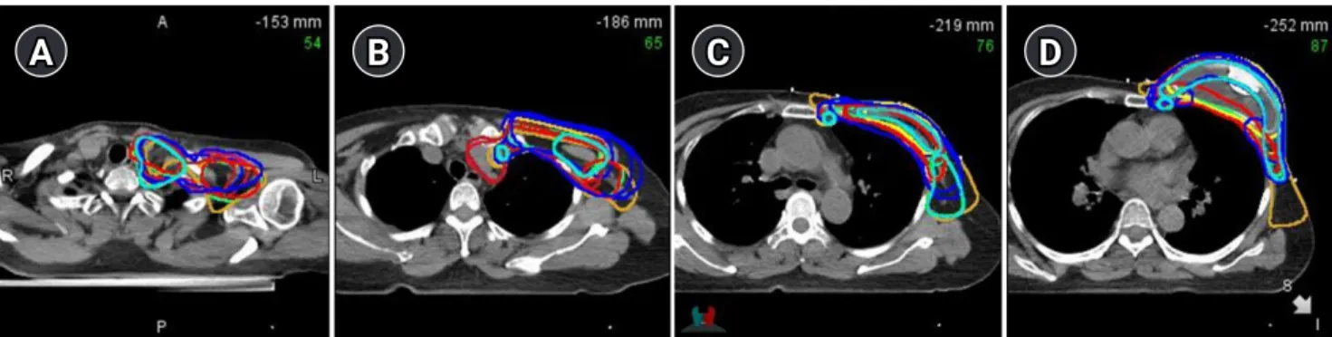
In terms of target volume delineation, the structures included in
the target volume varied significantly between physicians, even within the same clinical scenario (Figs. 4 and 5; Supplementary Fig.
S1). In a case where the patient was diagnosed with initial clinical internal mammary node metastasis (case 3), 13/15 physicians includ- ed the internal mammary node in the target volume. In a case of the patient who underwent pre-pectoral reconstruction instead of sub- pectoral reconstruction (case 4), 11/15 physicians included the tissue expander in the target volume. The dose-volume histograms were also analyzed. As for the planning target volume (PTV), it varied widely between physicians’ plans, even within the same clinical sce- nario. The median PTV volume was 563.67 mL (range, 278.84 to 1,100.77 mL; standard deviation [SD], 209.05 mL). Similarly, there was a wide difference in the mean lung dose; the median mean lung dose was 13.26 Gy (range, 5.33 to 18.80 Gy; SD, 4.60 Gy). There was also a wide variance in terms of lung V5 and V20; their median val- ues were 43.89 mL (range, 16.35 to 99.45 mL; SD, 26.64 mL), 23.65 mL (range, 6.7 to 34.03 mL; SD, 10.30 mL), respectively (Table 4).
100.0%
80.0%
60.0%
40.0%
20.0%
0.0%
100.0%
80.0%
60.0%
40.0%
20.0%
0.0%
50 40 30 20 10 0
A B C D E F G H I J K L M N O P A B C D E F G H I J K L M N O P
100 100
0 0 0 0 9 12
0
35
0
43
0
46
0
50
0
75
14
75
25
75
43
86
44
100
60
100
61
100
61
100 88
100
100100100100100100100 100100100100100100
25
86
25
75
25
57
14
56
0
40
0
39
0
39
12 ■ 1 stage 0 0
■ 2 stage
■ Prosthetic
■ Autologous
■ Flat
■ Full inflation
■ Partial deflation
■ Tissue expander
■ TRAM flap
■ Silicone
■ Tissue expander + silicone imp
■ DIEP flap
■ LD flap
■ Others
■ TUG flap
■ Silicone imp + LD flap
■ Saline
■ Local flap transposition
■ tissue expander + LD flap
■ IGAP flap
■ SIEA flap
■ PAP flap 11.15%
12.10%
23.89%
36.94%
0.32%
9.24%
3.82%
A B C D E F
B
D A
C
Fig. 1. Reconstruction details. (A) One-stage vs. two-stage reconstruction. (B) Prosthetic vs. autologous reconstruction. (C) Materials used for reconstruction. (D) Inflation status of the tissue expander at the time of radiation therapy. TRAM, transverse rectus abdominis myocutaneous;
DIEP, deep inferior epigastric perforator; LD, latissimus dorsi; TUG, transverse upper gracilis; IGAP, inferior gluteal artery perforator; SIEA, super-
ficial inferior epigastric artery; PAP, profunda artery perforator.
Table 3. Radiation treatments (n = 314)
Variable Value
Simulation position
Supine 305 (97.1)
Prone 2 (0.6)
Unknown 7 (2.2)
RT technique
Forward IMRT (field-in-field) 124 (39.5)
Arc-IMRT 90 (28.7)
Tangent, 3D 58 (18.5)
Static-IMRT 26 (8.3)
Helical tomo 8 (2.5)
2D (tangential) 1 (0.3)
Unknown 7 (2.2)
Fractionation
Conventional 209 (66.6)
Hypofractionation 105 (33.4)
Use of boost RT, yes 49 (15.6)
Boost RT dose (Gy) 9 ± 3.8
Expander status
Full deflation 4 (1.3)
Partial deflation 123 (39.2)
Full inflation 13 (4.1)
Use of bolus
No 146 (46.5)
Yes 168 (53.5)
Bolus off timing (Gy) 34.2 ± 14.6 Use of regional RT
No 18 (5.7)
AXL 7 (2.2)
SCL 52 (16.6)
IMN 1 (0.3)
AXL + SCL 74 (23.6)
AXL + IMN 1 (0.3)
SCL + IMN 4 (1.3)
AXL + SCL + IMN 157 (50.0)
Values are presented as number (%) or median ± standard deviation.
IMRT, intensity-modulated radiotherapy; RT, radiotherapy; AXL, axillary lymph node; SCL, supraclavicular lymph node; IMN, internal mammary node.
Discussion and Conclusion
According to the Korean Breast Cancer Society (KBCS) registry, the number of breast reconstruction surgeries has increased almost 3-fold between 2002 and 2013 [12]. In the 2017 KBCS report, the rate of reconstruction after mastectomy was 39.1%, 33.7%, and 9.4% in those in the age-group of < 40, 40–59, and ≥ 60 years, re- spectively [13]. In the present study, approximately one-third of patients who underwent PMRT underwent breast reconstruction and this rate is expected to increase in the future. The primary goal
100 80 60 40 20 0
100.00 90.00 80.00 70.00 60.00 50.00 40.00 30.00 20.00 10.00
Number of hospitals (%) Number of hospitals (%)
Dose (Gy) 40.05
No bolus Bolus applied Bolus
(around scar) Bolus
(Others) Bolus (Whole reconstructed
breast)
43.00 45.90 48.00 50.00 52.80 54.00
Case
■ 1
■ 2
■ 3
■ 4
■ 5
Case
■ 1
■ 2
■ 3
■ 4
■ 5
Fig. 2. Dose prescriptions according to different clinical scenarios.
Bolus Fig. 3. Details of bolus application.
in the treatment of women with breast cancer who undergo recon- struction and PMRT is to minimize the complications without com- promising the oncological outcomes. Therefore, understanding the practice patterns is crucial for the communication between the pa- tients and surgeons. To our knowledge, this is the first study to re- port the practice patterns of PMRT in the setting of breast recon- struction in Korea.
Both RT techniques and breast reconstruction techniques have
evolved over time. In the present study, a majority of the patients
underwent immediate reconstruction; the rate of one-stage, two-
stage, autologous-based, and prosthetic-based reconstruction was
51%, 49%, 23%, and 60.5%, respectively. The selection of the re-
construction material is individualized for each patient based on
several patient, anatomical, and disease-specific factors, such as
comorbidities, shape and size of the breast, availability of tissue
around the breast and other donor sites, and planned adjuvant
therapy. However, as presented in Fig. 1, we observed wide in-
100
80
60
40
20
0
Number of hospitals (%)
Target
Tissue expander Skin
surface Pectoralis m. Intercostal
m. Rib Axl Lv 1-2 Axl Lv 3 SCL IMN
Case
■ 1
■ 2
■ 3
■ 4
■ 5