N-Nitrosodiethylamine과 사염화탄소로 유발된 흰쥐의 간암발생에 대한 인진호탕 추출액의 효과
윤중식․김정상† 동신대학교 한의과대학 해부학교실
The Effect of the Injinhotang Extract on the Hepatocarcinogenesis Induced by N-Nitrosodiethylamine and Carbon Tetrachloride in Rats
Jung Sik Yoon and Jeong Sang Kim†
Dept. of Anatomy, College of Oriental Medicine, Dongshin University, Jeonnam 520-714, Korea
Abstract
In order to examine the effect of Injinhotang extract on the hepatocarcinogenesis induced by N-nitrosodiethyl- amine (NDEA) and carbon tetrachloride (CCl4) in 8 week-old rats. Experimental rats were subdivided into three groups; normal group (Nor), hepatic cancer inducing control group (Con), and control group administered Injinhotang extract 260 mg/kg/day (IJH). The body weight decreased significantly (p<0.05) in the Con compared with the Nor. The body weight of IJH group more increased than Con. Rats intoxicated with NDEA had sig- nificantly (p<0.05) increased levels of serum AST, ALT, LDH, ALP, and AFP. On the contrary, group treated with Injinhotang extract had inhibited levels of serum AST, ALT, LDH, ALP, and AFP. The bcl-2 mRNA ex- pression levels in rat liver were more increased in the IJH than Con, but these levels of c-myc mRNA were more decreased in the IJH than Con. Also, cytoplasmic vacuolizations in the liver of NDEA-administrated rats were inhibited by the treatment of Injinhotang extract. These results suggest that administration of Injinhotang extract suppresses or retards NDEA and CCl4-induced hepatocarcinogenesis in rats.
Key words: Injinhotang, N-nitrosodiethylamine, hepatocarcinogenesis, AFP, bcl-2
†Corresponding author. E-mail: [email protected]
†Phone: 82-61-330-3512, Fax: 82-61-330-3499
서 론
인진호탕은 인진, 치자, 대황으로 구성되어 있으며 황달, 항 염증, 해열, 그리고 이뇨작용 등을 가지는 것으로 알려져 왔다.
인진호(Artemisia capillaris Herba)는 사철쑥(Artemisia capillaris Thunberg)의 과수를 포함하는 지상부를 채취한 것을 말하며, 생약명으로는 인진, 인진호, 추호라 불린다. 인 진호탕은 현대에서 항염증제, 해열제, 이뇨제로서 담즙분비 를 촉진시키는 동시에 담낭 수축작용이 있고 아울러 간염에 대한 간 보호 작용이 있다고 알려져 있다(1-3). 인진호탕을 구성하는 세 가지 성분 중 하나인 인진은 capillarisin, cap- illien, capillin, capillarin과 scoparone 등이 함유되어 있고, 이 성분들은 담즙 분비를 촉진시켜 간 기능 개선 효과, 항암, 항염증, 항균, 항바이러스 작용, 혈압 강하 등의 활성을 갖는 것으로 알려져 있다(4-6). 치자의 주성분인 genipin, gar- deniside, crosin, crocetin과 gardenin 등은 항균, 항바이러스 작용, 소염작용, 해열작용, 이담작용이 있으며, 또한 genipo- side는 간, 신장과 심장에 항종양인자로서의 기능을 할 수
있는 것으로 보고되었다(7,8). 대황은 sennosides A-F, rha- tannin, rhein 그리고 lindleyin 등이 있으며 주로 항균, 이담, 지혈, 항암, 간 기능 보호, 면역조절, 소화효소분비억제 등의 기능이 있는 것으로 알려져 있다(9,10).
국내외의 인진호탕에 대한 연구를 보면 항고지혈증(11), 간 기능 개선효과(12), 항균활성(13), 항산화 효과(14) 등이 주로 연구되어지고 있으며, 인진호탕의 투여가 trans- forming growth factor-β1(TGF-β1)에 의해 유도된 흰쥐의 간 세포사(apoptosis)에 대한 보호효과가 있음을 보고하였 고(15), Yamashiki 등(16)은 concanavalin A로 유도한 생쥐 의 간염 모델에서 혈청의 염증성 cytokine인 IL-2, IL-12와 IFN-γ의 수준을 낮추고, 또한 AST, ALT와 LDH 등의 수준 을 낮추는데 탁월한 효과가 있다고 보고하였다. 현재까지 인진호탕의 항암활성에 관한 보고는 있으나(17), 직접적인 간암에 대한 연구는 거의 진행되지 않고 있는 실정이다.
간암의 발생과정을 연구하기 위해 이용되는 화학성 발암 물질중 하나인 nitrosamine은 탄 단백질식품, 절임육류가공 품과 같은 음식물에서 발견되는 발암성물질로서 알코올성
음료, 화장품, 절삭기 윤활유, 고무, 담배 등 주변 환경에서 흔히 인체에 오염될 수 있다(18). 본 실험에서 사용된 N-ni- trosodiethylamine(NDEA)은 nitrosodialkylamine에 속하는 (CH3CH2)2N-NO의 분자식을 가지는 무취의 황색 액체이다.
주로 사람과 동물의 간장이나 신장에 궤양 또는 암을 일으키 는 물질로 잘 알려져 있다. 근래에는 NDEA의 투여로 개시기 동안 DNA에 변이를 보이는 저항성 간세포를 유발하고, 이차 적으로 사염화탄소(CCl4), 2-acetylaminofluorene(2-AAF), phenobarbital, 간 부분 절제술 등(19-21)을 병행하여 많은 간암 유발 연구에 활용되고 있다.
최근에는 cyclooxygenase(COX) 억제제, 레티노이드 유 사체, 비타민 K2, glycyrrhizin, 녹차, 인삼 등의 간암 예방효 과(22-26)와 인체에 대한 부작용을 최소화하면서 항암 효과 를 발현하는 천연물질을 이용한 치료제 개발 연구가 이루어 지고 있다(27-29).
이에 본 연구에서는 간암의 발생이 진행되는 과정에서 인 진호탕 추출액의 항암효과를 규명하기 위하여 체중, 혈청 중 효소 활성도 측정, 암이 유발된 간세포 사멸 유전자의 발현 정도 그리고 조직학적 관찰을 실시하여 유의성 있는 결과를 얻었기에 보고하는 바이다.
재료 및 방법
실험재료
실험동물은 체중 210±10 g 내외의 Sprague Dawley계 웅성 흰쥐를 (주)샘타코에서 구입하였다. 실험동물은 동신 대학교 한의과대학 동물사육실에서 일정한 조건(온도:
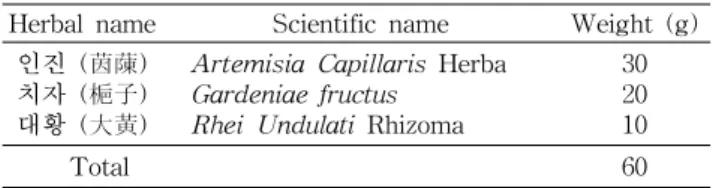
21±2oC, 습도: 50~60%, 명암: 12시간 주기)하에서 일반 고 형사료와 물을 충분히 공급하면서 1주 동안 적응시킨 후 실 험에 사용하였다. 인진호탕(Injinhotang, 茵蔯蒿湯)의 구성 과 용량은 아래와 같다(Table 1). 인진호탕 4첩을 물 600 mL 에 넣고 약탕기로 3시간 동안 가열한 다음 농축한 후 저온순 환수조(CA-1500, EYELA, Japan)에서 1차 동결한 다음 동 결건조기(SFDSMO06, Samwon, Korea)로 동결건조 하여 31.8 g의 분말을 얻었다.
실험동물 사육 및 간암 유발
정상군을 제외한 모든 실험동물은 N-nitrosodiethyl- amine(NDEA, Sigma Chemical Co., USA) 200 mg/kg을 0.9% 식염수 1 mL에 용해하여 1주 간격으로 2회 복강투여 하였다. NDEA투여 1주 후부터 사염화탄소(CCl4, Sigma
Table 1. Prescription of Injinhotang
Herbal name Scientific name Weight (g)
인진 (茵蔯) 치자 (梔子) 대황 (大黃)
Artemisia Capillaris Herba Gardeniae fructus
Rhei Undulati Rhizoma
30 20 10
Total 60
Chemical Co., USA)와 corn oil을 1:1로 희석한 약물 1 mL을 1주 간격으로 3회 복강투여 하여 간암을 유발하였다. 모든 실험군의 실험동물은 5마리씩으로 하였다. 정상군(Nor)은 통상적인 사료와 물을 공급하였으며, 대조군(Con) 흰쥐는 간암이 유발된 후부터 식염수 0.5 mL을 실험기간 동안 매일 구강투여 하였다. 실험군(IJH)은 NDEA을 투여한 2주 후부 터 8주 동안 인진호탕추출액 건조분말을 생리식염수에 0.5 mL에 용해하여 체중 60 kg인 사람의 1일 투약량인 260 mg/kg을 구강투여 하였다.
체중 측정
체중은 실험기간 동안 매주 1회 정해진 시간에 측정하였 고, 실험시작 후 2주, 4주, 6주 및 8주 결과를 이용하였다.
생화학적 분석
약물 투여 8주 후 흰쥐를 urethane으로 마취한 다음 복부 의 정중선을 따라 절개하고 심장 채혈을 시행하였다. 혈액은 시험관에 넣어 실온에서 30분간 방치하여 혈액을 응고시킨 다음 3,000 rpm에서 원심분리하고 상등액을 회수하여 -80oC 초저온 냉동고에 보관하면서 실험에 사용하였다. Aspartate aminotransferase(AST)와 alanine aminotransferase(ALT), lactate dehydrogenase(LDH), alkaline phosphatase(ALP) 활성도는 자동분석기용 kit 시약(아산제약)을 사용하여 생 화학자동분석기(photometer 5010, Germany)에서 측정하였 다. 그리고 alpha-fetoprotein(AFP)은 AFP ELISA kit (Sierra Resources International, Inc., Valrico, FL)를 이용 하여 혈장내 AFP 농도를 제조사의 실험방법에 따라 측정하 였다.
RT-PCR을 이용한 bcl-2와 c-myc의 mRNA 정량
흰쥐의 간을 획득한 후 RNA-spin total RNA extraction kit를 이용하여 총 RNA를 얻었다. 총 RNA는 분광광도계를 이용하여 OD값을 측정하고 총 RNA양을 정량한 후 -70oC 냉동고에 보관하였다. Reverse transcription은 DEPC-DW 로 최종부피를 50 μL로 맞춘 후 total RNA에 RT-pre- mix(Bioneer, Korea) 혼합용액을 사용하여 70oC에서 5분 동 안 RNA를 변성시키고, 42oC에서 1시간 동안 역전사하여 cDNA를 합성하고, 94oC에서 5분 동안 M-MLV reverse transcriptase를 불활성화 시키는 과정으로 진행되었다. 합 성된 cDNA는 PCR-PreMix(Bioneer, Korea)와 각각의 pri- mer(Table 2)를 사용하여 GAPDH는 94oC 60초, 57oC 60초, 72oC 60초의 30 cycle로, bcl-2는 94oC 60초, 48oC 60초, 72oC 60초의 35 cycle로, c-myc은 94oC 60초, 59oC 60초, 72oC 60 초의 30 cycle로 PCR을 수행하였다. 이렇게 얻어진 PCR product는 1.5% agarose gel에서 전기영동 하여 UV(Kodak, USA)에서 관찰하였다.
조직학적 관찰
각 군에서 부검한 흰쥐의 간 조직을 적출한 후 Bouin 용액
Table 3. Effect of Injinhotang extract on body weight of rats (g)
Group1) 0 week 2 weeks 4 weeks 6 weeks 8 weeks
Nor Con IJH
224±0.752) 223±3.08 223±1.05
283±3.12 264±7.40 282±7.37
337±7.92 300±11.10* 320±7.82
382±12.35 335±10.36* 357±10.48
427±15.12 370±10.85* 395±14.05
1)Nor, not administrated with NDEA, CCl4 and Injinhotang extract; Con, administrated with NDEA and CCl4; IJH, administrated with NDEA, CCl4 and Injinhotang extract.
2)All values are mean±SE (n=5).
*Significant differences were compared with normal at p<0.05.
Table 4. Effect of Injinhotang extract on plasma AST, ALT, LDH, ALP, AFP activities in rats
Group1) AST (U/L) ALT (U/L) LDH (U/L) ALP (U/L) AFP (ng/mL)
Nor Con IJH
95±5.862) 179±7.03* 96±14.16#
60±5.32 82±3.62* 74±5.39
2724±206.79 5293±282.00* 2838±274.86#
227±9.66 348±28.79* 285±17.09*
2.78±0.29 5.51±0.44* 3.61±0.52#
1)Nor, not administrated with NDEA, CCl4 and Injinhotang extract; Con, administrated with NDEA and CCl4; IJH, administrated with NDEA, CCl4 and Injinhotang extract.
2)All values are mean±SE (n=5).
Significant differences were compared with normal/control at *p<0.05/#p<0.05.
Table 2. Primers designed for amplification of the mRNA in RT-PCR
Primer Sequence Product
size 1. bcl-2 F: 5'-CGGGAGAACAGGGTATGA-3'
R: 5'-CAGGCTGGAAGGAGAAGAT-3' 149 bp 2. c-myc F: 5'-CTCTTCTTCCTCGGACTCGC-3'
R: 5'-GCGCAGGGCAAAAAAGCTAC-3'484 bp 3. GAPDH F: 5'-AATGCATCCTGCACCACCAA-3'
R: 5'-GTAGCCATATTCATTGTCAT-3'600 bp
을 사용하여 24시간 동안 고정시켰다. 고정된 조직은 파라핀 포매 과정을 거쳐 microtome으로 5 μm 두께로 조직절편을 만들어 Hematoxylin & Eosin(H-E) 염색을 시행하여 광학 현미경(Olympus BX51, Japan)으로 관찰하였다.
통계처리
실험 결과는 mean±SE로 표시하였고, 대조군과 실험군 과의 평균의 차이를 검정할 때에는 Student's test로 검정하 여 p값이 0.05 미만일 때 통계적으로 유의한 차이가 있는 것으로 판정하였다.
결과 및 고찰
체중 변화
일반적으로 종양 환자의 경우 현저한 식욕부진과 함께 탈 수증상이 수반되며, 이로 인한 만성적인 체중감소가 초래되 는데, 본 실험에서는 2주째에 정상군(283±3.12 g)에 비하여 대조군(264±7.40 g)은 약 20 g정도 감소하였으나, 인진호탕 추출물을 투여한 IJH군(282±7.37 g)은 약간 감소하였다. 4 주군에서부터 대조군(300±11.10 g)은 정상군(337±7.92 g) 에 비하여 유의성(p<0.05) 있게 감소하여 실험 종료 시점인
8주째까지 동일한 결과를 보였으나, 인진호탕 추출물이 투 여된 실험군의 체중은 대조군에 비하여 높은 경향을 나타냈 다(Table 3). Flavonoid의 일종인 morin(3,5,7,2',4'-penta- hydroxyflavone)의 효과를 보기 위한 실험에서 NDEA으로 유도된 군은 정상군에 비해 체중이 유의적으로 감소되는 반 면에 morin과 병합투여한 군은 정상군에 가까운 체중을 유 지하였다고 보고하였는데(30) 이는 본 실험의 결과와 일치 하였다.
혈청 중 효소의 변화
AST와 ALT는 심장근과 간세포내에 있는 특성효소로서 조직세포의 손상에 의해 혈청중의 활성도가 증가되어지며, 이러한 활성도의 상승은 간담도 질환, 심장질환에서 일어나 고 특히 만성간염의 경우에는 현저하게 증가된다. 본 실험에 서 AST의 활성(Table 4)은 대조군(179±7.03 U/L)이 정상 군에(95±5.86 U/L) 비하여 유의적인(p<0.05) 증가를 보였 으며, IJH군은 96±14.16 U/L로 나타내어 대조군에 비하여 유의성(p<0.05) 있게 감소하였다.
ALT의 활성(Table 4)은 대조군이 82±3.62 U/L인데 비 하여 IJH군에서는 74±5.39 U/L으로 나타나 증가억제 현상 은 보였으나 유의성은 없었고, 정상군(60±5.32 U/L)에 대해 서는 유의성(p<0.05) 있게 증가하였다.
LDH는 악성 종양세포에서 다량 생성되므로 암의 존재를 파악하는데 이용되어진다. LDH의 활성은 정상군(2724±
206.79 U/L)에 비하여 대조군(5293±282.00 U/L)에서 유의 적인(p<0.05) 증가를 보였다. 그러나 인진호탕 추출물을 투 여한 IJH군(2838±274.86 U/L)은 대조군에 비하여 유의성 (p<0.05) 있게 감소하였다(Table 4).
ALP는 만성간염이나 간경변증에서도 올라갈 수 있으나, 만들어진 담즙이 간세포에서 잘 배출되지 못하거나 담도가 막혔을 때 현저히 증가한다. ALP의 활성은 정상군(227±9.6
Fig. 2. Histological sections of liver of rats in response to Injinhotang ex- tracts in the NDEA and CCl4-induced liver injury. ⓐ: Normal group, ad- ministered saline. ⓑ: Control group, ad- ministered saline after NDEA treatment.
ⓒ: IJH group, administered Injinhotang extract (260 mg/kg/day) after NDEA treatment. The control group showing vacuolated cytoplasm. CV, central vein.
Haematoxylin-Eosin stain. Original magnification ×400.
U/L)에 비하여 대조군(348±28.79 U/L)에서 유의적인(p<
0.05) 증가를 나타냈고, 인진호탕 추출물을 투여한 IJH군 (285±17.09 U/L)은 대조군에 비해 감소하는 경향을 나타냈 다(Table 4). 콩과식물인 Bauhinia racemosa L.의 NDEA에 대한 효과 실험에서 GOT, GPT와 ALP의 수치가 NDEA를 투여한 군에서는 현저히 증가한다는 것을 보고하였다(31).
Bansal 등(32)은 비타민 E를 전처치한 다음 NDEA로 간 손 상을 유발한 실험에서 AST, ALT, ALP와 LDH의 수치를 감소시키는 효과가 있다고 보고하였다. 본 실험에서도 AST, ALT, ALP와 LDH의 활성이 대조군에서는 증가하였 으나 인진호탕 추출물을 투여한 군에서는 감소하는 경향을 보여 효과가 있는 것으로 생각된다.
AFP는 원래 임신초기의 태아에서 나타나는 혈청 단백의 일종으로 건강한 성인의 혈액에서는 볼 수 없으나 원발성간 암 환자의 95%에서 증가하기 때문에 간암의 종양 marker로 서 이용되고 있다(33). 인진호탕 추출액을 투여한 다음 혈청 AFP의 농도를 측정한 결과 IJH군(3.61±0.52 ng/mL)은 대 조군에 비하여 유의성(p<0.05) 있게 감소하였다. 그리고 정 상군(2.78±0.29 ng/mL)에 비하여 대조군(5.51±0.86 ng/mL) 에서는 약 98% 증가하여 유의성(p<0.05)이 있었다(Table 4). Flavonoid의 일종인 morin(3,5,7,2'4'-pentahydroxy- flavone)의 효과를 보기 위한 실험에서 NDEA으로 유도된 군은 유의적으로 증가되었는데, morin과 병합투여한 군은 AFP 수준이 유의적으로 감소되었다고 보고하였다(30).
인진호탕에 대한 bcl-2와 c-myc의 mRNA 발현 수준 Bcl-2계 단백질 중 Bcl-2는 칼슘의 항상성 유지, 항산화 효소계의 강화 및 미토콘드리아에서 cytochrome C 방출을 억제하여 apoptosis를 억제한다(34,35). 본 연구에서 bcl-2 mRNA 발현 정도를 살펴본 결과, 간암 유발 후 8주간 인진호 탕을 투여한 실험군의 bcl-2 mRNA 발현은 대조군에 비해 증가되었다(Fig. 1).
C-myc oncogene은 전립선암종(36), 간세포암종(37) 및 골수성 백혈병(38) 등 여러 종양의 세포주에서 c-myc 유전 자의 증폭과 산물이 증가한다는 것이 보고되었다. Morris 간암(39)과 2-amino-3-methylimidazo-quinoline(40)으로 유도된 간암에서도 c-myc oncogene의 중폭 및 과다발현이
Fig. 1. Effect of Injinhotang extract on bcl-2 and c-myc mRNA in rat liver by RT-PCR. The effects of Injinhotang ex- tract on gene expression in the NDEA and CCl4-induced liver injury. Nor, not administrated with NDEA, CCl4 and Injinhotang extract; Con, administrated with NDEA and CCl4; IJH, adminis- trated with NDEA, CCl4 and Injinhotang extract.
일어난다는 보고가 있었으며, 3-methyl-4-dimethylami- noazobenzene(41)에 의해 유발된 흰쥐의 원발성 간종양에 서도 c-myc oncogene의 전사체가 증가되었다는 보고가 있 다. 본 실험에서도 c-myc mRNA의 경우는 대조군이 실험군 에서보다 더 증가됨이 관찰되었다(Fig. 1).
간 조직의 관찰
적출한 간조직을 광학현미경으로 관찰한 결과에서 정상 군의 간 조직은 뚜렷한 둥근 핵을 갖고 있었고 세포질은 거 의 일정하게 eosin으로 염색되었으며, 간소엽의 구조도 잘 배열되어 있었다(Fig. 2a). NDEA와 사염화탄소를 투여한 대조군은 간소엽의 구조 및 핵의 형태가 불완전하고, 일부에 서 세포질 공포화가 유발된 것을 관찰할 수 있었으며, 이로 인해 세포질의 염색성은 정상군이나 IJH군보다 현저히 감소 되었다(Fig. 2b). 그러나 인진호탕을 투여한 IJH군은 간소엽 의 구조가 비교적 잘 유지되어 있었고, 핵 형태가 비교적 뚜렷하였다. 또한, 세포질의 염색성은 정상군과 유사하게 관 찰되었으며 세포질의 공포화 현상은 관찰되지 않았다(Fig.
2c). Mourão 등(42)의 NDEA로 유도된 간암에 대한 in- dole-3-acetic acid(IAA)의 실험에서 NDEA만을 투여한 군 에서 간 손상의 가장 두드러진 특징은 정상대조군과 비교하 여 간세포의 팽대성 증가와 세포질 공포화 그리고 공포 형성 의 유도라고 하였는데, 이는 본 실험의 결과와 일치하였다.
요 약
본 실험은 N-nitrosodiethylamine(NDEA)와 사염화탄소 (CCl4)로 유발된 흰쥐의 간암 생성에 대한 인진호탕의 효과 를 조사하였다. 실험은 8주 동안 실시하였고, 세 군으로 분류 하였다. 정상군(Nor), 간암을 유발한 대조군(Con), 간암 유 발 후 인진호탕 추출액(260 mg/kg/day)을 투여한 실험군 (IJH)으로 구분하였다. 체중은 정상군에 비하여 대조군에서 유의적으로 감소하였지만 실험군에서는 증가하였다. NDEA 로 유발된 흰쥐의 혈청 AST, ALT, LDH, ALP와 AFP의 수준은 증가하였다. 반대로, 인진호탕 추출액을 처리했을 때, 혈청 AST, ALT, LDH, ALP와 AFP의 수준이 감소하였 다. 흰쥐의 간에서 bcl-2 mRNA의 수준은 대조군에 비해 실험군에서 증가되었다. 그러나 c-myc mRNA의 수준은 대 조군에 비해 감소하였다. 또한 NDEA로 손상된 간세포 조직 의 공포화가 인진호탕 추출액의 투여로 감소하는 것을 관찰 할 수 있었다. 이상의 결과로 보아 인진호탕 추출액 투여가 NDEA와 사염화탄소로 유도된 흰쥐의 간암 생성을 억제 또 는 지연하는 것으로 사료된다.
문 헌
1. Seo1 HC, Suzuki M, Ohnishi-Kameyama M, Oh MJ, Kim HR, Kim JH, Nagata T. 2003. Extraction and identification of antioxidant components from Artemisia capillaris herba.
Plant Foods for Human Nutrition 58: 1-12.
2. Tan RX, Zheng WF, Tang HQ. 1998. Biologically active substances from the genus Artemisia. Planta Med 64:
295-302.
3. Yamahara J, Yamada T, Kimura H, Sawada T, Fujimura H. 1982. Biologically active principles of crude drugs. II.
Anti-allergic principles in "Shoseiryu-To" anti-inflam- matory properties of paeoniflorin and its derivatives. J Pharmacobiodyn 5: 921-929.
4. Yamahara J, Kobayashi G, Matsuda H, Katayama T, Fujimura H. 1989. The effect of scoparone, a coumarin de- rivative isolated from the Chinese crude drug Artemisiae capillaris Flos, on the heart. Chem Pharm Bull 37: 1297- 1299.
5. Mori H, Xu QA, Sakamoto O, Uesugi Y, Ono Y, Koda A, Nishioka I. 1988. Immunological mechanisms of antitumor activity of some kinds of Chinese herbs: Meth A-induced delayed type hypersensitivity. Jpn J Pharmacol 48: 37-46.
6. Kiso Y, Ogasawara S, Hirota K, Watanabe N, Oshima Y, Konno C, Hikino H. 1984. Antihepatotoxic principles of Artemisia capillaris buds. Planta Med 50: 81-85.
7. Peng CH, Huang CN, Wang CJ. 2005. The anti-tumor effect and mechanisms of action of penta-acetyl geniposide. Curr Cancer Drug Targets 5: 299-305.
8. Kimura Y, Okuda H, Arichi S. 1982. Effects of geniposide isolated from Gardenia jasminoides on metabolic alter- ations in high sugar diet-fed rats. Chem Pharm Bull 30:
4444-4447.
9. Jin H, Sakaida I, Tsuchiya M, Okita K. 2005. Herbal medi- cine Rhei rhizome prevents liver fibrosis in rat liver cir- rhosis induced by a choline-deficient L-amino acid-defined
diet. Life Sci 76: 2805-2816.
10. Matsuda H, Tomohiro N, Hiraba K, Harima S, Ko S, Matsuo K, Yoshikawa M, Kubo M. 2001. Study on an- ti-Oketsu activity of rhubarb II. Anti-allergic effects of stilbene components from Rhei undulati Rhizoma. Biol Pharm Bull 24: 264-267.
11. Lee J, Chae K, Ha J, Park BY, Lee HS, Jeong S, Kim MY, Yoon M. 2008. Regulation of obesity and lipid disorders by herbal extracts from Morus alba, Melissa officinalis, and Artemisia capillaris in high-fat diet-induced obese mice.
J Ethnopharmacol 115: 263-270.
12. Han KH, Jeon YJ, Athukorala Y, Choi KD, Kim CJ, Cho JK, Sekikawa M, Fukushima M, Lee CH. 2006. A water extract of Artemisia capillaris prevents 2,2'-azobis(2-ami- dinopropane) dihydrochloride-induced liver damage in rats.
J Med Food 9: 342-347.
13. Luo H, Lin S, Ren F, Wu L, Chen L, Sun Y. 2007. Antioxi- dant and antimicrobial capacity of Chinese medicinal herb extracts in raw sheep meat. J Food Prot 70: 1440-1445.
14. Chu CY, Tseng TH, Hwang JM, Chou FP, Wang CJ. 1999.
Protective effects of capillarisin on tert-butylhydroper- oxide-induced oxidative damage in rat primary hepato- cytes. Arch Toxicol 73: 263-268.
15. Yamamoto M, Ogawa K, Morita M, Fukuda K, Komatsu Y. 1996. The herbal medicine Inchin-ko-to inhibits liver cell apoptosis induced by transforming growth factor beta 1.
Hepatology 23: 552-559.
16. Yamashiki M, Mase A, Arai I, Huang XX, Nobori T, Nishimura A, Sakaguchi S, Inoue K. 2000. Effects of the Japanese herbal medicine 'Inchinko-to' (TJ-135) on con- canavalin A-induced hepatitis in mice. Clin Sci 99: 421-431.
17. Hu YQ, Tan RX, Chu MY, Zhou J. 2000. Apoptosis in hu- man hepatoma cell line SMMC-7721 induced by wa- ter-soluble macromolecular components of Artemisia ca- pillaris Thunberg. Jpn J Cancer Res 91: 113-117.
18. Bartsch H, Ohshima H, Shuker DE, Pignatelli B, Calmels S. 1990. Exposure of humans to endogenous N-nitroso compounds: implication in cancer etiology. Mutat Res 238:
255-267.
19. Farber E. 1984. The multistep nature of cancer develop- ment. Cancer Res 44: 4217-4223.
20. Peraino C, Fry RJ, Staffeldt E, Kisieleski WE. 1973. Effects of varying the exposure to phenobarbital on its enhance- ment of 2-acetylaminofluorene-induced hepatic tumori- genesis in the rat. Cancer Res 33: 2701-2705.
21. Solt DB, Cayama E, Tsuda H, Enomoto K, Lee G, Farber E. 1983. Promotion of liver cancer development by brief ex- posure to dietary 2-acetylaminofluorene plus partial hep- atectomy or carbon tetrachloride. Cancer Res 43: 188-191.
22. Cervello M, Montalto G. 2006. Cyclooxygenases in hep- atocellular carcinoma. World J Gastroenterol 12: 5113- 5121.
23. Habu D, Shiomi S, Tamori A, Takeda T, Tanaka T, Kubo S, Nishiguchi S. 2004. Role of vitamin K2 in the develop- ment of hepatocellular carcinoma in women with viral cir- rhosis of the liver. JAMA 292: 358-361.
24. Ikeda K, Arase Y, Kobayashi M, Saitoh S, Someya T, Hosaka T, Sezaki H, Akuta N, Suzuki Y, Suzuki F, Kumada H. 2006. A long-term glycyrrhizin injection therapy reduces hepatocellular carcinogenesis rate in patients with interfer- on-resistant active chronic hepatitis C: a cohort study of 1249 patients. Dig Dis Sci 51: 603-609.
25. Nakachi K, Matsuyama S, Miyake S, Suganuma M, Imai K. 2000. Preventive effects of drinking green tea on cancer and cardiovascular disease: epidemiological evidence for
multiple targeting prevention. Biofactors 13: 49-54.
26. Takai K, Okuno M, Yasuda I, Matsushima-Nishiwaki R, Uematsu T, Tsurumi H, Shiratori Y, Muto Y, Moriwaki H.
2005. Prevention of second primary tumors by an acyclic retinoid in patients with hepatocellular carcinoma. Updated analysis of the long-term follow-up data. Intervirology 48:
39-45.
27. Davis ID. 2000. An overview of cancer immunotherapy.
Immunol Cell Biol 78: 179-195.
28. Han SB, Lee CW, Jeon YJ, Hong ND, Yoo ID, Yang KH, Kim HM. 1999. The inhibitory effect of polysaccharides isolated from Phellinus linteus on tumor growth and metastasis. Immunopharmacology 41: 157-164.
29. Wang HX, Liu WK, Ng TB, Ooi VE, Chang ST. 1995.
Immunomodulatory and antitumor activities of a poly- saccharide-peptide complex from a mycelial culture of Tricholoma sp., a local edible mushroom. Life Sci 57:
269-281.
30. Sivaramakrishnan V, Shilpa PN, Praveen Kumar VR, Niranjali Devaraj S. 2008. Attenuation of N-nitrosodiethyl- amine-induced hepatocellular carcinogenesis by a novel flavonol-Morin. Chem Biol Interact 171: 79-88.
31. Kumar RS, Sunderam RS, Sivakumar T, Sivakumar P, Sureshkumar R, Kanagasabi R, Vijaya M, Perumal BP, Gupta M, Mazumdar UK, Kumar MS, Kumar KA. 2007.
Effect of Bauhinia racemosa stem bark on N-nitro- sodiethylamine-induced hepatocarcinogenesis in rats. Am J Chin Med 35: 103-114.
32. Bansal AK, Bansal M, Soni G, Bhatnagar D. 2005. Protec- tive role of vitamin E pre-treatment on N-nitrosodiethyl- amine induced oxidative stress in rat liver. Chem Biol Interact 156: 101-111.
33. Wepsic HT, Kirkpatrick A. 1979. Alpha-fetoprotein and its relevance to human disease. Gastroenterology 77: 787-796.
34. Stähelin BJ, Marti U, Zimmermann H, Reichen J. 1999. The interaction of Bcl-2 and Bax regulates apoptosis in biliary epithelial cells of rats with obstructive jaundice. Virchows Arch 434: 333-339.
35. Finucane DM, Bossy-Wetzel E, Waterhouse NJ, Cotter TG, Green DR. 1999. Bax-induced caspase activation and apop- tosis via cytochrome c release from mitochondria is in- hibitable by Bcl-xL. J Biol Chem 274: 2225-2233.
36. Rijnders AW, van der Korput JA, van Steenbrugge GJ, Romijn JC, Trapman J. 1985. Expression of cellular onco- genes in human prostatic carcinoma cell lines. Biochem Biophys Res Commun 132: 548-554.
37. Huber BE, Thorgeirsson SS. 1987. Analysis of c-myc ex- pression in a human hepatoma cell line. Cancer Res 47:
3414-3420.
38. Wolman SR, Lanfrancone L, Dalla-Favera R, Ripley S, Henderson AS. 1985. Oncogene mobility in a human leuke- mia line HL-60. Cancer Genet Cytogenet 17: 133-141.
39. Hayashi K, Makino R, Sugimura T. 1984. Amplification and over-expression of the c-myc gene in Morris hepatomas.
Gann 75: 475-478.
40. Ishikawa F, Takaku F, Nagao M, Ochiai M, Hayashi K, Takayama S, Sugimura T. 1985. Activated oncogenes in a rat hepatocellular carcinoma induced by 2-amino-3- methylimidazo[4,5-f]quinoline. Jpn J Cancer Res 76: 425- 428.
41. Cote GJ, Lastra BA, Cook JR, Huang DP, Chiu JF. 1985.
Oncogene expression in rat hepatomas and during hepato- carcinogenesis. Cancer Lett 26: 121-127.
42. Mourão LR, Santana RS, Paulo LM, Pugine SM, Chaible LM, Fukumasu H, Dagli ML, de Melo MP. 2009. Protective action of indole-3-acetic acid on induced hepatocarcinoma in mice. Cell Biochem Funct 27: 16-22.
(2009년 2월 17일 접수; 2009년 3월 17일 채택)