23(2) : 132-138 (2017)
https://doi.org/10.20307/nps.2017.23.2.132
132
Synergetic Hepatoprotective Effects of Korean Red Ginseng and Pueraria Radix on the Liver Damaged-Induced by Carbon Tetrachloride (CCl 4 ) in Mice
Seung Hwan Hwang
1, Zhiqiang Wang
1, Il-Jun Kang
1, and Soon Sung Lim
1,2,3*
1
Department of Food Science and Nutrition, Hallym University, 1 Hallymdeahak-gil, Chuncheon 24252, Republic of Korea
2
Institute of Korean Nutrition, Hallym University, 1 Hallymdeahak-gil, Chuncheon 24252, Republic of Korea
3
Institute of Natural Medicine, Hallym University, 1 Hallymdeahak-gil, Chuncheon 24252, Republic of Korea
Abstract − This study was designed to investigate the synergetic hepatoprotective effects from a mixture of Korean Red Ginseng and Pueraria Radix on carbon tetrachloride (CCl
4)-induced hepatotoxicity in mice. Liver toxicity was induced by intraperitoneal administration of CCl
4(0.6 mg/kg) in 12 groups of ICR mice. The negative control group was given CCl
4without test samples and the normal group was given no treatment. Among treatment groups, the RGAP treatment (Korean Red ginseng acetic acid extract : Pueraria radix water extract, w/
w, 38.4:57.6) decreased CCl
4-elevated ALT (101.60 IU/L), AST (833.89 IU/L), and LDH (365.02 IU/L) levels in the serum, and increased the SOD (11.03 unit/mg protein) and CAT (0.37 unit/mg protein) levels and the LPO levels (59.09 µM/g tissue) more than that in the mice group with CCl
4-induced control group hepatotoxicity.
These results suggest that administration of a mixture of Korean Red ginseng and Pueraria radix decreases CCl
4- induced liver damage and enhances antioxidant activity in mice and imply that administration of the mixture in a certain ratio is more effective than single administration of either Korean Red ginseng or Pueraria radix alone.
Keywords − Carbon tetrachloride, Red Ginseng, Pueraria Radix, Hepatoprotective, Synergetic
Introduction
Although the modern world benefits from rapid development of industries and the convenience of auto- mation, the side effects and consequences of industrialism such as exposure to risks in industrial fields, fatigue from change of life patterns, oxidative stress, nutritional imbalance, improper diet, and lack of exercise, damage the liver function, resulting in liver disorders, liver cirrhosis, chronic hepatitis, and liver cancer.
1In particular, liver disease has a high prevalence rate in Korea and poses a threat to the health of its citizens. Thus, studies are actively being conducted on natural substances with therapeutic hepatic effects.
2Currently employed treatment methods of liver disease are largely classified into dietary therapy and medicinal therapy, and in most cases, these two methods are combined. In medicinal therapy, various medicines with different working mechanisms are used, based on the cause and type of liver disease. Typical medicines used in
clinical settings are regeneration promoters of hepatocytes, liver function supplements such as silymarin, biphenyl dimethyl dicarboxylate, urusodeoxycholic acid, glutathione, and glycyrrhizin extracted and separated from natural materials, antiviral medicines such as acyclovir, immuno- suppressants such as corticosteroids, 6-mercaptopurine, and azathioprine, and vitamin B complex.
3Ginseng (Panax ginseng) is a medicinal plant that grows in the far Eastern regions, including Korea, China, and Japan. Red ginseng (RG) is defined as steamed and dried red-colored ginseng that is over 4 years old. RG has a tough tissue and a red outer surface because of a non- enzymatic-browning reaction during the drying process.
4The major ingredients of RG are saponin-family ginseno- side, polyacetylene-family panaxytriol, panaxadiol, and acidic polysaccharides, which have been reported to have effects such as promotion and enhancement of liver regeneration
5, detoxification of toxic materials and alcohol
6, enhancement of immunity
7, antioxidant functions
8, and anti-cancer functions.
9,10Pueraria radix (PR) belongs to the family Leguminosae and is the root of Kudzu (Pueraria lobata Ohwi) with its periderm eliminated. It has been used as food and medicine in folk remedies for colds, headaches, appease-
*Author for correspondence
Soon Sung Lim, Department of Food Science and Nutrition, Hallym University, 1 Hallymdeahak-gil, Chuncheon 24252, Republic of Korea
Tel: +82-33-248-2133; E-mail: [email protected]
ment of thirst, diabetes, and diarrhea.
11PR contains isoflavonoid-family daidzein, daidzin, genistein, and puerarin, and triterpenoid-family soyasapogenol A and kuzusapogenol B. The presence of substances such as polysaccharides has also been reported. These substances have been reported to have effects such as sedation, hypotensive functions
12, extension of coronary arteries
13, suppression of lipid peroxidation
14, detoxification of alcohol
15, and liver protective functions.
16While there have been many studies on the hepatoprotective effect of separate administration of RG and PR, there is a lack of studies on comparison and analysis of the effect of combined or mixed adminis- tration of RG and PR. Therefore, in order to develop a medicine to prevent and treat liver disease, this study investigated the effect of the administration of a mixture of RG, PR, Meretrix lusoria, Artemisia asiatica, shell calcium extract, milk thistle (including 80% silymarin), and vitamin complex in a certain ratio in liver tissues damaged by CCl
4in ICR mice. Additionally, this study compared treatment groups to investigate the synergistic effect of PR and RG and the reduction of liver damage.
Experimental
Plant materials – Among the test samples, 4-year-old ginseng was purchased from a large supermarket in the city of Chuncheon and PR was produced by and purchased from Young-cheon in the Gyeongbuk, Korea.
Vitamin complex, shell calcium, M. lusoria, and A.
asiatica were supplied by JS F&B Inc (Chuncheon, Korea). Milk thistle (including 80% silymarin) and product “Urusa” of Daewoong Pham. Co., Ltd (Ursodeo- xycholic acid content: 100 mg) were generally used as positive controls in Korea. 2 kg of ginseng were put into an RG maker (Korea Maker, Korea), fumigated for 24 h, and dried in a 65
oC dry oven. This yielded 1.98 kg of reddish-black RG. Then, 1 L of distilled water was added to 100 g of RG, and reflux extraction was conducted at 100
oC for 3 h. The mixture was freeze-dried to yield 39 g of RG water extract (RGWE). RG acidic extract (RGAE) was refluxed for extraction using 0.1% acetic acid, and evaporated under reduced pressure to yield a residue (42 g). PR, M. lusoria, and A. asiatica were extracted using the same method with distilled water, and 40 g, 5 g, and 35 g of extracts were obtained, respectively.
Animals and treatment – Experimental animals used in this study were ICR mice with an average body weight of 37 ± 3 g, purchased from Joong-Ang Experimental Animal Center Inc. They were adapted for 1 week and
separated into 13 groups. The breeding environment for animal experiments was an automatically-controlled animal breeding room with a temperature of 22 ± 2
oC, humidity of 55 ± 5%, odor of less than 20 ppm, illumi- nation of 150-300 lux, and a 12-h light and shade cycle.
Solid feed (Samyang Oil and Feed Corp., Korea) and water were freely supplied. The experimentation, breeding, and management of animals were conducted according to the ‘Guide for the Care and Use of Laboratory Animals’, and experiments were performed with the authorization of the Ethics Committee of Hallym University (Hallym- 2009-78).
Liver toxicity was induced by intraperitoneal adminis- tration of CCl
4(0.6 mg/kg, CCl
4: Olive oil, 3:2, v/v; final concentration = 1.0 mg/kg) in 12 groups of mice (all groups except for the normal group, or N group). The negative control group (C group) was given CCl
4without test samples and the N group was given no treatment. The two positive control groups were administered product
“Urusa” of Daewoong Pham. Co., Ltd in Korea (U group, 70 mg/kg) and milk thistle (including 80% silymarin, S group, 50 mg/kg). The RGAP (RGAE : PWE, 38.4:57.6%), RGW (RAWE 96.0%), RGA (RGAW 96.0%), and PW (PWE 96.0%) were orally administered 200 mg/kg (b.w) for 10 days (n = 7) with vitamin complex (4.0%). In addition, other groups were orally administered 200 mg/
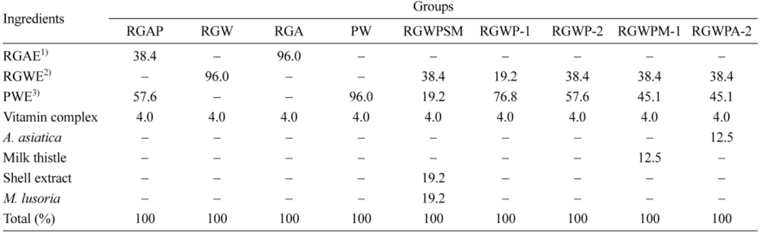
kg (b.w) for 10 days (n = 7) with various RGAE and PWE ratio (%) and A. asiatica (12.5%), milk thistle (12.5%), shell extract (19.2%) and M. lusoria (19.2%) including vitamin complex (4.0%). After completion of the experiments, the animals were fasted for 18 h and anesthetized with diethyl ether. The compositions of the oral administration experimental diets are shown in Table 1.
Blood samples were drawn from an orbit vein.
Collected blood was separated by centrifugation (350 × g at 4
oC for 20 min), and serum was used for blood biomarkers analysis. Right after blood collection, livers were removed and washed in physiological saline solution (0.9% NaCl solution). Water was removed from the livers, weights were measured, and they were stored in −75
oC until the time of analysis.
Measurement of alanine aminotransferase (ALT),
aspartate aminotransferase (AST) and lactate dehy-
drogenase (LDH) activities in serum – The activity of
alanine aminotransferase (ALT) and aspartate aminotrans-
ferase (AST) was measured with a kit (Asan Pharmaceutical
Inc., Korea) manufactured according to Reitman and
Frankel method
17, by estimating the absorbance at 505 nm
using a spectrophotometer (EL 800 Universal Microplate
Reader, BIO-TEK Instrument, Inc., Winooski, VT, USA).
According to King method
18, the activity of lactate dehydrogenase (LDH) was measured as follows: substrate that contained 2.31 g of lithium lactate and 2.42 g of tris- hydroxy-methylamino-methane per 100 mL and chromo- genic reagent that contained 574 mg of NAD and 3.4 mL of 1-methylphenassium metalsulfate were mixed at a ratio of 1:1 (v/v), maintained for 5 min at 37
oC, and reacted for 10 min at 37
oC by adding test sample. The last reaction was terminated by HCl, and activity was measured at a wavelength of 570 nm.
Measurement of activities of superoxide dismutase (SOD), catalase (CAT) and lipid peroxidase (LPO) in liver tissue – After 0.1 M phosphate buffer (pH 7.4) was added to 1 g of removed liver four times for homogeni- zation, centrifugation (600 × g) was performed for 15 min.
Using the cytosol fraction acquired from the ultracentrifu- gation of supernatant for 1 h (105,000 × g), the activity of superoxide dismutase (SOD) was measured according to Cropo et al.
19method and activity of catalase (CAT) was measured according to Lee and Shin method
20at 420 and 240 nm, respectively. According to Ohkawa et al. method
21, activity of lipid peroxidase (LPO) was measured as follows: 20% acetate buffer (pH 3.5) and 8.1% sodium dodecyl sulfate were placed into homogenized triturated liquid, and the color reagent 0.8% thiobarbituric acid was added. Then, the reaction was conducted for 1 h at 95
oC and cooled to room temperature, and n-BuOH : Pyridine (15:1, v/v) was added. After 15 min of centrifugation, the red layer was removed and measured at a wavelength of 540 nm.
Histopathological observation of liver tissue – Liver tissue of ICR mice administered CCl
4was fixed in 10%
formalin solution, embedded in paraffin at a thickness of
5 μm, treated with hematoxylin-eosin staining, and observed with an optical microscope.
22Statistical analysis – The results of this study were expressed as the mean ± standard error (SE) for each experimental group (n = 7) by using SAS package, and comparisons among data were carried out using Student’s unpaired t-tests (p < 0.05).
Results
Measurement of increase in body weight and liver weight – Increases in the body weight of the experimental animals are presented in Fig. 1. Significant differences in weight increases of N group, comparison group, and experimental groups were not observed. Weight increase was somewhat greater in the C group administered CCl
4than in the N group, while the liver damage in S, U, and experimental groups restricted their body weight increase.
Table 1. Composition of oral administration experimental diets (%)
Ingredients Groups
RGAP RGW RGA PW RGWPSM RGWP-1 RGWP-2 RGWPM-1 RGWPA-2
RGAE
1)38.4 − 96.0 − − − − − −
RGWE
2)− 96.0 − − 38.4 19.2 38.4 38.4 38.4
PWE
3)57.6 − − 96.0 19.2 76.8 57.6 45.1 45.1
Vitamin complex 4.0 4.0 4.0 4.0 4.0 4.0 4.0 4.0 4.0
A. asiatica − − − − − − − − 12.5
Milk thistle − − − − − − − 12.5 −
Shell extract − − − − 19.2 − − − −
M. lusoria − − − − 19.2 − − − −
Total (%) 100 100 100 100 100 100 100 100 100
1)
RGAE : Red ginseng acid extract
2)
RGWE : Red ginseng water extract
3)
PWE : Pueraria radix water extract
Fig. 1. Body weight change after 10-day oral administration of Red ginseng and Pueraria radix extract
1)
Refer to Table 1.
2)
Data are expressed as the mean ± SE (n = 7)
In addition, liver weight per body weight of 100 g was measured to examine the effect of various ways of ingestion of RG and PR against CCl
4and CCl
4adminis- tration on the organ weight (Fig. 2). Significant differences in liver weight increase of N group and experimental groups were not observed.
Change in the activities of alanine aminotransferase (ALT), aspartate aminotransferase (AST) in serum – The results of the examination of the marker enzymes of liver function alanine aminotransferase (ALT) and aspartate aminotransferase (AST) after inducing liver toxicity with CCl
4are presented in Fig. 3. The activities of ALT and AST were 41.04 and 300.23 IU/L in the N group and 455.33 and 6887.74 IU/L in the C group, respectively.
This demonstrates the induction of liver toxicity by CCl
4, and that the experimental group administered CCl
4also had increased activity, verifying that liver damage was induced by CCl
4. ALT activity decreased in both the S group (22.80 IU/L) and in the U group (154.78 IU/L). A significant decrease in ALT activity was also seen in experimental groups, and among them, RGWPSM, RGWP1, 2, RGWPM-1, and RGWPA-2 (130.67-148.00 IU/L) had 1.04-1.18-fold decreased activity compared to the U group (p < 0.05). Among experimental groups, the RGAP group, in which the treatment was a mixture of acetic acid extract of RG and PR (101.60 IU/L), showed a 1.52-fold decrease in ALT activity compared to the U group, exhibiting the greatest decrease in activity. However, no experimental group decreased to the level of the N or S group in terms of ALT activity. In the case of AST, the S and U groups exhibited a decrease in activity of 788.78 and 1006.22 IU/L, respectively. As seen in Fig. 3, the RGAP group (833.89 IU/L) experienced a decrease in
activity that was 1.21 times greater than that in the U group. Other experimental groups did not show a distinct decrease in activity of AST, and AST activity was not decreased to the level of the N or S groups.
Changes in the activity of lactate dehydrogenase (LDH) in serum − Lactate dehydrogenase (LDH) in serum is an enzyme of the glycolytic pathway, which is distributed in the heart, muscles and liver.
23Activity of LDH markedly increased in C group with CCl
4-induced liver toxicity and no administration of test samples (C group, 5072.56 IU/L), while its activity significantly decreased in the S group (192.22 IU/L), U group (577.89 IU/L), and the experimental groups (364.67-3206.44 IU/
L) (p < 0.05). In particular, among experimental groups, the RGWPSM and RGAP groups showed decreases 1.02 and 1.58 times greater, respectively, than the U group.
However, these groups showed less of a decrease than the S group did.
Fig. 2. Liver weight per 100 g of body weight, to examine the effect of CCl
4and administration of extract of Red ginseng and Pueraria radix on the weight of the organ
1)
Refer to Table 1.
2)
Data are expressed as the mean ± SE (n = 7)
Statistical significance of differences was calculated between C group and experiment groups. *P < 0.05, **P < 0.01, ***P < 0.001.
Fig. 3. Effect of Red ginseng and Pueraria radix on the activity of serum alanine transaminase (a; ALT), aspartate transaminase (b; AST), and lactate dehydrogenase (c; LDH) in CCl
4-treated ICR mice
1)
Refer to Table 1
2)
Data are expressed as the mean ± SE (n = 7)
Statistical significance of differences was calculated between C
group and experiment groups. *P < 0.05, **P < 0.01, ***P < 0.001.
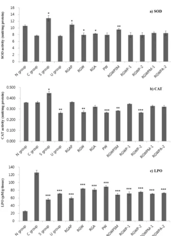
Change in activities of superoxide dismutase (SOD) and catalase (CAT) in liver tissue − Change in the activity of antioxidant enzyme superoxide dismutase (SOD) and catalase (CAT), which eliminate superoxide and hydrogen peroxide after induction of liver toxicity with CCl
4, is presented in Fig. 4. Activity of SOD decreased in C group (7.74 U/mg) with CCl
4-induced liver toxicity and no administration of test samples, while its activity increased in S and experimental groups.
Judging from the fact that administration of S group (13.01 U/mg) significantly increases CCl
4-induced SOD, the effect of silymarin was also recognized. No activation of SOD was confirmed in U group (7.88 U/mg), which showed lower activity than experimental groups. On the contrary, it was confirmed that the RGAP (11.32 U/mg), RGA (8.32 U/mg), and RGWPSM groups (9.81 U/mg) had significantly improved activity, with a level similar to that of the S group (p < 0.05), even though it still falls short of the level of the S group. Other groups had higher SOD activity than the U group, though their levels did not reach those of the S group. In most experimental groups, CAT activity recovered to the level of the N group (0.36 U/mg), which is equal to or higher than that of the U group (0.26 U/mg), though not significant.
Change in activity of lipid peroxidase (LPO) in liver tissue − Changes in lipid peroxidase (LPO) levels generated by toxic substances that attack cell membranes are presented in Fig. 4. Induction of toxicity was judged to be performed well as the activity of LPO increased in CCl
4- administered C group. In this study, however, LPO decreased more significantly in the S (56.01 μM/mg), U Fig. 4. Effect of Red ginseng and Pueraria radix on the activity
of superoxide dismutase (a; SOD), catalase (b; CAT), and lipid peroxidase (c; LPO) in CCl
4-treated ICR mice
1)
Refer to Table 1
2)