저작자표시-비영리-변경금지 2.0 대한민국 이용자는 아래의 조건을 따르는 경우에 한하여 자유롭게 l 이 저작물을 복제, 배포, 전송, 전시, 공연 및 방송할 수 있습니다. 다음과 같은 조건을 따라야 합니다: l 귀하는, 이 저작물의 재이용이나 배포의 경우, 이 저작물에 적용된 이용허락조건 을 명확하게 나타내어야 합니다. l 저작권자로부터 별도의 허가를 받으면 이러한 조건들은 적용되지 않습니다. 저작권법에 따른 이용자의 권리는 위의 내용에 의하여 영향을 받지 않습니다. 이것은 이용허락규약(Legal Code)을 이해하기 쉽게 요약한 것입니다. Disclaimer 저작자표시. 귀하는 원저작자를 표시하여야 합니다. 비영리. 귀하는 이 저작물을 영리 목적으로 이용할 수 없습니다. 변경금지. 귀하는 이 저작물을 개작, 변형 또는 가공할 수 없습니다.
Doctoral Thesis of Philosophy
Hepatoprotective effects of norgalanthamine on
carbon tetrachloride (CCl
4
)-induced liver injury
in mice
Department of Veterinary Medicine
GRADUATE SCHOOL
JEJU NATIONAL UNIVERSITY
Nayeon Yang
CONTENTS
List of Abbreviation ---1
List of Figures ---2
List of Tables ---4
General Introduction ---5
References ---16
1 Abstract ---27
2 Introduction ---29
3 Materials and methods ---
31
4 Results ---
42
5 Discussion ---
63
References ---
68
Abstract in Korean ---
77
List of Abbreviations
ALT Alanine aminotransferase ap2 Adipocyte protein-2
α-SMA Alpha smooth muscle actin AST Aspartate aminotransferase
CAT Catalase
CCl4 Carbon tetrachloride
DPCs Dermal papilla cells HO-1 Heme oxygenase-1
Iba-1 Ionized calcium-binding protein-1 MCP-1 monocyte chemoattractant protein-1
NG Norgalanthamime
Nrf-2 Nuclear factor erythroid 2-related factor 2
IL Interleukin
PPARγ Peroxisome proliferator-activated receptor gamma ROS Reactive oxygen species
SOD Superoxide dismutase TNF Tumor necrosis factor
List of Figures
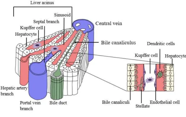
Figure 1. Three-dimensional structure of a liver lobule. --- 9
Figure 2. Pathobiochemical sequence of events during carbon
tetrachloride (CCl4)-induced liver damage --- 12
Figure 3. Schematic drawing of the experimental design --- 33
Figure 4. The effects of norgalanthamine on the serum levels of biochemical parameters in mice with CCl4-induced acute
liver injury --- 44
Figure 5. Protective effects of norgalanthamine on the histology of the liver in mice with CCl4-induced acute liver injury ---- 47 Figure 6. Oil red O staining of frozen sections of liver tissue from
treated and untreated mice reveals lipid accumulation --- 48
Figure 7. mRNA expression levels of CYPs in the liver --- 50 Figure 8. Antioxidant effect of norgalanthamine in mice with
CCl4-induced acute liver injury --- 52 Figure 9. Norgalanthamine pretreatment attenuates CCl4-induced
inflammatory responses in the liver at the transcriptional level --- 54
Figure 11. Real-time PCR analyses of PPAR-γ and aP2 expression in liver tissues --- 57
Figure 12. Representative immunoblots of Nrf-2 and HO-1 and Nrf-2 and HO-1 expression relative to the β-actin level in
CCl4-induced acute-liver-injury mice --- 59 Figure 13. Effects of norgalanthamine in CCl4-induced chronic liver
injury in mice --- 61
Figure 14. Real-time PCR analyses of αSMA and fibronectin
expression in liver tissues --- 62
Figure 15. Schematic diagram of the proposed molecular effects of norgalanthamine in mice with CCl4-induced liver injury -- 66
List of Tables
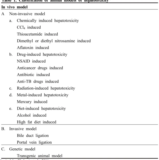
Table 1. Classification of animal models of hepatotoxicity --- 10
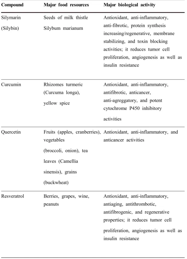
Table 2. Most frequently studied hepatoprotective phytochemicals --- 14
Table 3. Grading score for acute liver injury --- 36
Table 4. Primary antibodies used in the present study --- 38
General Introduction
Biological effects of Crinum asiaticum var. japonicum
Crinum asiaticum var. japonicum (Family: Amaryllidaceae), is an herbaceous
plant of small to moderate size. The plant is distributed on South Korea’s Jeju Island, and was designated Korea’s 19th Natural Treasure. Crinum asiaticum has been used as a rheumatic remedy and as an antipyretic and antiulcer agent, and for the alleviation of local pain and fever in Korea and Malaysia (Kim et al., 2006a).
Phytochemical studies on Crinum asiaticum var. japonicum have reported several phenanthridine alkaloids, triterpene alcohols, and flavonoids (Kim et al., 2006b). Alkaloids isolated from the bulbs of the tribe Amaryllidaceae have shown various pharmacological effects, such as anti-inflammatory activity (Kim et al., 2006a; Mahomoodally et al., 2020; Samud et al., 1999), antioxidant enzyme (Ghane et al., 2018; Goswami et al., 2020; Ilavenil et al., 2011; Indradevi et al., 2012; Mahomoodally et al., 2020), and anti-obesity activities (Jeong et al., 2016); prevention of hair loss and improvement of hair growth (Kang et al., 2017; Kim et
al., 2010; Yoon et al., 2019); cytotoxicity (Abdel-Halim et al., 2004; Likhitwitayawuid et al., 1993; Mahomoodally et al., 2020; Min et al., 2001; Weniger
et al., 1995); antiviral (Dominguez et al., 1992; Gabrielsen et al., 1992; Ieven et al.,
1979), antimalarial (Likhitwitayawuid et al., 1993), anti-platelet (Singh et al., 2011), and antineoplastic activities (Hyun et al., 2008; Mahomoodally et al., 2020; Pettit et
al., 1990); as well as effects on disease of the nervous system (Houghton et al.,
2004; Refaat J, 2013).
Amaryllidaceae alkaloids affect the central nervous system and have acetylcholinesterase-inhibitory, analgesic, anti-inflammatory, antiviral, antimalarial,
antitumor, or antineoplastic activity (Goswami et al., 2020; He et al., 2015; Hyun et
al., 2008; Kim et al., 2006b; Refaat J, 2013) .
The major components of this plant are crinamine from the aerial parts, together with lycorine, norgalanthamine, galanthamine, and epinorgalanthamine (Endo
et al., 2019; Mahomoodally et al., 2020; Park, 2000). Their medicinal properties
were appreciated after the discovery of pancratistatin as a promising
chemotherapeutic, as well as the commercialization of galanthamine for Alzheimer disease (AD) (Khumkhrong et al., 2019). Alkaloids isolated from the Amaryllidaceae family have a galanthamine-derived backbone and are potent acetylcholinesterase (AChE) inhibitors that have been used to treat the symptoms of AD (Lopez et al., 2002; Maelicke et al., 2001).
Crinamine has a potent anticancer effect in cervical cancer cells. Its cytotoxicity is selective to cervical cancer cell lines and it is more effective at inhibiting anchorage-independent tumor spheroid growth than several established chemotherapeutics. In addition, the compound activates DNA damage-independent apoptosis, reduces cell migration by downregulating key EMT inducers, and suppresses vascular endothelial growth factor-A secretion and in vivo angiogenesis (Khumkhrong et al., 2019). Crinamine inhibits nitric oxide production and induced
inducible nitric oxide synthase (iNOS) in lipopolysaccharide (LPS)-activated
macrophages (Kim et al., 2006b).
Lycorine is a natural alkaloid with immense therapeutic potential. Lycorine is active at a very low concentration and has high specificity against a number of cancers both in vivo and in vitro and against various drug-resistant cancer cells (Jiang and Liu, 2018; Liu et al., 2018; Roy et al., 2018; Shen et al., 2018; Wu et
protein synthesis was found in poliovirus-infected HeLa cells (Vrijsen et al., 1986). The main metabolite of lycorine degradation, ungeremine, and carbamate substitution at C-1 and C-2 of lycorine had stronger antibacterial activity toward the fish bacterial pathogen Flavobacterium columnare isolates than did lycorine. Lycorine shows significant inhibition of DNA topoisomerase-I activity, which is required for cell growth in parasites (Casu et al., 2011; Roy et al., 2018; Tan et al., 2011). The lycorine precursor norbelladine acts as an anti-inflammatory compound by inhibiting NF-kB signaling, which suppresses endplate-chondrocyte degeneration and prevents intervertebral disc degeneration (Wang et al., 2018). Degradation of acetylcholine (Ach) by AChE leads to brain cholinergic dysfunction in patients with AD. Lycorine also has analgesic, choleretic, and body-temperature lowering activity (Roy et al., 2018).
Norgalanthamine has hair-growth promoting effects, including increasing hair-fiber length in cultured rat vibrissa follicles and increasing dermal papilla cell (DPC) proliferation (Kim et al., 2010; Yoon et al., 2019). Norgalanthamine can stimulate the anagen phase of the hair cycle in DPCs via activation of the ERK 1/2, PI3K/AKT, and Wnt/β-catenin pathways (Yoon et al., 2019).
These studies revealed that a component norgalathamine possesses a variety of biological activity through cell activation signals. Little is known on the effect of norgalanthamine in oxidative stress induced in vivo models including CCl4 induced
liver injury. The biological effects of norgalathamine remains to be further studied.
Liver is one of the most important metabolic organs
multiple functions. It receives oxygenated blood from the heart via the hepatic artery, and nutrient-rich blood from the gastrointestinal tract via the portal vein (Malarkey et
al., 2005). The blood flows through liver sinusoids (terminal vessels between
hepatocyte cords and lined with Kupffer cells and endothelial cells), empties into the central vein, and exits the liver via the hepatic veins (Hollander et al., 1987). Hepatocytes make up 70–85% of all liver cells (Baratta et al., 2009). They are key functional cells with important metabolic, secretory, and endocrine functions (Treuting
et al., 2017). Kupffer cells, another type of liver cell, are specialized macrophages on
the walls of the liver sinusoids (Baratta et al., 2009). Hepatic stellate cells (HSCs) are pericytes in the space of Disse, which is located between the hepatocytes and the sinusoidal endothelium (Friedman, 2008). Quiescent HSCs can be activated in response to liver damage, leading to collagen formation, fibrosis, or cirrhosis (Friedman, 2008; Li et al., 2018; Solis-Herruzo et al., 2003; Zhao et al., 2014). Bile is secreted by hepatocytes, and drains into biliary ductules, which are lined with epithelial cells, and leaves the liver via the bile duct (Wang et al., 2017).
Animal models of hepatotoxicity
The liver filters toxic substances from the body. Hepatic damage may occur when accumulation of toxins is faster than the ability of the liver to remove them (Bigoniya P. et al., 2009). Hepatotoxicity may imply chemically driven liver damage. Some medical agents when taken at very high doses but also in therapeutic ranges can injure the liver (McGill and Jaeschke, 2019). Chemicals that cause liver injury are termed hepatotoxins (Bhakuni et al., 2016). Several other factors may also cause hepatotoxicity, including herbal remedies, viral infections, autoimmune disorders, intoxication, and an imbalanced diet, among others (Pandit et al., 2012).
Figure 1. Three-dimensional structure of a liver lobule. Blood in the branches of the hepatic artery and portal vein enters the sinusoids, between the cords of liver cells, and courses toward the central vein, which is a tributary of the hepatic vein. Bile flows in the opposite direction, from the center out, toward the tributaries of the bile duct. Bile canaliculi are tiny channels which exist between the cell surfaces of neighboring hepatic parenchymal cells. They are supported by a delicate connective tissue stroma. Hepatic sinusoids are lined by two cell types: (1) discontinuous endothelial cells and (2) phogocytic cells of kupffer. Modified from Springer Nature Publishing AG, Adams et al., Nat. Rev. Immunol., 2006.
1) cited by Bhakuni et al., 2016
Table 1. Classification of animal models of hepatotoxicity1) In vivo model
A Non-invasive model
a. Chemically induced hepatotoxicity
CCl4 induced
Thioacetamide induced
Dimethyl or diethyl nitrosamine induced Aflatoxin induced
b. Drug-induced hepatotoxicity
NSAID induced
Anticancer drugs induced Antibiotic induced
Anti-TB drugs induced
c. Radiation-induced hepatotoxicity
d. Metal-induced hepatotoxicity Mercury induced
e. Diet-induced hepatotoxicity Alcohol induced
High fat diet induced
B. Invasive model
Bile duct ligation Portal vein ligation
C. Genetic model
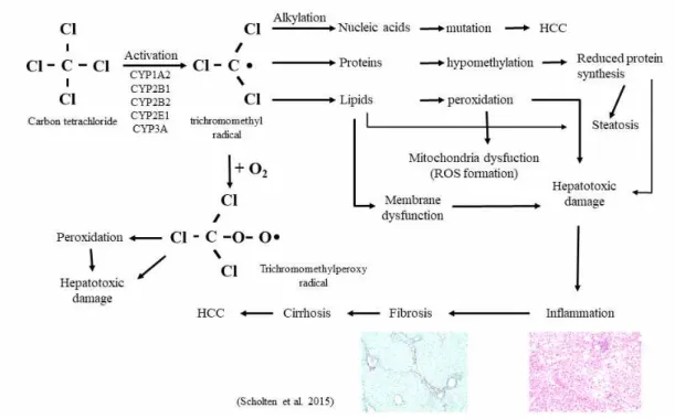
Mechanisms of carbon tetrachloride-induced liver injury
Carbon tetrachloride is an inorganic compound with the chemical formula CCl4.
It is a clear liquid that evaporates easily and has a sweet odor. It is a common industrial solvent and is hepatotoxic (Abraham et al., 1999). It is the most widely used hepatic toxicant in studies of liver injury involving laboratory animals (Bhakuni
et al., 2016). CCl4 is metabolized in the liver by the cytochrome P450 superfamily
of monooxygenases (CYP family) to the trichloromethyl radical (CCl3*). Subsequently,
this radical reacts with nucleic acids, proteins, and lipids, thereby impairing key cellular processes and resulting in altered lipid metabolism (fatty degeneration and steatosis) and lower levels of proteins. Adduct formation between CCl3* and DNA
triggers mutations and the formation of HCC. The formation of trichloromethylperoxy radicals (CCl3OO*) by oxygenation of CCl3* initiates lipid peroxidation and the
destruction of polyunsaturated fatty acids. Consequently, the membrane permeability in
all cellular compartments (mitochondria, endoplasmic reticulum, and plasma
membrane) is lowered and generalized hepatic damage occurs, characterized by inflammation, fibrosis, cirrhosis, and HCC. A comprehensive summary of the pathogenic events that occur in the liver during CCl4-induced damage is given
elsewhere. Besides its hepatotoxicity, minor acute systemic toxicity of CCl4 has been
described, particularly in the peritoneum, mucosa, respiratory tract, and central nervous system (Brattin et al., 1985; Recknagel et al., 1989; Scholten et al., 2015; Starkel and Leclercq, 2011).
Figure 2. Pathobiochemical sequence of events during carbon tetrachloride (CCl4)-induced liver damage. In the liver, CCl4 is metabolized by cytochrome P450
(CYP) enzymes to a trichloromethyl radical that can be further oxygenated to the trichloromethylperoxy radical. Both radicals are highly reactive and induce complex cellular alterations that result in hepatotoxic damage, inflammation, fibrosis, cirrhosis and hepatocellular carcinoma (HCC). Modified from Scholten et al., 2015.
Hepatoprotective effects of medicinal plants
There are approximately 75,000 higher-plant species, about 10% of which have been used in traditional remedies. However, perhaps only about 1% of these have been recognized through scientific studies to have therapeutic value when used in extract form. One of the most common causes of liver disease is inflammation, often resulting from abuse of alcohol, poor diet, or malnutrition. Drug-induced liver damage or liver dysfunction is an important public-health challenge. According to the United States Acute Liver Failure Study Group, drug-induced liver injury accounts for more than 50% of acute liver failure, including hepatotoxicity caused by overdose of acetaminophen (39%) and idiosyncratic liver injury triggered by other drugs. Hepatic cell injury is caused by various toxic chemicals (certain antibiotics, chemotherapeutic agents, carbon tetrachloride, and thioacetamide), excessive alcohol consumption, and microbes. It is clear that medicinal plants play an important role in a variety of diseases. Different medicinal herbs and plants extracts have potent hepatoprotective activity in animal models. Extracts of leaves and some medicinal plants have therapeutic potential for hepatic diseases (Roy et al., 2014). Use of substances from natural sources for phytotherapy of various diseases, including liver diseases, is desirable because they are relatively inexpensive and widely available. Silymarin and resveratrol are two of many examples of natural substances with marked hepatoprotective potential as a result of their antioxidant, anti-inflammatory, and liver-regenerative capabilities (Farghali et al., 2015; Shakya, 2020).
The CCl4-injured liver has been used to test the efficacy of anti-inflammatory
Table 2. Most frequently studied hepatoprotective phytochemicals2)
Compound Major food resources Major biological activity
Silymarin (Silybin)
Seeds of milk thistle Silybum marianum
Antioxidant, anti-inflammatory, anti-fibrotic, protein synthesis increasing/regenerative, membrane stabilizing, and toxin blocking activities; it reduces tumor cell proliferation, angiogenesis as well as insulin resistance
Curcumin Rhizomes turmeric
(Curcuma longa), yellow spice
Antioxidant, anti-inflammatory, antifibrotic, anticancer,
anti-agreggatory, and potent cytochrome P450 inhibitory activities
Quercetin Fruits (apples, cranberries),
vegetables
(broccoli, onion), tea leaves (Camellia sinensis), grains (buckwheat)
Antioxidant, anti-inflammatory, and anticancer activities
Resveratrol Berries, grapes, wine,
peanuts
Antioxidant, anti-inflammatory, antiaging, antithrombotic, antifibrogenic, and regenerative properties; it reduces tumor cell proliferation, angiogenesis as well as insulin resistance
2) cited by Farghali et al., 2015; Shakya, 2020
Glycyrrhizin Root of Indian licorice
(Glycyrrhiza glabra)
Antioxidant, anti-inflammatory, anticancer, antiviral, and immunomodulatory activities
Colchicine Plants of the genus
Colchicum (autumn crocus, Colchicum autumnale)
Anti-inflammatory, antifibrotic, and immunomodulatory activities
References
Abdel-Halim, O.B., Morikawa, T., Ando, S., Matsuda, H., Yohikawa, M., 2004. New crinine-type alkaloids with inhibitory effect on induction of inducible nitric oxide synthase from Crinum yemense. J Nat Prod 67(7), 1119-1124.
Abraham, P., Wilfred, G., Cathrine, 1999. Oxidative damage to the lipids and proteins pf the lungs, testis and kidney of rats during carbon tetrachloride intoxication. Clin Chim Acta 289(1-2), 177-179.
Adams, D.H., Eksteen, B., 2006. Aberrant homing of mucosal T cells and extra-intestinal manifestations of inflammatory bowel disease. Nat Rev Immunol 6(3), 244-251.
Baratta, J.L., Ngo, A., Lopez, B., Kasabwalla, N., Longmuir, K.J., Robertson, R.T., 2009. Cellular organization of normal mouse liver: a histological, quantitative immunocytochemical, and fine structural analysis. Histochem Cell Biol 131(6), 713-726.
Bhakuni, G.S., Bedi, O., Bariwal, J., Deshmukh, R., Kumar, P., 2016. Animal models of hepatotoxicity. Inflamm Res 65(1), 13-24.
Bigoniya, P., Singh, C.S., Shukla, A., 2009. A Comprehensive Review of Different Liver Toxicants Used in Experimental Pharmacology. IJPSDR 1(3), 124-135.
Brattin, W.J., Glende Jr, E.A., Recknagel, R.O., 1985. Pathological mechanisms in carbon tetrachloride hepatotoxicity. J Free Radic Biol Med 1(1), 27-38.
Casu, L., Cottiglia, F., Leonti, M., De Logu, A., Agus, E., Tse-Dinh, Y.C., Lombardo, V., Sissi, C., 2011. Ungeremine effectively targets mammalian as well as bacterial type I and type II topoisomerases. Bioorg Med Chem Lett 21(23), 7041-7044.
Dominguez, X.A., B, G.E., Rombold, C., Utz, W., Achenbach, H., 1992. Neolignans, Norneolignans, and Other Components from Krameria sonorae1. Planta Med 58(4), 382-383.
Endo, Y., Sugiura, Y., Funasaki, M., Kagechika, H., Ishibashi, M., Ohsaki, A., 2019. Two new alkaloids from Crinum asiaticum var. japonicum. J Nat Med 73(3), 648-652.
Farghali, H., Canova, N.K., Zakhari, S., 2015. Hepatoprotective properties of extensively studied medicinal plant active constituents: possible common mechanisms. Pharm Biol 53(6), 781-791.
Friedman, S.L., 2008. Hepatic stellate cells: protean, multifunctional, and enigmatic cells of the liver. Physiol Rev 88(1), 125-172.
Gabrielsen, B., Monath, T.P., Huggins, J.W., Kefauver, D.F., Pettit, G.R., Groszek, G., Hollingshead, M., Kirsi, J.J., Shannon, W.M., Schubert, E.M., et al., 1992.
Antiviral (RNA) activity of selected Amaryllidaceae isoquinoline constituents and synthesis of related substances. J Nat Prod 55(11), 1569-1581.
Ghane, SG., Attar, UA., Yadav, PB., Lekhak, MM., 2018. Antioxidant, anti-diabetic, acetylcholinesterase inhibitory potential and estimation of alkaloids (lycorine and galanthamine) from Crinum species: An important source of anticancer and anti-Alzheimer drug. Ind Crops Prod 125, 168-177.
Goswami, S., Das, R., Ghosh, P., Chakraborty, T., Barman, A., Ray, S., 2020. Comparative antioxidant and antimicrobial potentials of leaf successive extract fractions of poison bulb, Crinum asiaticum L. Ind Crops Prod 154, 112667.
He, M., Qu, C., Gao, O., Hu, X., Hong, X., 2015. Biological and pharmacological activities of amaryllidaceae alkaloids. RSC Adv 5(21), 16562-16574.
Hollander, C.F., van Bezooijen, C.F., Solleveld, H.A., 1987. Anatomy, function and aging in the mouse liver. Arch Toxicol Suppl 10, 244-250.
Houghton, P.J., Agbedahunsi, J.M., Adegbulugbe, A., 2004. Choline esterase inhibitory properties of alkaloids from two Nigerian Crinum species. Phytochemistry 65(21), 2893-2896.
Hyun, J.H., Kang, J.I., Kim, S.C., Kim, E., Kang, J.H., Kwon, J.M., Park, D.B., Lee, Y.J., Yoo, E.S., Kang, H.K., 2008. The Effects of Crinum asiaticum on the Apoptosis Induction and the Reversal of Multidrug Resistance in HL-60/MX2. Toxicol Res 24(1), 29-36.
Ieven, M., Vanden Berghe, D.A., Mertens, F., Vlietinck, A., Lammens, E., 1979. Screening of higher plants for biological activities. I. Antimicrobial activity. Planta Med 36(4), 311-321.
Ilavenil, S., Kaleeswaran, B., Sumitha, P., Tamilvendan, D., Ravikumar, S., 2011. Protection of human erythrocyte using Crinum asiaticum extract and lycorine from oxidative damage induced by 2-amidinopropane. Saudi J Biol Sci 18(2), 181-187.
Indradevi, S., Ilavenil, S., Kaleeswaran, B., Srigopalram, S., Ravikumar, S., 2012. Ethanolic extract of Crinum asiaticum attenuates hyperglycemia-mediated oxidative stress and protects hepatocytes in alloxan induced experimental diabetic rats. J King Saud Univ Sci 24(2), 171-177.
Jeong, Y.J., Sohn, E.H., Jung, Y.H., Yoon, W.J., Cho, Y.M., Kim, I., Lee, S.R., Kang, S.C., 2016. Anti-obesity effect of Crinum asiaticum var. japonicum Baker extract in high-fat diet-induced and monogenic obese mice. Biomed Pharmacother 82, 35-43.
Jiang, Q.Q., Liu, W.B., 2018. Lycorine inhibits melanoma A375 cell growth and metastasis through the inactivation of the PI3K/AKT signaling pathway. Med Sci (Paris) 34 Focus issue F1, 33-38.
Kang, J.I., Choi, J.H., Lee, J.G., Yoo, E.S., Kim, Y.H., Kang, H.K., 2017. The Mechanism of Crinum asiaticum var. japonicum on the Activation of Anagen. Kor J Pharmacogn 48(2), 148-154.
Khumkhrong, P., Piboonprai, K., Chaichompoo, W., Pimtong, W., Khongkow, M., Namdee, K., Jantimaporn, A., Japrung, D., Asawapirom, U., Suksamrarn, A., Iempridee, T., 2019. Crinamine Induces Apoptosis and Inhibits Proliferation, Migration, and Angiogenesis in Cervical Cancer SiHa Cells. Biomolecules 9(9), 494.
Kim, S.C., Kang, J.I., Kim, M.K., Hyun, J.H., Boo, H.J., Park, D.B., Lee, Y.J., Yoo, E.S., Kim, Y.H., Kim, Y.H, Kang, H.K., 2010. Promotion effect of norgalanthamine, a component of Crinum asiaticum, on hair growth. Eur J Dermatol 20(1), 42-48.
Kim, Y.H., Kim, K.S., Park, S.H., Lee, S.H., Kim, Y.J., Kim, Y.S, Kim, J.H., Kim, K.H., 2006a. Anti-inflammatory Activity of Crinum asiaticum Linne var.
japonicum Extract and its Application as a Cosmeceutical Ingredient. J Soc
Cosmet Sci Korea 32(1), 59-64.
Kim, Y.H., Park, E.J., Park, M.H., Badarch, U., Woldemichael, G.M., Beutler, J.A., 2006b. Crinamine from Crinum asiaticum var. japonicum Inhibits Hypoxia Inducible Factor-1 Activity But Not Activity of Hypoxia Inducible Factor-2. Biol Pharm Bull 29(10), 2140-2142.
Li, Y., Zhu, M., Huo, Y., Zhang, X., Liao, M., 2018. Anti-fibrosis activity of combination therapy with epigallocatechin gallate, taurine and genistein by regulating glycolysis, gluconeogenesis, and ribosomal and lysosomal signaling pathways in HSC-T6 cells. Exp Ther Med 16(6), 4329-4338.
Likhitwitayawuid, K., Angerhofer, C.K., Chai, H., Pezzuto, J.M., Cordell, G.A., Ruangrungsi, N., 1993. Cytotoxic and antimalarial alkaloids from the bulbs of
Crinum amabile. J Nat Prod 56(8), 1331-1338.
Liu, W., Zhang, Q., Tang, Q., Hu, C., Huang, J., Liu, Y., Lu, Y., Wang, Q., Li, G., Zhang, R., 2018. Lycorine inhibits cell proliferation and migration by inhibiting ROCK1/cofilin-induced actin dynamics in HepG2 hepatoblastoma cells. Oncol Rep 40(4), 2298-2306.
Lopez, S., Bastida, J., Viladomat, F., Codina, C., 2002. Acetylcholinesterase inhibitory activity of some Amaryllidaceae alkaloids and Narcissus extracts. Life Sci 71(21), 2521-2529.
Maelicke, A., Samochocki, M., Jostock, R., Fehrenbacher, A., Ludwig, J., Albuquerque, E.X., Zerlin, M., 2001. Allosteric sensitization of nicotinic receptors by galantamine, a new treatment strategy for Alzheimer's disease. Biol Psychiatry 49(3), 279-288.
Mahomoodally, M.F., Sadeer, N.B., Suroowan, S., Jugreet, S., Lobine, D., Rengasamy, K.R.R., 2020. Ethnomedicinal, phytochemistry, toxicity and pharmacological benefits of poison bulb – Crinum asiaticum L. S Afr J Bot 00, 1-14.
Malarkey, D.E., Johnson, K., Ryan, L., Boorman, G., Maronpot, R.R., 2005. New insights into functional aspects of liver morphology. Toxicol Pathol 33(1), 27-34.
McGill, M.R., Jaeschke, H., 2019. Animal models of drug-induced liver injury. Biochim Biophys Acta Mol Basis Dis 1865(5), 1031-1039.
Min, B.S., Gao, J.J., Nakamura, N., Kim, Y.H., Hattori, M., 2001. Cytotoxic alkaloids and a flavan from the bulbs of Crinum asiaticum var. japonicum. Chem Pharm Bull (Tokyo) 49(9), 1217-1219.
Pandit, A., Sachdeva, T., Bafna, P., 2012. Drug-Induced Hepatotoxicity: A Review. J Appl Pharm Sci 02(05), 233-243.
Park, M.H., 2000. Chemical Constituents and Biological Activity of Crinum asiaticum var. japonicum. Graduated School of Chungnam National University for the degree of Master of Pharmaceutical Science.
Pettit, G.R., Cragg, G.M., Singh, S.B., Duke, J.A., Doubek, D.L., 1990. Antineoplastic agents, 162. Zephyranthes candida. J Nat Prod 53(1), 176-178.
Recknagel, R.O., Glende, E.A., Dolak, J.A., Waller, R.L., 1989. Mechanisms of carbon tetrachloride toxicity. Pharmacol Ther 43(1), 139-154.
Refaat J, M.S.K., Mahmoud A.R., Ahmed A.A., 2013. Crinum; An endless source of bioactive principles; A review. Part V. Biological Profile. IJPSR 4(4), 1239-1252.
Roy, A., Bhoumik, D., Sahu, R.K., Dwivedi, J., 2014. Medicinal plants used in liver protection-a review. UK J Pharm Biosci 2(1), 23-33.
Roy, M., Liang, L., Xiao, X., Feng, P., Ye, M., Liu, J., 2018. Lycorine: A prospective natural lead for anticancer drug discovery. Biomed Pharmacother 107, 615-624.
Samud, A.M., Asmawi, M.Z., Sharma, J.N., Yusof, A.P., 1999. Anti-inflammatory activity of Crinum asiaticum plant and its effect on bradykinin-induced contractions on isolated uterus. Immunopharmacology 43(2-3), 311-316.
Scholten, D., Trebicka, J., Liedtke, C., Weiskirchen, R., 2015. The carbon tetrachloride model in mice. Lab Anim 49(S1), 4-11.
Shakya, A.K., 2020. Drug-induced Hepatotoxicity and Hepatoprotective Medicinal Plants: A Review. Indian J Pharm Educ Res 54(2), 234-250.
Shen, J., Zhang, T., Cheng, Z., Zhu, N., Wang, H., Lin, L., Wang, Z., Yi, H., Hu, M., 2018. Lycorine inhibits glioblastoma multiforme growth through EGFR suppression. J Exp Clin Cancer Res 37(1), 157.
Singh, K.A., Nayak, M.K., Jagannadham, M.V., Dash, D., 2011. Thrombolytic along with anti-platelet activity of crinumin, a protein constituent of Crinum
Solis-Herruzo, J.A., De La Torre, P., Munoz-Yague, M.T., 2003. Hepatic stellate cells (HSC): architects of hepatic fibrosis. Rev Esp Enferm Dig 95(6), 438-439.
Starkel, P., Leclercq, I.A., 2011. Animal models for the study of hepatic fibrosis. Best Pract Res Clin Gastroenterol 25(2), 319-333.
Tan, C.X., Schrader, K.K., Mizuno, C.S., Rimando, A.M., 2011. Activity of lycorine analogues against the fish bacterial pathogen Flavobacterium columnare. J Agric Food Chem 59(11), 5977-5985.
Treuting, P.M., Dintzis, S.M., Montine, K.S., 2017. Comparative anatomy and histology: a mouse, rat, and human atlas. Academic Press Second Edition, 229-239.
Vrijsen, R., Berghe, D.V., Vlietinck, A. Boeye, A., 1986. Lycorine: a eukaryotic termination inhibitor? J Biol Chem 261(2), 505-507.
Wang, G., Huang, K., Dong, Y., Chen, S., Zhang, J., Wang, J., Xie, Z., Lin, X.,
Fang, X., Fan, S., 2018. Lycorine Suppresses Endplate-Chondrocyte
Degeneration and Prevents Intervertebral Disc Degeneration by Inhibiting NF-kappaB Signalling Pathway. Cell Physiol Biochem 45(3), 1252-1269.
Wang, H., Liang, X., Gravot, G., Thorling, C., Crawford, D., Xu, Z., Liu, X., Roberts, M., 2017. Visualizing liver anatomy, physiology and pharmacology using multiphoton microscopy. J Biophotonics 10(1), 46-60.
Weniger, B., Italiano, L., Beck, J.P., Bastida, J., Bergonon, S., Codina, C., Lobstein, A., Anto, R., 1995. Cytotoxic activity of Amaryllidaceae alkaloids. Planta Med 61(1), 77-79.
Wu, S., Qiu, Y., Shao, Y., Yin, S., Wang, R., Pang, X., Ma, J., Zhang, C., Wu, B., Koo, S., Han, L., Zhang, Y, Gao, X., Wang, T., Yu, H., 2018. Lycorine Displays Potent Antitumor Efficacy in Colon Carcinoma by Targeting STAT3. Front Pharmacol 9, 881.
Yoon, H.S., Kagn, J.I., Kim, S.M., Ko, A., Koh, Y.S., Hyun, J.W., Yoon, S.P., Ahn, M., Kim, Y.H., Kang, J.H., 2019. Norgalanthamine stimulates proliferation of dermal papilla cells via anagen-activating signaling pathways. Biol Pharm Bull 42(1), 139-143.
Zhao, J., Tang, N., Wu, K., Dai, W., Ye, C., Shi, J., Zhang, J., Ning, B., Zeng, X., Lin, Y., 2014. MiR-21 simultaneously regulates ERK1 signaling in HSC activation and hepatocyte EMT in hepatic fibrosis. PLoS One 9(10), e108005, 1-10.
Hepatoprotective effects of norgalanthamine on
carbon tetrachloride (CCl
4
)-induced liver injury
1. Abstract
Norgalanthamine is a major component of Crinum asiaticum var.
japonicum that exhibits several biological activities. This study evaluated the
anti-inflammatory and anti-oxidative properties of norgalanthamine in mice with carbon tetrachloride (CCl4)-induced acute liver injury. Norgalanthamine (1
and 10 mg/kg) was orally administered to mice for 7 days, after which liver injury was induced by CCl4 (1.5 ml/kg, i.p.). The vehicle and positive
controls consisted of phosphate-buffered saline and silymarin (100 mg/kg), respectively. The mice were euthanized 24 h after CCl4 administration.
In CCl4-injured mice, norgalanthamine pretreatment significantly reversed
the increases in serum alanine aminotransferase, aspartate aminotransferase, and total bilirubin levels, and the decrease in the serum glucose level. In the liver, norgalanthamine restored the activities of the antioxidant enzymes superoxide dismutase and catalase, while reducing lipid accumulation and, concurrently, the expression of genes involved in lipid synthesis, including peroxisome proliferator-activated receptor γ and adipocyte protein-2. Norgalanthamine also ameliorated inflammation by down-regulating the expression of the pro-inflammatory mediators TNF-α, IL-1β, and MCP-1, and up-regulating the Nrf2/HO-1 pathway. The hepatoprotective effect of norgalanthamine in CCl4-injured mice was also reflected in the histopathologic scores. In addition,
norgalanthamine decreased collagen deposition in liver tissue as shown on picrosirius red staining by down-regulating expression of the fibrosis-related genes αSMA and fibronectin. The findings indicate that norgalanthamine attenuates liver fibrosis and could be a novel approach in its prevention.
Collectively, these findings imply that norgalanthamine mitigates CCl4-induced
hepatic injury by increasing anti-oxidative activity, down-regulating pro-inflammatory mediators and fibrosis-related genes in the liver.
2. Introduction
Carbon tetrachloride (CCl4) is a hepatotoxic substance that is used in animal
models of liver injury characterized by centrilobular hepatic necrosis (Ahn et al., 2014; Cao et al., 2017; Li et al., 2016; Rahman and Hodgson, 2000; Recknagel et
al., 1989), as well as pathophysiological changes, fatty changes (Unsal et al., 2020),
inflammation, and fibrosis (Scholten et al., 2015; Unsal et al., 2020). CCl4 injury
results from direct damage to tissue after injection and metabolism. CCl4 is converted
to trichloromethyl radicals (CCl3*) by cytochrome P450 (CYP), especially CYP2E1,
CYP2B1, CYP2B2, and CYP3A (Kim et al., 2010; Scholten et al., 2015). The CCl4
model of liver injury has been widely used to evaluate the therapeutic potential of drugs aimed at treating liver disease, and to study xenobiotic-induced liver injury, which resembles human liver disease both morphologically and in terms of the biochemical features of the cellular lesions(McGill and Jaeschke, 2019; Scholten et
al., 2015). CCl4 induces hepatotoxicity after metabolite activation in the liver (McGill
and Jaeschke, 2019; Scholten et al., 2015). The initial response to CCl4 consists of
the activation of hepatic macrophages, including Kupffer cells, the generation of free radicals, and the production of inflammatory mediators, including the cytokine tumor necrosis factor-alpha (TNF-α) and interleukins (ILs) (Bansal et al., 2005; Decker, 1990; Munakarmi et al., 2020; Zou et al., 2016), chemokines such as monocyte chemoattractant protein (MCP)-1 (Russmann et al., 2009), and pro-inflammatory mediators such as inducible nitric oxide synthase and cyclooxygenase, via the activation of nuclear factor kappa B (Liu et al., 2018a; Wu et al., 2018). Therefore, the CCl4-injured liver has been used to test the efficacy of anti-inflammatory agents
(Badger et al., 1996; Liu et al., 2018b; Ma et al., 2015; Rocha et al., 2014; Son et
al., 2007; Tipoe et al., 2010), with a particular focus on therapies based on a single
compound (Ahn et al., 2014; Hansen et al., 2017; Kim et al., 2010; Munakarmi et
Norgalanthamine, a major component of Crinum asiaticum var. japonicum, is a metabolite of galanthamine, a selective acetylcholinesterase inhibitor that promotes hair growth via the proliferation of dermal papilla, activation of the ERK 1/2, PI3K/AKT, and Wnt/β-catenin pathways (Yoon et al., 2019), and inhibition of 5α -reductase activity and of the TGF-β1-induced canonical pathway (Kang et al., 2017). In this study, we investigated the hepatoprotective effects of norgalanthamine in the context of its effects on biochemical indicators of liver damage, the activities of oxidative stress enzymes, and the levels of inflammatory mediators.
3. MATERIALS AND METHODS
Chemical and regents
Norgalanthamine (D292035) was obtained from Toronto Research Chemicals (Toronto, Canada). Silymarin, used as a positive control, was purchased from Sigma-Aldrich (St. Louis, MO, USA). Commercial colorimetric assay kits for the measurement of superoxide dismutase (SOD) and catalase (CAT) activity were purchased from Abcam (Cambridge, UK). Polyclonal antibodies targeting Nrf-2 and HO-1 were purchased from Cell Signaling Technology (Beverly, MA, USA), and ionized calcium binding adapter molecule 1 (Iba-1) was provided by Wako Pure Chemical Industries, Ltd. (Osaka, Japan). The ABC Elite kit and diaminobenzidine (DAB) substrate were purchased from Vector Laboratories (Burlingame, CA, USA).
Animals
Six-week-old male C57BL/6 mice weighing 20–22 g (DBL, Chungbuk, Korea) were used for all experiments. The animals were maintained at a controlled temperature of 25–28℃ with a 12-h light/dark cycle, fed a standard diet and provided water ad libitum. All experimental procedures were conducted in accordance with the guidelines for the Care and Use of Laboratory Animals of Sangji University in Wonju City, Korea (permit number: 2020-11). The animal protocols conformed to current international laws and to the policies of the National Institutes of Health (NIH) Guide for the Care and Use of Laboratory Animals (NIH Publication no. 85– 23, 1985, revised 1996). Every effort was made to minimize the number of animals used in this study and to reduce their pain and suffering.
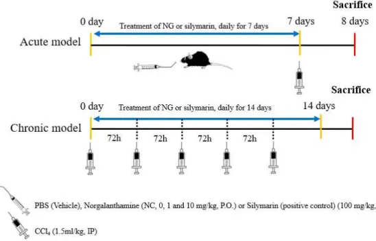
Experimental design
In the acute model, the mice were randomly divided into five groups of six animals each. The norgalanthamine-treated CCl4-injured group was orally administered
norgalanthamine (1 or 10 mg/mL [NG1 and NG10, respectively], diluted in phosphate-buffered saline [PBS], pH 7.4) for 7 consecutive days. In both the acute and chronic models, PBS, which served as the vehicle control and silymarin served as the positive control (100 mg/kg; (Ni and Wang, 2016)). Both were administered following the same protocol. Liver injury was induced with a 1:1 (v/v) mixture of CCl4 and sterile olive oil injected intraperitoneally (i.p., 1.5 mL/kg). CCl4 was given
once 24 h after the last dose of test substance. In the chronic model, the mice were randomly divided into four groups of six animals each. PBS (pH 7.4) was administered orally to normal controls. The norgalanthamine-treated CCl4-injured group
was orally administered norgalanthamine (10 mg/mL [NG10], diluted in PBS, pH 7.4) for 14 consecutive days. To induce liver injury, a 1:1 (v/v) mixture of CCl4 and
sterile olive oil was injected intraperitoneally (i.p., 1.5 mL/kg). CCl4 was given every
Figure 3. Shematic drawing of the experimental design used to evaluate the effects of norgalanthamine on CCl4-induced liver injury in mice. Acute model (top), the
norgalanthamine-treated CCl4-injured group was orally administered norgalanthamine (1
or 10 mg/mL [NG1 and NG10, respectively], diluted in phosphate-buffered saline [PBS], pH 7.4) for 7 consecutive days. CCl4 was given once (i.p., 1.5 mL/kg) 24 h
after the last dose of test substance. Chronic model (bottom), the
norgalanthamine-treated CCl4-injured group was orally administered norgalanthamine
(10 mg/mL [NG10], diluted in PBS, pH 7.4) for 14 consecutive days. CCl4 was
given every 72 h for 14 days (i.p., 1.5 mL/kg). In both the acute and chronic models, PBS, which served as the vehicle control and silymarin served as the positive control (100 mg/kg).
Sample collection and blood biochemistry analysis
The mice were fasted after treatment and euthanized 24 h after induction of CCl4 injury in the acute model, or the last dose of the drug in the chronic model.
After anesthetization of the mice by isoflurane inhalation (Hana Pharm Co., Ltd, Seoul, Korea), blood was collected from the inferior vena cava for serum analysis the liver was dissected for histopathology, gene expression, and immunochemistry studies (Fig. 3). Liver pieces were fixed in 10% neutral buffered formalin for histopathology or immediately frozen and stored for RNA extraction. Blood samples were centrifuged at 13,000 rpm for 10 min at 4℃ to separate the serum. The serum levels of alanine aminotransferase (ALT), aspartate aminotransferase (AST), total bilirubin (T-bil), and glucose were measured using a Beckman Coulter AU680 analyzer (Beckman Coulter, Tokyo, Japan) according to the manufacturer’s instructions.
Histopathological examination of liver damage
Sections (4 µm) of paraffin-embedded liver were deparaffinized and then stained with hematoxylin and eosin (H&E) for light microscopy examination. The degree of necrosis after acute liver injury was evaluated using an injury score based on the severity of necrotic lesions in the liver parenchyma (Table 3). In brief, each sample was independently scored by three pathologists blinded to the group assignment. The scoring system was as follows: grade 0, no pathological change; grade 1, degenerated hepatocytes with rare foci of necrosis; grade 2, small areas of mild centrilobular necrosis around the central vein; grade 3, mild centrilobular necrosis, but more severe than grade 2; grade 4, centrilobular necrosis more severe than grade 3 (Dai et al., 2018; Li et al., 2013).
Necrotic areas were evaluated in paraffin-embedded sections stained with toluidine blue, and the severity of fatty changes was assessed in fixed frozen sections stained with oil red O, which detects neutral triglycerides and lipids. Picrosirius red (Polysciences, Inc., PA, USA) staining was used for fibrillary collagen . The proportions of lipid (red-stained area as a percentage of the total area of the liver section), necrotic (light-blue-stained area as a percentage of the total area of the liver section) area, and collagen deposition (red-stained area as a percentage of the total area of the liver section) were determined using Aperio eSlide Manager software (Leica Biosystems, Buffalo Grove, IL, USA).
Table 3. Grading score for Acute liver injury3)
3) cited by Dai et al., 2018; Li et al., 2013
Grade Criteria
0 No pathological change
1 Presence of degenerated hepatocytes with only rare foci of necrosis
2 Small area of mild centrilobular necrosis a round the central vein
3 Area of mild centrilobular necrosis severer than Grade 2
Assays of antioxidant enzyme activities in liver
Liver pieces from the mice were immediately frozen until use. The tissue was then homogenized in a pestle homogenizer, and SOD and CAT activities were determined according to the instructions supplied with the commercial assay kits (Abcam).
Western blot analysis
Protein expression was analyzed quantitatively by western blot, performed as described in a previous study (Kim et al., 2017). The cytosolic and nuclear fractions were separated using NE-PER® nuclear and cytoplasmic extraction reagents, as recommended by the manufacturer (Thermo Scientific, Rockford, IL, USA). Proteins (40 μg/sample) were subjected to 10% (w/v) sodium dodecyl sulfate or sodium lauryl sulfate polyacrylamide gel electrophoresis (SDS- and SLS-PAGE, respectively) and transferred to a nitrocellulose membrane (Schleicher and Schuell, Keene, NH, USA).
Target proteins were immunodetected by incubation of the membrane for 2 h with specific primary antibodies (Table 4). The bound proteins were detected using a chemiluminescent substrate (Miracle-Star; iNtRON Biotech, Gyeonggi, Korea). The blots were then imaged and the densities of the bands were analyzed using ImageJ software (NIH, Bethesda, MD, USA). β-actin served as the internal control.
The optical density (OD; per mm2) of each band was measured and the
density ratios relative to the β-actin band were determined using ImageJ. The data are presented as the means ± standard error of the mean (SEM).
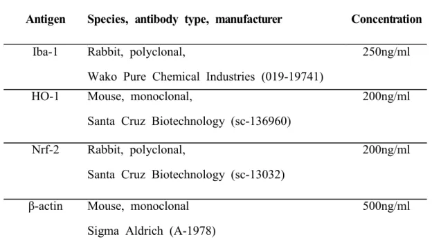
Table 4. Primary antibodies used in the present study
Abbreviations: HO-1, hemeoxygenase-1; Iba-1, Ionized calcium binding protein-1; IgG, immunoglobulin;.Nrf-2, nuclear factor erythroid 2-related factor-2.
Antigen Species, antibody type, manufacturer Concentration
Iba-1 Rabbit, polyclonal,
Wako Pure Chemical Industries (019-19741)
250ng/ml
HO-1 Mouse, monoclonal,
Santa Cruz Biotechnology (sc-136960)
200ng/ml
Nrf-2 Rabbit, polyclonal,
Santa Cruz Biotechnology (sc-13032)
200ng/ml
β-actin Mouse, monoclonal Sigma Aldrich (A-1978)
Quantitative real-time PCR
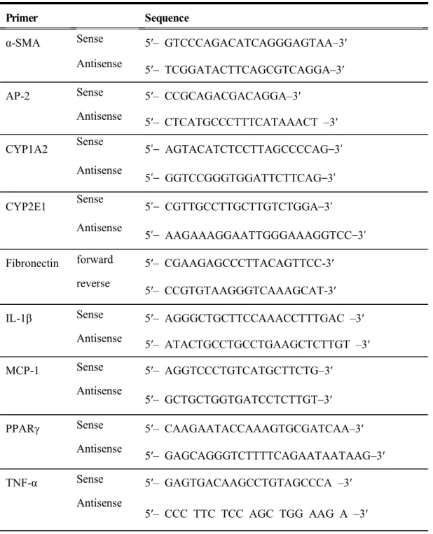
Gene-expression levels were analyzed in mRNA extracted from mouse liver using TRIzol® reagent (Ambion, Austin, TX, USA), CellScripttm all-in-one 5× first-strand cDNA, and cDNA Synthesis Master Mix (CellSafe, Gyeonggi-do, Korea). Real-time PCR was performed using Luna® Universal qPCR Master Mix (New England BioLabs, Ipswich, MA, USA). mRNA expression levels were calculated according to the 2-ΔΔCt method, with GAPDH as the internal control. The primers used in the PCRs are listed in Table 3 (Gavish et al., 2008; Nelson et al., 2004; Tipoe et al., 2010; Truong et al., 2016).
Table 5. Primer sequences used in the present study
Primer Sequence
α-SMA Sense 5′– GTCCCAGACATCAGGGAGTAA–3′
Antisense
5′– TCGGATACTTCAGCGTCAGGA–3′
AP-2 Sense 5′– CCGCAGACGACAGGA–3′
Antisense
5′– CTCATGCCCTTTCATAAACT –3′
CYP1A2 Sense 5′– AGTACATCTCCTTAGCCCCAG–3′
Antisense
5′– GGTCCGGGTGGATTCTTCAG–3′
CYP2E1 Sense 5′– CGTTGCCTTGCTTGTCTGGA–3′
Antisense
5′– AAGAAAGGAATTGGGAAAGGTCC–3′
Fibronectin forward 5′– CGAAGAGCCCTTACAGTTCC-3′
reverse
5′– CCGTGTAAGGGTCAAAGCAT-3′
IL-1β Sense 5′– AGGGCTGCTTCCAAACCTTTGAC –3′
Antisense
5′– ATACTGCCTGCCTGAAGCTCTTGT –3′
MCP-1 Sense 5′– AGGTCCCTGTCATGCTTCTG–3′
Antisense
5′– GCTGCTGGTGATCCTCTTGT–3′
PPARγ Sense 5′– CAAGAATACCAAAGTGCGATCAA–3′
Antisense
5′– GAGCAGGGTCTTTTCAGAATAATAAG–3′
TNF-α Sense 5′– GAGTGACAAGCCTGTAGCCCA –3′
Antisense
Immunohistochemistry
The liver sections were incubated with rabbit anti-Iba-1 antibody. The peroxidase reaction was visualized using a DAB kit (Vector Laboratories, Burlingame, CA, USA). The slides were counterstained with hematoxylin and then mounted. Iba-1-immunostained areas were semi-quantitatively analyzed using ImageJ (n=6 animals per group). Five different sections from each liver were assessed and the positive area was calculated as a percentage of the total area of each section (n=5 animals per group).
Measurement of liver hydroxyproline content
Hepatic hydroxyproline content was quantified colorimetrically in flash frozen liver samples using a hydroxyproline kit (Abcam, Cambridge, UK) according to the manufacturer’s instructions. The experimental results were quantified by comparison to a standard curve of known hydroxyproline concentrations and expressed as mg hydroxyproline/mg liver.
Statistical anaysis
All results are presented as the means ± SEM. Statistical significance was defined as a P value < 0.05 based on the results of one-way analysis of variance followed by Bonferroni’s multiple comparison post-hoc test.
4. Results
Evaluation of biochemical parameters in serum
Serum levels of the enzymes ALT, AST, and T-bil were measured to determine the protective role of norgalanthamine against CCl4-induced hepatic injury
(Fig. 4). In the control mice, serum ALT, AST and T-bil levels were in the normal range (31.8 ± 2.20, 42.2 ± 2.29, and 0.17 ± 0.00 U/L, respectively; Fig. 4) whereas they were significantly higher in the vehicle-treated CCl4-injured mice (6,088.0 ±
697.47 U/L, p < 0.001) mice they were significantly higher than in the normal control mice (0.27 ± 0.02 U/L).
Pretreatment with 1 mg norgalanthamine/kg significantly reduced levels of ALT (4,176.0 ± 441.97 U/L, p <0.05) and AST (2,952.0 ± 358.07 U/L, p < 0.05) in
mice with CCl4-induced liver injury. At the higher dose of 10 mg
norgalanthamine/kg, significant dose-dependent reductions compared to vehicle-treated CCl4-injured mice were determined for ALT (3,588.00 ± 281.13 U/L, p < 0.01),
AST (2,236.00 ± 158.83 U/L, p < 0.001), and T-bil (0.22 ± 0.01 U/L, p < 0.05).
In the positive control (silymarin 100), serum ALT (3,588.00 ± 225.73 U/L, p < 0.01), AST (1,840.00 ± 185.66 U/L, p < 0.001), and T-bil (0.21 ± 0.01 U/L, p < 0.05) levels decreased significantly compared to the vehicle-treated mice. The absence of a significant difference between the silymarin and norgalanthamine groups implies that the effects on serum biomarkers of CCl4-induced liver injury were similar.
(296.60 ± 17.87 U/L, p < 0.001), but significantly higher in NG10 mice (104.40 ± 6.95 U/L) than in vehicle-treated mice (32.80 ± 3.53 U/L, p < 0.05).
Figure 4. The effects of norgalanthamine on the serum levels of biochemical parameters in mice with CCl4-induced acute liver injury. (A) The inhibitory effects of norgalanthamine (1 and 10 mg/kg) on serum aspartate aminotransferase (AST), (B) alanine aminotransferase (ALT), (C) total bilirubin, and (D) glucose in mice with CCl4-induced acute liver injury. Silymarin (positive control) dose: 100 mg//kg. Values
are mean ± SEM, n = 5 per group. *** P <0.001 vs. normal control; # P <0.05, ##
Norgalanthamine improves histopathological changes in the liver
The histopathological studies demonstrated protective effects of norgalanthamine against CCl4-induced liver injury (Fig. 5). In the normal control mice, the liver
showed a normal histological architecture, including hepatic cells with well-preserved cytoplasm, a prominent nucleus, and a central vein (Fig. 5A). In the livers of CCl4-induced mice, however, large areas of pericentral necrosis were seen together
with a loss of hepatic architecture, an inflammatory cell infiltrate, and cell swelling (Fig. 5B). The hepatic lesions were less severe in the livers of the NG1, NG10, and silymarin 100 CCl4-injured mice (Fig. 5C, 5D) than in those of the vehicle-treated
CCl4 control mice (Fig. 5E).
Liver-injury grades were evaluated based on H&E staining. Based on the histological scores, pretreatment with either 1 or 10 mg norgalanthamine significantly inhibited acute hepatic injury compared to vehicle treatment (p < 0.001). Similar effects were observed in the silymarin 100 group (Fig. 5F).
The extent of centrilobular necrosis around the central vein was assessed by toluidine blue staining (Fig. 5G). The semi-quantitative results showed a larger necrotic area in the vehicle-treated CCl4 control group, but a reduction in the extent
of necrosis was seen in the NG10 and silymarin 100 groups.
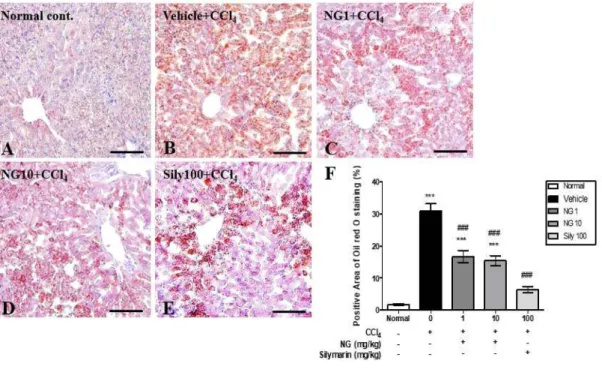
The liver tissue of the mice was also examined for fatty changes (Fig. 6). In the normal control, lipid droplets were not detected by oil red O staining (Fig. 6A), whereas in the vehicle-treated CCl4-injured group staining revealed diffuse fatty
degeneration throughout the liver (Fig. 6B). By contrast, in the NG1 and NG10 groups (Fig.6C, 6D), and in the silymarin 100 group (Fig. 6E), there was a relative reduction in the extent of fatty changes in the livers of mice with CCl4-induced
injury. The percentage of oil-red-O-positive areas was significantly higher in the vehicle-treated group than in the normal control group (p < 0.001), but was significantly reduced in the NG1 and NG10 groups (p < 0.001), as well as the silymarin 100 group (p < 0.001) (Fig. 6F). These results imply that norgalanthamine treatment can mitigate CCl4-induced histopathological changes in mouse liver.
Figure 5. Protective effects of norgalanthamine on the histology of the liver in mice with CCl4-induced acute liver injury. Liver tissues were stained with hematoxylin and eosin (H&E) and their histological characteristics were assessed by three blinded observers. (A) Normal control group; (B) CCl4 vehicle group; (C) CCl4 injury + 1
mg norgalanthamine /kg; (D) CCl4 injury + 10 mg norgalanthamine/kg; (E) CCl4
injury + 100 mg silymarin/kg; (F), histological score (0–4); (G) quantitative analysis of toluidine blue-negative areas in mouse liver. Values represent mean ± SEM, n = 5 per group. ** p<0.01 and *** p<0.001 vs. normal control group; ## p<0.01 and ### p<0.001 vs. CCl4-induced acute injury with vehicle treatment. Scale bar = 100
Figure 6. Oil red O staining of frozen sections of liver tissue from treated and untreated mice reveals lipid accumulation. (A) Normal control; (B), CCl4-induced
vehicle-treated group; (C), CCl4 injury +1 mg norgalanthamine/kg; (D) CCl4 injury
+10 mg norgalanthamine/kg; (E) positive control: CCl4 injury +100 mg silymarin/kg;
(F) quantification of oil-red-O-positive areas. Values represent means ± SEM, n = 5 per group. *** P <0.001 vs. normal control; ### P <0.001 vs. CCl4-induced acute
Norgalanthamine regulation of expression of hepatic CYP1A2 gene
The hepatic mRNA expression of CYP1A2 and CYP2E1 was determined by qPCR analysis (Fig. 7). CYP2E1 mRNA expression was significantly decreased in all
administration groups compared to the normal control group (vehicle-treated CCl4
group and NG10, p <0.01; NG 1 and silymarin, p <0.05). However, in all the treatment groups, expression levels showed no changes. On the other hand, CYP1A2 mRNA expression significantly decreased in the vehicle-treated CCl4 group compared
to the normal control group, but increased significantly in the NG10 groups (p <0.05) and silymarin (p <0.001) groups.
Figure 7. mRNA expression levels of CYPs in the liver (A–B). The mRNA expression levels of CYP2E1 and CYP1A2 were measured using the quantitative reverse transcription polymerase chain reaction. Gapdh was used as the housekeeping gene. The data are presented as the means± SEM. * p <0.05, ** p <0.01 and *** p <0.001 vs. the normal control group; # p <0.05 and ### p <0.001 vs. the
CCl4-induced acute injury group with vehicle treatment. mRNA: messenger RNA;
Norgalanthamine up-regulates SOD and catalase activities
The antioxidant effects of norgalanthamine on CCl4-induced liver injury were
investigated by measuring the activities of the antioxidant enzymes SOD and CAT in the liver tissues of the five groups of mice (Fig. 8). In the CCl4-induced mice, SOD
and CAT activities decreased significantly (80.37 ± 5.24 and 0.46 ± 0.04 U/L, respectively; both p < 0.05). Pre-treatment with norgalanthamine prior to CCl4 injury
significantly up-regulated the levels of both enzymes compared to the vehicle-treated mice, with a significant effect on SOD seen in the NG10 group (102.22 ± 2.26 U/L, p < 0.01; Fig. 8A), and on CAT in both the NG1 and NG10 groups (0.62 ± 0.03, p < 0.01; 0.98 ± 0.21, p < 0.05, respectively; Fig. 8B). This implies that the hepatoprotective effect of norgalanthamine is derived from its antioxidant activity.
Fig. 8. Antioxidant effect of norgalanthamine in mice with CCl4-induced acute liver
injury. Injury is as indexed by the enzyme activities of (A) superoxide dismutase and (B) catalase. The data are expressed as means ± SEM, n = 5 per group. * P <0.05 vs. the normal control; # P < 0.05 and ## P < 0.01 vs. the CCl4-induced acute injury
Norgalanthamine reduced inflammation in the liver
The anti-inflammatory properties of norgalanthamine were further evaluated by assessing TNF-α, IL-1β, and MCP-1 mRNA levels in the livers of the mice (Fig. 9). TNF-α and IL-1β mRNA expression increased significantly in the vehicle-treated CCl4-injured group (3.41 ± 0.56 and 2.37 ± 0.27 U/L, respectively) versus the normal control group (0.71 ± 0.26 and 1.42 ± 0.45 U/L, respectively; both p < 0.01) but were significantly reduced in the norgalanthamine groups (2.30 ± 0.27 and 1.47 ± 0.02 U/L, respectively, both p < 0.05; Fig. 9A, B). In addition, the mRNA levels of the inflammatory chemokine MCP-1 were significantly higher in the vehicle-treated CCl4-injured group than in the normal control group (12.37 ± 1.00 vs. 1.31 ± 0.27
U/L; p < 0.01) and significantly lower in the NG1 and NG10 groups than in the vehicle treatment group (6.36 ± 1.33-fold change, 6.94 ± 0.77-fold change, respectively, both p < 0.01; Fig. 9).
Kupffer cells/macrophages are activated during liver injury (Ahn et al., 2016). In the mouse livers in the normal control group, Iba-1-positive Kupffer cells were detected along the sinusoids (Fig. 10A), but no infiltration of inflammatory cells was observed. By contrast, in the livers of CCl4-injured mice, activated Kupffer cells and
an inflammatory cell infiltrate were detected (Fig. 10B). Pretreatment with 10 mg norgalanthamine/kg (Fig. 10D) or 100 mg silymarin/kg (Fig. 10E) reduced the numbers of Iba1-positive cells. Furthermore, while the percentages of Iba-1-positive areas were significantly higher in the vehicle-treated group than in the normal control group (p < 0.01), they were significantly lower in the NG10 (p < 0.01) and silymarin 100 (p < 0.01) groups than in the vehicle-treated group (Fig. 10F).
Figure 9. Norgalanthamine pretreatment attenuates CCl4-induced inflammatory
responses in the liver at the transcriptional level. (A) TNFα mRNA; (B), IL1β mRNA; and (C), MCP-1 mRNA. Data are presented as means ± SEM, n = 5 per group. * P <0.05 and ** P <0.01 vs. the normal control; # P <0.05 and ## P
Figure 10. (A–E) Immunohistochemical staining of Iba-1 in liver sections. (F) Bar graph shows the semi-quantitative analysis of Iba-1-positive areas. Scale bar = 100 μ m. Values in (F) are means ± SEM, n = 5 per group. *** P < 0.001 vs. the normal control group; ## P < 0.01, ### P <0.001 vs. the CCl4-induced
Norgalanthamine down-regulates the expression of mRNAs linked to adipogenesis in the liver
The molecular mechanisms underlying the anti-adipogenic effects of
norgalanthamine were investigated by analyzing the mRNA levels of genes involved in lipid uptake and metabolism, including proliferator-activated receptor-γ (PPAR-γ) and adipocyte fatty acid-binding protein-2 (AP2) (Fig. 11). The results showed that the two genes were expressed at significantly higher levels in the vehicle-treated group (p < 0.01) whereas norgalanthamine treatment caused significant reductions in both genes (p < 0.05).
Figure 11. Real-time PCR analyses of PPAR-γ and AP2 expression in liver tissues.
Bar graph shows the expression of PPAR-γ and AP2 in CCl4-induced
acute-liver-injury mice relative to that of the normal control mice. Data are presented as means ± SEM, n = 5 per group. * P < 0.05, ** P < 0.01 vs. the normal control group; # P < 0.05 and # P < 0.01 vs. the CCl4-induced acute injury with
Norgalanthamine up-regulates Nrf-2 and HO-1 levels in mice with CCl4-induced liver injury
Western blotting was performed to determine the effect of norgalanthamine on Nrf-2 levels in the cytosol and nucleus of liver cells in the treated mice. Compared to the vehicle-treated group, norgalanthamine significantly increased both the cytoplasmic levels of Nrf-2 and the extent of the protein’s nuclear translocation (Fig. 12A). Consistent with these findings, HO-1 protein levels were significantly higher in the NG10 group than in the vehicle-treated group (Fig. 12B).
Figure 12. Representative immunoblots of (A) Nrf-2 and HO-1 and (B) Nrf-2 and HO-1 expression relative to the β-actin level in CCl4-induced acute-liver-injury mice. Data are presented as mean ± SE, n = 5 per group. * P < 0.05 vs. the normal control group; # P < 0.05 and # P < 0.01 vs. the CCl4-induced acute injury with
Effects of norgalanthamine on CCl4-induced chronic liver injury
Liver sections were stained with Picrosirius red stain. Collagen fibers were detected around perivenular regions of the normal control group (Fig. 13A). However, collagen tissue proliferation by fibrosis was detected in the vehicle-treated group (Fig. 13B). The norgalanthamine and silymarin treatments significantly reduced collagen tissue proliferation in the liver (Fig. 13C and 13D) compared with the vehicle-treated group. The positive areas with Picrosirius red staining were significantly higher in the vehicle-treated group (3.77 ± 0.27%, p < 0.01) than in the normal control group (1.60 ± 0.14%). The 10-mg/kg norgalanthamine group (3.00 ± 0.02%, p < 0.05) showed significantly lower percent areas of collagen tissue compared with the liver tissue in the vehicle-treated group (Fig. 13F). In liver tissue, hydroxyproline levels were significantly lower in the 10-mg/kg norgalanthamine group (189.26 ± 35.53 g, p < 0.05) than in the vehicle-treated group (33.87 ± 5.81g) (Fig. 13G). Changes in the
biochemical serum and histopathological parameters of CCl4-induced
chronic-liver-injury mice were similar to those in CCl4-induced acute-liver-injury mice
(data not shown). In addition, the expression of α-SMA and fibronectin mRNA decreased significantly in the 10-mg/kg norgalanthamine group (3.33 ± 1.76-fold change, p < 0.01; 1.03 ± 0.15-fold change, p < 0.05) compared to the vehicle-treated group (5.24 ± 1.75-fold change; 1.48 ± 0.07, both p < 0.05) (Fig. 14).
Figure 13. Effects of norgalanthamine in CCl4-induced chronic liver injury in mice. (A–D) Picrosirius red staining of liver sections. (F) Positive area of Picrosirius red staining. (G) Quantitative hydroxyproline in CCl4-induced chronic liver injury mice.
Data are presented as means ± SEM, n = 5 per group. * P < 0.05 and ** P < 0.01 vs. the normal control group; # P < 0.05 vs. the CCl4-induced acute injury