- 37 -
서 론
2012년에 발표된 중앙암등록본부 자료에 갑상선암은 남녀 를 합쳐서 연 평균 36,021건 발생하여 전체 암 발생 비율의 17.8%
로 1위를 차지했다.1) 이 중 34,869건(96.8%)이 분화 갑상선암 으로, 분화 갑상선암은 다른 조직에서 발생한 암에 비해 비교 적 천천히 자라고 예후가 양호한 것으로 알려져 있으나, 10~
15%에서 원격전이가 발생되고 20년 누적재발률이 30%에 이 른다.1,2)
분화 갑상선암의 대다수를 차지하는 유두암에서, 진단 시 중앙 림프절 전이율은 저자에 따라 40~65%까지 보고되고 있 다.3-5) 갑상선 유두암에서 림프절 전이는 생존율에 영향을 주 지 않는다고 보고되고 있으나, 림프절 전이에 대한 평가는 국 소 재발의 가능성을 예측하는데 중요한 인자로 알려져 있다.6-9) 경부 전산화단층촬영(CT), 자기공명영상(MRI), 양전자방출 단층촬영(PET) 검사를 통해 중앙 림프절 전이를 발견 할 수 있는 확률은 30% 정도로,10) 미국 갑상선 학회(American Thy- roid Association, ATA)의 개정된 분화 갑상선암의 치료 지침 을 살펴보면, 초음파 검사 이외의 영상 의학적 검사는 추천되 지 않고 있다.11) 그러나 경부 전산화단층촬영은 갑상선 결절과
Received : September 15, 2013 / Revised : October 24, 2013
Accepted : October 25, 2013
교신저자 : 안순현, 463-707 경기도 분당구 성남시 구미동 300 서울대학교 의과대학 분당서울대학교병원 이비인후과학교실 전화 : (031) 787-7403 ・ 전송 : (031) 787-4057
E-mail : [email protected] 대한 두경부 종양 학회지 제 29 권 제 2 호 2013
갑상선 유두암의 외측 경부림프절 전이에 대한 수술 전 평가로서 CT의 역할
서울대학교 의과대학 분당서울대학교병원 이비인후과학교실
석준걸·김형규·김윤중·한규희·안순현
=
Abstract
=The Role of CT as a Preoperative Evaluation of Lateral Cervical Lymph Node Metastasis in Papillary Thyroid Carcinoma
Jungirl Seok, MD, Hyung gu Kim, MD, Yoonjoong Kim, MD, Kyu-Hee Han, MD, Soon-Hyun Ahn, MD, PhD
Department of Otorhinolaryngology-Head and Neck Surgery, Seoul National University Bundang Hospital, Seongnam, Korea
Background and Objectives:To assess the usefulness of computed tomography image before papillary thy-
roid cancer surgery, focus on confirmation of lateral cervical lymph node metastasis not detected by ultrasonogra- phy. Material and Methods:From January 2008 to May 2009, total 150 patients who had undergone thyroid sur- gery and been confirmed papillary thyroid cancer pathologically were enrolled. They had taken neck computed tomography following the ultrasonography. Results:Computed tomography had found suspicious metastatic lat- eral neck lesion in 13 patients. After the image study, lateral neck lymph node dissection had been included in their surgical plan. Of these, only 7 cases were confirmed pathologically lateral neck lymph node metastasis(positive predictive value=0.54). Taken as whole 150 patients, additionally 4.7% of patients confirmed lateral neck lymph node metastasis by preoperative computed tomography.Conclusion:If preoperative ultrasonography was per-
formed precisely, additional benefits that could be achieved by computed tomography were not much.KEY WORDS
:Thyroid neoplasmsㆍTomographyㆍX-ray computedㆍLymph node excision.online©MLComm
- 38 -
종격 종양의 감별이 필요할 때 유용하며 악성결절의 림프절 전 이 여부와 기도침범여부 등의 감별에 이용된다.12,13)분화 갑상선암의 림프절 전이에 대한 초음파 및 CT의 진단 적 가치에 대한 몇몇 연구가 이루어졌으며, 김 등14)은 술 전 초 음파와 CT를 함께 검사했을 때 초음파 단독 검사 보다 경부 림프절 전이 진단에 대한 민감도가 유의하게 높으며, 특이도는 비슷한 정도로 보고하였나 최 등15)은 CT가 초음파 보다 경부 림프절 전이 발견에 대한 민감도가 높으나 그 진단의 정확도에 서 초음파, CT, 초음파와 CT를 함께 보았을 때 통계적으로 유 의한 수준은 아님을 보고하여 차이를 보였다.
이에 저자들은 수술 전 시행되는 CT의 유용성에 대한 저 자들의 경험을 분석해보고, 수술계획의 변경 여부 및 최종 병 리학적 소견을 비교 고찰을 통해 그 의미를 찾아보고자 했다.
대상 및 방법
2008년 1월부터 2009년 5월까지 분당서울대학교병원에서 수술 후 갑상선 유두암으로 진단받고 3년 이상 추적관찰을 시 행한 환자들을 대상으로, 수술 전 초음파 및 세침검사(fine needle aspiration, FNA)를 시행한 후 수술이 계획된 후, 수 술 전 경부 CT를 촬영했던 환자 총 150명을 대상으로 후향적 의무기록 분석을 시행하였다. 병리 결과, CT촬영 일자와 수술 일자, 초음파에서 림프절 전이 여부, CT에서 림프절 전이 여부, 수술 초기 계획 및 검사 이후 변경된 수술 계획, 전이된 림프 절의 수 및 위치, 최종 병기 등을 조사하였다. 초음파 및 CT 검 사는 영상의학과에서 판독하였다. 수술 계획 변경 여부는 CT 촬영 후에 외측 경부 림프절에 대한 술식이 포함된 것을 의미
하는 것으로, 갑상선 유두암의 예후에 영향을 주지 않는 것으 로 알려진 중앙 림프절 절제술(central lymph node dissection) 이 추가되거나 제외된 경우는 제외하였다.
통계학적 유의성은 SPSS 12(Chicago, IL, USA) 소프트웨 어를 이용하여 chi-square와 Fisher’s exact test를 통해 확인 하였다.
결 과
수술 후 갑상선 유두암으로 진단받은 총 150명 중 남성 42명 (28.0%), 여성 108명(72.0%)이었고 남성의 평균 연령은 48.5±
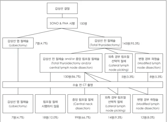
10.5세, 여성의 평균 연령은 47.8±11.1세였다. 추적 관찰 기간 은 37개월에서 최대 54개월까지였다. 이 중 48명은 외부에서 시행한 초음파 검사였으며, 102명은 본원 영상의학과에서 시 행하였으며 150명 모두 본원에서 전산화단층촬영을 시행하였 다. 초음파 검사 결과를 통해 수술 방법을 계획 한 후 전산화 단층촬영의 결과를 통해 수술 방법을 확정하였다(Fig. 1).
수술 방법은 갑상선 결절의 크기 및 개수, 양측성 여부에 따 라 일측 갑상선 절제술(thyroid lobectomy), 갑상선 전 절제술 (total thyroidectomy), 중앙 림프절 절제를 포함한 갑상선 전 절제술(total thyroidectomy with central lymph node dissec- tion), 갑상선 전 절제술과 중앙 림프절 및 외측에 의심되는 림 프절의 선택적 제거(total thyroidectomy with central lymph node dissection and lateral lymph node picking), 갑상선 전 절제술과 중앙 림프절 및 외측 경부 절제술(total thyroidecto- my with central lymph node dissection and lateral neck dis- section)으로 총 5가지 술식을 시행하였다(Table 1). 일측 갑상
갑상선 결절
SONO & FNA 시행 150명
갑상선 엽 절제술
(Lobectomy) 7명(4.7%)
7명(4.7%) 18명(12.0%) 99명(66.7%) 14명(9.3%) 12명(8.0%)
143명(95.3%)
130명(86.7%) 5명(3.3%) 8명(5.3%)
갑상선 엽 절제술 (Lobectomy)
중앙 림프절 절제 (Central neck dissection)
외측 경부 림프절 선택적 절제 (Lateral lymph node picking)
변형 경부 곽청술 (Modified lymph node dissection) 림프절 절제
시행하지 않음
갑상선 전 절제술 and/or 중앙 림프절 절제술 (Total thyroidectomy and/or central lymph node dissecton)
외측 경부 림프절 선택적 절재 (Lateral lymph node picking)
변형 경부 곽청술 (Modified lymph node dissection) 갑상선 전 절제술
(Total thyroidectomy)
수술 전 CT 촬영
Fig. 1. Flow chart of surgical plans of patients.
- 39 -
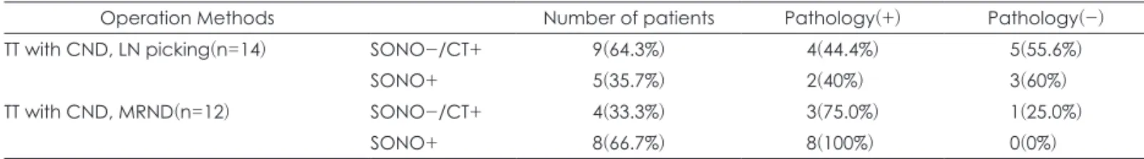
선 절제술은 총 7명(4.7%)이 시행 받았으며, 이 중 2예에서 병 리 검사상 종양의 외막 침범이 관찰되어 잔존 갑상선 절제술 (completion thyroidectomy)를 시행 받았다. 갑상선 전 절제 술만 시행 받은 환자는 총 18명(12.0%)였으며 이 중 재발한 예 는 없었다. 갑상선 전 절제술 및 중앙 림프절 절제술을 시행 받 은 환자는 총 99명(66.7%)으로, 이 중 절제된 중앙 림프절에서 전이가 발견되지 않았던 1예에서 경과관찰 중 재발되어 경부 절제술을 시행 받았다. 갑상선 전 절제술 및 중앙 림프절 절제 술, 외측 림프절의 선택적 제거 수술을 받은 환자는 총 14명(9.3%) 였으며, 이 중 9명(64.3%)이 경부 전산화단층촬영 후에 외측 림프절 제거술이 추가된 환자였다. 14명 중 6명에서 절제된 외 측 경부절에 림프절 전이가 관찰되었으며, 경부 전산화단층촬 영 후에 외측 림프절 제거술이 추가된 9명 중에서는 4명이 해 당되었다. 14명 모두에서 재발은 관찰되지 않았다. 갑상선 전 절 제술 및 중앙 림프절 절제술, 경부 절제술을 시행 받은 환자는 총 12명(8.0%)였으며, 이 중 4명(33.3%)이 경부 전산화단층촬 영 후에 경부 절제술이 추가된 환자였다. 12명 중 11명에서 절 제된 외측 경부절에 림프절 전이가 관찰되었으며, 경부 전산화 단층촬영 후에 경부 절제술이 추가된 4명 중에서는 3명이 해 당되었다. 14명 중 2명이 경과관찰 중 재발하여 재수술을 시 행 받았다(Table 2).외측 경부 림프절 절제에 해당하는 두 가지 술식을 합쳐서 보면 총 26명(17.3%)이 외측 경부 림프절 절제에 해당하는 수 술을 시행 받았고 17명(65.4%)에서 외측 경부 림프절에 전이 가 확인되었다. 26명 중 13명(50.0%)이 경부 전산화단층촬영 후에 외측 경부 림프절 절제가 수술 범위에 포함되었다. 이 중 실제 외측 경부 림프절에 전이가 확인된 환자는 13명 중 7명 (53.8%)이었다.
환자군 전체로 비교하였을 때 전산화단층촬영으로 외측 경
부절 절제 수술이 추가된 환자는 150명 중 13명으로 8.6%에 해당하였으며 13명 중 7명에서 외측 경부 림프절 전이가 확인 되어 양성 예측도(positive predictable value)는 0.54였다.
고 찰
분화 갑상선암, 특히 갑상선 유두암은 진단시 이미 20~50%
의 환자가 중앙 림프절을 포함한 경부림프절의 전이가 발견된
다.16-20) 림프절 전이를 확인하는 것은 분화 갑상선암의 치료 방
법 결정 및 예후를 판단함에 있어 중요한 의미가 있으나, 전이 된 병변이 RAI(Radioactive iodine)치료에 잘 반응하기 때문 에, 림프절 전이 여부가 다른 종양과 다르게 원발 부위를 수 술적으로 제거하려는 계획에 있어 큰 영향을 주지 못한다.21) 또한 CT, MRI, PET 등의 초음파 이외의 검사들이 경부 림프 절 전이를 발견할 수 있는 정도가 30~40%로 상대적으로 낮 기 때문에,10) 미국 갑상선 학회의 진료 지침에 따르면 수술 전 초음파 이외의 영상의학적 검사를 권하지 않는다.11)
그러나 실제 임상에서는 갑상선 주변의 구조물 확인 등의 이유로 CT를 촬영하게 되는 경우가 있고, 초음파에서 발견하 지 못하는 외측 경부 림프절 전이가 발견되는 경우가 있었다.
본 연구에서는 수술 전 경부 전산화단층촬영을 한 150예의 갑 상선 유두암 환자들 중에서 총 13명(8.7%)에서 수술 전 초음 파에서 발견되지 않았던 전이가 의심되는 외측 경부 병변이 발 견되었는데, 수술 후 병리학적으로 전이가 진단된 환자는 총 7명으로 0.54의 양성 예측도를 보였다. 전체 150명에서 보면 7명(4.7%)에 해당하는 추가적인 외측 경부 림프절 전이 환자 를 진단 할 수 있었던 것이다.
여러 연구로부터 경부 전산화단층촬영이 초음파 검사에 비 해 경부 림프절 전이의 진단에 대해 뒤지지 않는 다는 보고가 있으나, 초음파가 갑상선 암의 진단에 있어 항상 시행된다고 가 정할 때 4.7%의 비율이 결코 낮지는 않지만 초음파에서 발견 되지 않았던 외측 경부 림프절 전이를 진단하기 위해 전산화 단층촬영을 하는 것이 어느 정도의 이득이 있을지 술자들의 고 민이 필요할 것이라고 생각된다. 또한 초음파에서 확인되지 않 은 경부 림프절 전의 의심 소견에 대한 양성예측도가 0.54 정 도로, 불필요한 추가 절개, 경부 절제술로 인해 발생하는 합병 증 등을 고려한다면, 큰 이득을 얻지 못할 가능성이 있다.
Table 1. Operation methods
Operation method Number of patients(%)
Lobectomy 7( 4.7%)
Total thyroidectomy 18(12.0%)
TT with CND 99(66.7%)
TT with CND, LN picking 14( 9.3%)
TT with CND, MRND 12( 8.0%)
TT : Total thyroidectomy, CND : Central neck dissection, LN : Lymph node, MRND : Modified radical neck dissection
Table 2. Pathological detection rate between two lateral neck lymph node operation methods
Operation Methods Number of patients Pathology(+) Pathology(-)
TT with CND, LN picking(n=14) SONO-/CT+ 9(64.3%) 4(44.4%) 5(55.6%)
SONO+ 5(35.7%) 2(40%) 3(60%)
TT with CND, MRND(n=12) SONO-/CT+ 4(33.3%) 3(75.0%) 1(25.0%)
SONO+ 8(66.7%) 8(100%) 0(0%)
SONO+ : Operation method decided from ultrasonography findings only, SONO-/CT+ : Operation method was changed after
CT study. In case of CT detected suspicious metastatic lesion which was not detected by ultrasonography, Pathology+ : Patho-
logically confirmed lateral neck lymph node metastasis, except central node metastasis
- 40 -
전산화단층촬영 검사의 유용성을 초음파와 비교하기 위해 서는 검사의 민감도와 특이도를 구해서 비교해봐야 하나, 본 검사의 대상자들 중에 경부 림프절 절제술을 시행 받지 않은 환자들에게서 림프절 전이여부를 알 수 있는 방법이 없기 때 문에 민감도와 특이도를 구할 수 없었다는 것이 본 연구의 한 계점으로 볼 수 있다.위의 연구 결과로부터 분화 갑상선, 특히 갑상선 유두암에 대한 절제술 전에 시행되는 관습적인 경부 전산화단층촬영을 지양하고 선택적으로 단층 촬영 검사를 시행하거나, 초음파 검 사의 정확도를 높이는 방향을 모색하는 것이 환자에게 불필요 한 수술 범위의 확장과 합병증을 예방할 수 있는 방법일 것으 로 본다.
중심 단어 : 갑상선 유두상암 ・측경부 림프절 전이 ・양성예측 도 ・전산화단층촬영.