2004; 9(1): 26-35
서 론
Apoptosis는 개체의 발생단계나 DNA 손상, 바이
러스 감염 등에 의한 유전적 조절 하에서 일어나 는 정교한 개체의 방어기전이란 점에서 necrosis와 구별된다.1) 또한 apoptosis는 개체보존수준에서 손 상된 세포들의 제거를 위한 중요한 수단이며, 세
Apoptosis 유발에 의한 β-lapachone의 인체폐암세포 증식 억제에 관한 연구
동의대학교 한의과대학 1생화학교실 및 2해부학교실, 3부산대학교 자연과학대학 생물학과
이재훈
1․박 철
1․최병태
2․이원호
3․최영현
1Growth Inhibition of Human Lung Carcinoma Cells by
β-lapachone through Induction of Apoptosis
Jae Hun Lee1, Cheol Park1, Byung Tae Choi2, Won Ho Lee3 and Yung Hyun Choi1 Departments of 1Biochemistry and 2Anatomy, Dong-Eui University College of Oriental Medicine, Busan 614-052, Korea, 3Department of Biology, Pusan National University, Busan 609-735, Korea
The DNA topoismerase I inhibitor β-lapachone, the product of a lapacho tree (Tabebuia avellanedae) from South America, activates a novel apoptotic response in a number of cell lines. In the present report, we investigated the effects of β-lapachone on the growth of human lung in human non-small-cell-lung-cancer A549 cells. Upon treatment with β-lapachone, a concentration-dependent inhibition of cell viability and cell proliferation was observed as measured by hemocytometer counts and MTT assay. The β-lapachone- treated cells developed many of the hallmark features of apoptosis, including membrane shrinking, condensation of chromatin and DNA fragmentation. These apoptotic effects of β-lapachone in A549 cells were associated with a marked induction of pro-apoptotic Bax expression, however the levels of anti-apoptotic Bcl-2 expression were decreased in a dose-dependent manner. Accordingly, elevated amount of cyclin- dependent kinase inhibitor p21 expression accompanied by up-regulation of tumor suppressor p53 was observed. By RT-PCR analyses, decrease in gene expression level of telomerase reverse transcriptase and telomeric repeat binding factor were also observed.
Thus, these findings suggest that β-lapachone may be a potential anti-cancer therapeutics for the control of human lung cancer cell model.
ꠏꠏꠏꠏꠏꠏꠏꠏꠏꠏꠏꠏꠏꠏꠏꠏꠏꠏꠏꠏꠏꠏꠏꠏꠏꠏꠏꠏꠏꠏꠏꠏꠏꠏꠏꠏꠏꠏꠏꠏꠏꠏꠏꠏꠏꠏꠏꠏꠏꠏꠏꠏꠏꠏꠏꠏꠏꠏꠏꠏꠏꠏꠏꠏꠏꠏꠏꠏꠏꠏꠏꠏꠏ Key Words: β-lapachone, Lung carcinoma, Apoptosis, Bax, Bcl-2
책임저자:최영현, ꂕ 614-052, 부산광역시 진구 양정동 산 45번지, 동의대학교 한의과대학 생화학교실 Tel: 051-850-7413, Fax: 051-853-4036, E-mail: [email protected]
접수일:2004년 1월 16일, 게재승인일:2004년 3월 2일
포주기 의존적 또는 비의존적으로 동시에 일어날 수 있으나, 정상적인 세포주기의 이탈이나 세포주 기조절에 중요한 cyclin-dependent kinases (Cdks) 활성의 변화가 apoptotic cell death의 주원인이 될 수 있다.2) Apoptosis의 유발에 p53이나, Bcl-2 및 Bax와 같은 유전자가 관여한다는 사실이 알려지 면서 apoptosis와 연관된 분자적 기전이 최근 많이 밝혀지고 있다.3,4) 그중 대표적인 것으로 Bcl-2는 apoptosis를 억제하는 반면, Bax는 과발현되었을 때 apoptosis를 유도하는데, 두 단백질은 서로 di- mer를 형성하면서 cytochrome c와 같은 apoptosis 유발에 관여하는 인자들의 조절에 관여한다.5,6) 또 한 caspases라고 이름 붙여진 ICE/CED-like pro- tease family 역시 apoptosis 유발에 중요한 역할을 하는데, 이들은 proenzyme 형태로 존재하다가 Bax 를 포함한 apoptosis 유도를 활성화시키는 신호에 의해 활성화된 cysteine-related proteases로 되어 직 접 또는 간접적으로 세포 내 존재하는 많은 표적 단백질의 분해에 관여한다.7,8) 따라서 항암제 개발 을 위한 후보물질의 암세포 apoptosis 조절관련 기 전해석은 필수적으로 선행되어져야 할 분야로 인 식되어지고 있다.
DNA topoisomerase 활성의 억제제로 알려진 β- lapachone (3,4-dihydro-2,2-dimenthyl-2H-napthol [1,2- b]pyran-5,6-dione)은 남미지역에 서식하는 lapacho (Tabebuia avellanedae)라는 나무의 수피에서 처음 동정된 천연 quinone계 물질의 하나이다.9) 이미 잘 알려진 DNA topoisomerase I 억제제인 campto- thecin과는 달리, 이 물질은 topoisomerase I-cleava- ble complex의 유도 없이 DNA topoisomerase I의 촉매 활성을 억제하는 것으로 알려져 있다.10,11) β-lapachone은 anti-carcinogenic, anti-bacterial, anti- fungal, anti-trypanocidal 및 cytotoxic activities를 포 함한 많은 약리학적 작용을 가지는데,12∼16) 이는 reactive oxygen species (ROS)의 생성과 연관성이 있는 것으로 알려져 있다.11,17,18) 이를 바탕으로 한 인체암세포 증식억제 가능성과 연관된 β-lapa- chone을 이용한 최근 연구보고들에서 다양한 종 류의 표적 세포에서 암세포의 성장을 억제시키고 apoptosis를 일으키는 것으로 보고되어져 오고 있
다.16,18∼31) 그러나 현재까지 몇몇 보고가 꾸준히
있어 왔음에도 불구하고 세포증식에서 β-lapa-
chone에 의한 성장억제의 분자생물학적 기전은 여전히 많은 부분이 알려져 있지 않다.
본 연구에서는 이러한 β-lapachone의 항암기전 해석의 일환으로 인체 폐암세포의 성장에 미치는 β-lapachone의 영향을 조사하였다. 본 연구의 결 과에 의하면 β-lapachone의 처리 농도 의존적으로 A549 폐암세포의 증식은 억제되었으며, 핵 내 apoptotic body의 형성과 DNA fragmentation 증가 현상이 뚜렷하여 apoptosis의 과정에 의하여 암세 포의 사멸이 유도되는 것임을 알 수 있었다. 아울 러 Bax의 발현 증가에 따른 Bcl-2의 발현 감소 및 종양억제 유전자 p53의 발현 증가와 연관된 Cdk inhibitor p21의 발현 증가 현상이 β-lapachone에 의한 증식억제 및 apoptosis 유발에 주요한 요인으 로 작용함을 알 수 있었다.
재료 및 방법
1) 암세포의 배양 및 β-lapachone의 처리 본 실험에 사용한 A549 인체 폐암세포는 Amer- ican Type Culture Collection (Rockville, MD, USA) 에서 구입하였으며 RPMI-1640 배지를 이용하여 5% CO2, 37oC의 조건하에서 배양하였다. β-lapa- chone은 Biomol (Plymouth Meeting, PA, USA)에서 구입하였으며 이를 순수 alcohol에 녹여 10 mM의 stock 용액으로 제조한 뒤 -20oC에 보관하여 적 정 농도로 배지에 희석하여 처리하였다.
2) 암세포의 성장억제 및 형태적 변화 관찰 준비된 암세포를 분주하여 24시간 동안 안정화 시킨 후, β-lapachone을 농도별로 처리하여 48시 간 동안 배양하였다. 48시간 처리 후, trypsin을 처 리하고 phosphate-buffered saline (PBS)로 수세하였 다. 이를 trypan blue로 염색한 뒤 hemacytometer를 이용하여 살아 있는 세포의 수를 정상 배지에서 자란 암세포와 비교하였다. 또는 β-lapachone이 처리된 배지를 제거하고 tetrazolium bromide salt (MTT, Sigma Chemical Co., St. Louis, MO, USA) 시약을 0.5 mg/ml 농도가 되도록 성장배지로 희석 하여 2 ml씩 분주하여 3시간 동안 배양한 다음, MTT 시약을 제거하고 dimethylsulfoxide (DMSO, Sigma)를 첨가하여 well에 생성된 formazin을 녹인
후 ELISA reader (Molecular Devices, Sunnyvale, CA, USA)로 540 nm에서 흡광도를 측정하였다.
3) DAPI 염색을 통한 핵의 관찰
β-lapachone에 의한 암세포의 apoptosis 유발 여 부를 확인하기 위한 핵의 형태변화 관찰을 위하 여 β-lapachone이 처리된 세포들을 PBS로 수세하 고 3.7% paraformaldehyde로 상온에서 10분간 고 정시킨 후 형광 염색물질인 4,6-diamidino-2-pheny- lindole (DAPI, Sigma) 용액을 이용하여 10분간 염 색하였다. 이들 세포를 다시 PBS로 2회 수세한 후 fluorescence microscope를 이용하여 핵의 형태 변화를 정상군과 비교하였다.
4) DNA fragmentation 분석
준비된 세포를 PBS로 수세한 후 apoptotic DNA Ladder Kit (Roche Molecular Biochemical, Man- heim, Germany)를 사용하여 DNA fragmentation 현 상 여부를 조사하였다. 즉 binding/lysis buffer를 이 용하여 세포 내의 단백질과 지질을 녹여낸 다음, ethanol이 함유된 washing buffer를 이용하여 수차 례 씻어내고, elution buffer로 DNA fragments를 얻 어내었다. 얻어낸 DNA는 적당량의 DNA loading buffer와 섞어 1% agarose (Sigma) gel을 이용하여 120 V 조건하에서 전이시킨 후, gel을 ethidium bromide (EtBr, Sigma) 용액으로 염색하여 UV- transiluminator로 DNA fragment의 여부를 관찰하 였다.
5) Reverse transcription-PCR 분석
동일한 조건에서 준비된 암세포를 대상으로 RNAzol B (TEL-TEST, Inc., Texas, USA)를 이용하 여 total RNA를 분리하였다. 분리된 RNA를 정량 한 후, oligo dT primer와 AMV reverse transcriptase 를 이용하여 2μg의 RNA에서 ss cDNA를 합성하 였다. 이 cDNA를 template로 사용하여 관찰 대상 유전자를 polymerase chain reaction (PCR) 방법으 로 증폭하였다(Table 1). 이때 housekeeping 유전자인 glyceraldehyde-3-phosphate dehydrogenase (GAPDH)를 internal control로 사용하였다. 각 PCR 산물들을 1% agarose gel을 이용하여 전기영동하고 EtBr로 염색한 후 UV 하에서 확인하였다.
6) 전기영동 및 Western blotting
정상 및 β-lapachone이 처리된 배지에서 자란 세포들을 lysis buffer로 용해한 후, 고속원심분리 로 세포 내 잔사물을 분리시킨 후 동량의 단백질 을 SDS-polyacrylamide gel 전기영동으로 분리하였 다. 분리된 단백질을 함유한 acrylamide gel을 ni- trocellulose membrane (Schleicher and Schuell, Keene, NH, USA)으로 electroblotting에 의해 전이 시킨 후, 10% skin milk를 함유한 PBS-T (0.1%
Tween 20 in PBS)에 4oC에서 1시간 이상 incuba- tion하면서 비특이적인 단백질들에 대한 blocking 을 실시하였다. 그리고 특정 단백질에 대한 항체 를 membrane에 적용시켜 항원 항체 반응을 일으 킨 후, PBS-T로 씻어내고 특정 항체에 대한 이차 항체 반응을 실시한 후 enhanced chemilumine- scent (ECL) 용액(Amersham Life Science Corp., Arlington Heights, IL, USA)을 적용시킨 다음 X- ray film에 감광시켜 특정 단백질의 양을 분석하 였다. 본 실험에 사용된 항체들은 Santa Cruz Bio- technology Inc. (Santa Cruz, CA, USA) 및 Calbio- chem (Cambridge, MA, USA)에서 구입하였으며, 이차 항체로 사용된 peroxidase-labeled donkey anti- rabbit immunoglobulin 및 peroxidase-labeled sheep anti-mouse immunoglobulin은 Amersham Corp.
(Arlington Heights, IL, USA)에서 구입하였다.
결과 및 고찰
1) 암세포의 성장에 미치는 β-lapachone의 영향
A549 인체 폐암세포의 성장에 미치는 β-lapa- chone의 영향을 알아보기 위하여 48시간동안 β- lapachone을 처리한 후 살아있는 세포를 hemacy- tometer를 이용하여 조사해본 결과, Fig. 1A에서처 럼 β-lapachone의 처리농도 의존적으로 암세포의 증식이 억제되었다. 이는 Fig. 1B에 나타낸 봐와 같은 MTT assay에 의한 결과와도 유사한 경향성 이었음을 알 수 있었다. 특히 3μM 이상의 β- lapachone 처리군에서는 50% 이상의 세포증식 억 제 효과가 있었으며, 이는 다른 종류의 전립선 암
세포,16,19∼22) 신경교종,23) 간암세포,24) 백혈병세
Table 1. Gene-specific primers for RT-PCR
ꠧꠧꠧꠧꠧꠧꠧꠧꠧꠧꠧꠧꠧꠧꠧꠧꠧꠧꠧꠧꠧꠧꠧꠧꠧꠧꠧꠧꠧꠧꠧꠧꠧꠧꠧꠧꠧꠧꠧꠧꠧꠧꠧꠧꠧꠧꠧꠧꠧꠧꠧꠧꠧꠧꠧꠧꠧꠧꠧꠧꠧꠧꠧꠧꠧꠧꠧꠧꠧꠧꠧꠧꠧꠧꠧꠧꠧꠧꠧꠧꠧꠧꠧꠧꠧꠧꠧꠧꠧꠧꠧꠧꠧꠧꠧꠧꠧꠧꠧꠧ
Gene name Sequence
ꠏꠏꠏꠏꠏꠏꠏꠏꠏꠏꠏꠏꠏꠏꠏꠏꠏꠏꠏꠏꠏꠏꠏꠏꠏꠏꠏꠏꠏꠏꠏꠏꠏꠏꠏꠏꠏꠏꠏꠏꠏꠏꠏꠏꠏꠏꠏꠏꠏꠏꠏꠏꠏꠏꠏꠏꠏꠏꠏꠏꠏꠏꠏꠏꠏꠏꠏꠏꠏꠏꠏꠏꠏꠏꠏꠏꠏꠏꠏꠏꠏꠏꠏꠏꠏꠏꠏꠏꠏꠏꠏꠏꠏꠏꠏꠏꠏꠏꠏꠏ
Bax Sence 5'-ATG-GAC-GGG-TCC-GGG-GAG-3'
Antisence 5'-TGG-AAG-AAG-ATG-GGC-TGA-3'
Bcl-XS/L Sence 5'-CAG-CTG-CAC-CTG-ACG-3'
Antisence 5'-GCT-GGG-TAG-GTG-CAT-3'
Bcl-2 Sence 5'-CAG-CTG-CAC-CTG-ACG-3'
Antisence 5'-GCT-GGG-TAG-GTG-CAT-3'
p53 Sence 5'-GCT-CTG-ACT-GTA-CCA-CCA-TCC-3'
Antisence 5'-CTC-TCG-GAA-CAT-CTC-GAA-GCG-3'
p21 Sence 5'-CTC-AGA-GGA-GGC-GCC-ATG-3'
Antisence 5'-GGG-CGG-ATT-AGG-GCT-TCC-3'
hTERTa Sence 5'-AGC-CAG-TCT-CAC-CTT-CAA-CC-3'
Antisence 5'-GTT-CTT-CCA-AAC-TTG-CTG-ATG-3'
hTEP-1b Sence 5'-TCA-AGC-CAA-ACC-TGA-ATC-TGA-G-3'
Antisence 5'-CCC-CGA-GTG-AAT-CTT-TCT-ACG-C-3'
hTRc Sence 5'-TCT-AAC-CCT-AAC-TGA-GAA-GGG-CGT-AG-3'
Antisence 5'-GTT-TGC-TCT-AGA-ATG-AAC-GGT-GGA-AG-3'
c-myc Sence 5'-AAG-ACT-CCA-GCG-CCT-TCT-CTC-3'
Antisence 5'-GTT-TTC-CAA-CTC-CGG-GAT-CTG-3'
GAPDHd Sence 5'-CGG-AGT-CAA-CGG-ATT-TGG-TCG-TAT-3'
Antisence 5'-AGC-CTT-CTC-CAT-GGT-GGT-GAA-GAC-3'
ꠏꠏꠏꠏꠏꠏꠏꠏꠏꠏꠏꠏꠏꠏꠏꠏꠏꠏꠏꠏꠏꠏꠏꠏꠏꠏꠏꠏꠏꠏꠏꠏꠏꠏꠏꠏꠏꠏꠏꠏꠏꠏꠏꠏꠏꠏꠏꠏꠏꠏꠏꠏꠏꠏꠏꠏꠏꠏꠏꠏꠏꠏꠏꠏꠏꠏꠏꠏꠏꠏꠏꠏꠏꠏꠏꠏꠏꠏꠏꠏꠏꠏꠏꠏꠏꠏꠏꠏꠏꠏꠏꠏꠏꠏꠏꠏꠏꠏꠏꠏ
atelomerase reverse transcriptase
btelomerase-associated protein
ctelomeric repeat binding factor
dglyceraldehyde-3-phosphate dehydrogenase
Fig. 1. Anti-proliferative effects of β-lapachone treatment in A549 human lung carcinoma cells. A549 cells were seeded and treated with β-lapachone at different concentrations for 48 h. (A) The viable cells were counted after β- lapachone treatment by hemacytometer counts of trypan blue-excluding cells. Results are expressed as average from two separate experiments. (B) After 48 h incubation with β-lapachone, MTT assay was performed. Results are expressed as average from two separate experiments.
포,18,25,26) 대장암세포27,28) 및 유방암세포29∼31) 등 대부분의 암세포에서 나타난 결과들과 유사하였 다.
2) β-lapachone에 의한 암세포의 형태변화 및 apoptosis의 유발
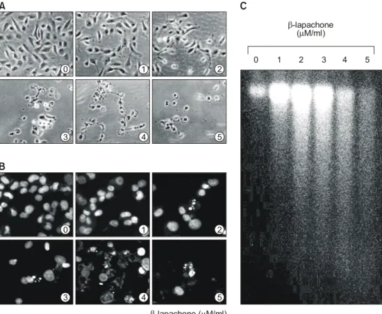
β-lapachone의 처리에 따른 A549 폐암세포의 증 식억제과정에서 암세포의 전체적인 형태변화를 관찰하기 위하여 48시간 동안 β-lapachone을 처리 한 후 위상차 현미경을 이용하여 조사한 결과는 Fig. 2A에 나타낸 바와 같다. 결과에서 알 수 있듯 이 β-lapachone이 함유된 배지에서 자란 세포는 β-lapachone 처리 농도 의존적으로 전체적으로 세
포질이 응축되면서 배양접시의 바닥에 낮게 부착 되는 듯한 모습을 보였다. 또한 세포의 모양이 길 어지면서 돌기와 같은 형태가 분지를 이루기 시 작하면서 membrane shrinking 및 세포의 rounding up 현상 등 매우 심한 세포의 형태적 변형을 초래 하였으며 3μM 농도에서 거의 모든 세포들이 부 착력을 상실하여 배지 위로 부유되는 현상을 관 찰할 수 있었다. 이는 정상 전립선 상피세포에서 관찰된 dendrite 형태와 비슷한 구조를 형성하는 것과는 매우 대조적인 양상이었으나,15) 선행 연구 의 결과들21,22,24,28)
과는 유사하여 암세포의 종류에 따라 β-lapachone에 의한 세포의 형태 변형은 다 소 차이가 있었음을 알 수 있었다.
Fig. 2. Morphological changes, chromatin chondensation and DNA fragmentation by β-lapachone treatment in A549 human lung carcinoma cells. (A) Cells were untreated or treated with β-lapachone for 48 h, stained and then photographed by microscope (Magnification, ×200). (B) The untreated cells and β-lapachone treated cells for 48 h were harvested, and spun down. After fixing, the cells were stained with DAPI solution. Stained nuclei were then observed under fluorescent microscope using blue filter (Magnification, ×400). (C) Genomic DNA was extracted and analyzed by 1% agarose gel electrophoresis in the presence of EtBr.
β-lapachoneµ ( M/ml)
β-lapachone ( M/ml)µ
0 1 2 3 4 5
A C
B
3 3
0 0
4 4
1 1
5 5
2 2
β-lapachone에 의한 이상의 암세포 세포성장 억 제에 따른 심한 형태적 변형이 apoptosis 유발과 연관성이 있는지를 조사하기 위하여 먼저 DAPI 염색에 의한 암세포의 핵 형태를 조사하였다. Fig.
2B에 나타낸 바와 같이 정상 배지에서 자란 암세 포에서는 핵의 형태가 뚜렷하게 정상으로 염색이 되었으나 β-lapachone이 함유된 배지에서 자란 암 세포에서는 apoptosis의 전형적인 특징인 chro- matin condensation 현상에 의한 apoptotic body를 쉽게 관찰할 수 있었다.32,33) 한편 apoptotic body 형성과 함께 apoptosis 유발의 직접적인 증거에 해 당하는 DNA fragmentation 여부를 agarose gel 전 기영동으로 조사하였다. 이를 위하여 정상 및 β- lapachone이 함유된 배지에서 자란 세포를 대상으 로 총 DNA를 추출하여 조사한 결과는 Fig. 2C에 나타낸 바와 같다. 즉 β-lapachone이 처리된 암세 포에서는 apoptosis가 일어난 세포들에서 볼 수 있 는 전형적인 DNA laddering을 관찰할 수 있었다.
이는 결국 β-lapachone의 처리에 의하여 endonu- clease가 활성화되어 chromosomal DNA가 단편화 되었음을 의미하는 것이며,32,33) β-lapachone 처리 에 의한 이상의 두 가지 현상은 간암세포,24) 백혈
병세포,18,25) 유방암세포29) 및 대장암세포28) 등에서
관찰된 결과들과 일치하였다. 따라서 이상의 결과 에서 β-lapachone 처리에 의한 A549 폐암세포의 증식 억제가 apoptosis 유발과 밀접한 관련이 있음 을 알 수 있었다.
3) Bcl-2 family의 발현에 미치는 β-lapa- chone의 영향
Bcl-2 family에 속하는 몇 가지 중요한 인자들은 apoptosis 유발 조절에 가장 대표적인 유전자로 알 려져 있는데, 그중 Bcl-2는 anti-apoptotic 분자로서 apoptosis의 유발을 억제하는 기능을 가지며, Bax 는 pro-apoptotic 분자로 Bax 단백질 발현의 증가 는 apoptosis의 유발과 관계가 있다. 이들 두 유전 자는 세포 내 소기관 중 mitochondria로부터의 cytochrome c를 유리시켜 종양억제 유전자인 p53, caspases, DNA의 단편화와 연관된 endonuclease 등 의 활성을 조절한다.34,35) 이들은 서로 dimer의 형 태로 존재하며 그들의 발현 수준에 변화가 초래 되면 apoptosis가 유발되는 것으로 알려져 있다.5,6)
따라서 인체 폐암세포 A549에서 β-lapachone에 의한 apoptosis 유발에 이들 유전자가 관련되어 있 는지의 여부를 RT-PCR 및 Western blotting으로 조사한 결과는 Fig. 3에 나타낸 바와 같다. 결과에 서 알 수 있듯이 apoptosis를 유발하는 것과 관련 이 있는 Bax 유전자의 발현은 전사 및 번역 수준 에서 β-lapachone 처리 농도 의존적으로 매우 증 가된 반면, Bcl-2의 발현은 특히 단백질 수준에서 β-lapachone 처리 농도 의존적으로 매우 감소되었 다. 그리고 Bcl-2와 함께 apoptosis 유발 억제에 관 여하는 Bcl-xL의 경우 β-lapachone 처리에 따른
Fig. 3. Up-regulation of Bax and down-regulation of Bcl-2 by β-lapachone treatment in A549 human lung carcinoma cells. A549 cells were treated with indicated concentrations of β-lapachone. (A) After 48 h incuba- tion, total RNAs were isolated and RT-PCR was per- formed using Bax, Bcl-2 and Bcl-xL primers described in materials and methods. GAPDH was used as a house-keeping control gene. (B) After 48 h incubation, cells were lysed and equal proteins were resolved on 12% SDS-polyacrylamide gels and transferred onto nitrocellulose membranes. Western blots were detected with antibodies against Bax, Bcl-2 and Bcl-xL, and ECL detection.
0
0 1
1 2
2 3
3 4
4
Bax
Bax Bcl-2
Bcl-2 Bcl-xL
Bcl-xL GAPDH
Actin
A
B
발현의 큰 변화는 관찰할 수 없었으나, 저농도 처 리군에서부터 인산화의 정도가 매우 높게 나타남 을 알 수 있었다. 이는 결국 β-lapachone 처리에 의한 apoptosis이 유발에는 최소한 Bcl-2 family가 중요한 역할을 하고 있음을 의미하는 것이며, Bax 의 상대적 발현 증가로 인한 apoptosis 유발 관련 효소들의 활성화가 이루어지고 있음을 시사하여 주는 것이다. 본 연구의 결과는 Choi 등22,28)에 의 한 대장암세포와 전립선암세포, Wuerzberger 등29) 에 의한 유방암세포, Weller 등23)에 의한 신경교종 등에서 관찰된 β-lapachone에 의한 apoptosis 유발 에서 나타난 현상과 유사한 현상임을 알 수 있었 다. 또한 Bcl-2 유전자가 인위적인 과발현된 백혈 병세포에서 β-lapachone에 의한 apoptosis가 억제 되었음25)은 β-lapachone에 의한 apoptosis 유발에 Bcl-2의 발현감소에 따른 Bax의 발현증가가 중요 한 역할을 하고 있음을 뒷받침하여 주는 결과이 다.
4) p53 및 p21WAF1/CIP1의 발현에 미치는 β-lapachone의 영향
다음은 β-lapachone의 처리에 의한 폐암세포의 증식억제 현상이 종양억제 유전자 또는 세포주기 조절 억제인자들의 발현 변화와 상관성이 있는지 의 여부를 조사하기 위하여 현재까지 알려진 종 양억제 유전자 중 가장 중요한 p53 및 전체적인 세포주기의 진행에 중요한 역할을 하는 Cdk inhi- bitor p21의 발현에 미치는 β-lapachone의 영향을 조사하였다. Fig. 4의 결과에서 볼 수 있듯이 p53 및 p21 두 유전자의 발현은 β-lapachone의 처리에 따라 전사 및 번역 수준에서 모두 증가하였음을 알 수 있었다. Cdk inhibitor인 p21은 p53의 발현 증가에 의하여 전사 수준이 조절될 수 있으며,36∼
38) 암세포의 증식 억제, apoptosis 및 분화 유도에 중요한 역할을 하는 세포주기 전반에 걸친 가장 중요한 조절인자란 점에서 β-lapachone의 처리에 의하여 두 유전자가 동시에 발현이 증가되었다는 점은 매우 흥미로운 결과라고 사료된다. 그러나 Hueber 등39)의 결과에 의하면 인체 망막 상피세포 에서는 β-lapachone의 처리에 의한 apoptosis 유발 에서는 p53 및 p21의 유전자 발현이 관찰되지 않 았다고 보고한 바 있고, 인체 유방암세포에서도
이와 유사한 결과가 보고된 바 있다.29) 또한 Weller 등23)의 결과에 의하면, 정상 p53 유전자를 가진 신경교종세포에서는 β-lapachone의 처리에 의하여 p21의 발현이 p53과 동시에 증가하였으나, 동일조건에서 p53 유전자가 결여된 세포에서는 p21의 발현에 아무런 영향이 없었다. 그러나 최근 본 연구실의 결과에 의하면 p53 유전자가 결여된 인체 전립선암세포에서는 β-lapachone의 처리에 의하여 p53 비의존적으로 p21의 단백질 발현이 증가되었으며, p21 promoter 활성 또한 β-lapachone 의 처리 농도 및 시간 의존적으로 증가되었던 점
22)등을 고려해 볼 때 p53 비의존적인 p21 활성 조 절 가능성은 여전히 높을 것으로 예상되며, 세포 주에 따른 세포주기 조절 관련 유전자들에 관한
Fig. 4. Up-regulation of tumor suppressor p53 and Cdk inhibitor p21 by β-lapachone treatment in A549 human lung carcinoma cells. A549 cells were treated with indicated concentrations of β-lapachone. (A) After 48 h incubation, total RNAs were isolated and RT-PCR was performed using p53 and p21 primers described in materials and methods. GAPDH was used as a house- keeping control gene. (B) After 48 h incubation, cells were lysed and equal proteins were resolved on 10% or 12% SDS-polyacrylamide gels and transferred onto nitro- cellulose membranes. Western blots were detected with antibodies against p53 and p21, and ECL detection.
0
0 1
1 2
2 3
3 4
4
p53
p53 p21
p21 GAPDH
Actin
A
B
연구가 더 필요할 것으로 예상된다.
5) Telomere 조절관련 인자들의 발현에 미치 는 β-lapachone의 영향
한편 telomere는 염색체의 말단 부위에 repeat sequences [(TTAGGG)n]로 이루어져 있으며, 이런 반복구조의 형성 및 유지에 관여하는 효소가 telo- merase이다.40∼42) 정상 체세포에서는 telomerase의 활성이 없기 때문에 세포가 분열할수록 telomere 의 길이는 짧아지게 되지만, 암세포의 90% 이상 에서는 telomerase의 catalytic subunit 단백질을 cod- ing하는 hTERT가 과발현되어 있고 이로 인한 높 은 telomerase의 활성을 나타내고 있다. 따라서 노 화, 혈관신생 및 면역계질환 등에서 뿐만 아니라 암의 발생과 진행도 이들과 밀접한 연관성이 있 으며, 암의 진단과 진행의 정도를 나타내는 지표 로 사용될 수 있음을 시사하여 준다.40∼42) 노화의 개념에서 결국 telomere의 소실은 염색체의 안정 성이 상실되는 것이며 이는 DNA damaging agent 에 의한 p53 및 p21의 발현증가로 인한 세포주기 교란 유발로 설명되어지고 있다.43,44) 특히 암과 연관된 부분에서 telomerase의 활성은 hTERT 유 전자의 발현 조절에 의한 것이고, hTERT 유전자 의 promoter 부위에는 다른 유전자의 promoter 보 다 훨씬 더 많은 전사조절인자의 결합부위를 보 유하고 있어 activator 또는 repressor로 작용할 수
있다.40,42) 이러한 중요성을 고려하여 β-lapachone
의 처리에 의한 A549 폐암세포의 증식 억제가 염 색체 말단에 존재하는 telomere의 조절과 어떤 관 계가 있는지를 조사하였다. Fig. 5의 결과에서 알 수 있듯이 β-lapachone의 처리에 의하여 hTERT 및 hTR 유전자의 전사수준이 다소 감소되었으나, 또 다른 조절인자인 TEP-1 및 hTERT의 발현 조 절에 중요한 인자에 해당하는 c-myc의 발현은 β- lapachone의 처리에 의하여 큰 변화가 관찰되지 않았다. 그러나 β-lapachone 처리에 의한 암세포 의 증식억제 효과와 telomere 관련 분야에 관한 기존의 연구가 전혀 이루어져 있지 않았기 때문 에 이에 관한 보다 구체적이 후속 연구가 연관된 뒤 따라야 할 것으로 생각된다.
결 론
DNA topoismerase 활성 억제제로 알려진 β- lapachone은 남미지역에 서식하는 Tabebuia avel- lanedae의 수피에서 동정된 천연 quinone계 물질 로서 다양한 종류의 암세포에서 apoptosis를 유발 하는 것으로 알려져 왔다. 본 연구에서는 β- lapachone의 항암활성 기전 해석의 일환으로 인체 폐암세포 A549의 증식에 미치는 β-lapachone의 영향을 조사하였다. Hemocytometer count 및 MTT assay에 의한 결과에서, β-lapachone의 처리에 따 라 A549 폐암세포들은 β-lapachone 처리 농도 의 존적으로 증식이 억제되었음을 확인하였으며, β- lapachone이 처리된 세포에서 apoptosis가 유발된 세포에서 특징적으로 관찰되는 chromatin conden- sation 및 DNA fragmentation 현상을 관찰하였다.
이러한 β-lapachone의 apoptosis 유발에는 pro-apop- totic 인자인 Bax의 발현증가에 따른 anti-apoptotic 인자인 Bcl-2 발현의 감소와 직접적인 연관성이 있음을 관찰할 수 있었다. 그리고 세포의 성장 조 절에 중요한 역할을 하는 p53 및 p21의 발현 증가 및 telomere의 길이 조절과 관련이 있는 유전자의 Fig. 5. Effects of β-lapachone on the levels of telo- mere-regulatory genes expression in A549 human lung carcinoma cells. Cells were treated with various con- centrations of β-lapachone. After 48 h incubation, total RNAs were isolated, and RT-PCR was performed using indicated primers. The amplified PCR products were subjected to electrophoresis in a 1% agarose gel and visualized by EtBr staining. GAPDH was used as a house-keeping control gene.
0 1 2 3 4
hTERT
hTR
TEP-1
c-myc
GAPDH
발현 교란 역시 β-lapachone에 의한 암세포의 성 장억제와 어느 정도 관련이 있는 것으로 추정되 어진다. 그러나 본 연구의 결과만으로 β-lapa- chone의 항암작용 여부를 직접적으로 논하기에는 어렵기 때문에 다양한 추가적인 실험이 필수적으 로 수행되어야 할 것으로 생각된다.
참 고 문 헌
1) Evans VG. Multiple pathways to apoptosis. Cell Biol Int 1993; 17: 461-476.
2) Shi L, Nishioka WK, Th'ng J, Bradbury EM, Lit- chfield DW, Greenberg AH. Premature p34cdc2 activation required for apoptosis. Science 1994; 263:
1143-1145.
3) Chiarugi V, Magnelli L, Turchetti A, Cinelli M, Ca- vari S, Ruggiero M. Cell survival and death pro- grammes. Pharmacol Res 1994; 29: 101-110.
4) Nagata S. Apoptosis by death factor. Cell 1997; 88:
355-365.
5) Lenaz G, Bovina C, Formiggini G, Castelli GP.
Mitochondria, oxidative stress, and antioxidant de- fences. Acta Biochim Pol 1999; 46: 1-21.
6) Antonsson B, Martinou JC. The Bcl-2 protein fam- ily. Exp Cell Res 2000; 256: 50-57.
7) Martin SJ, Finucane DM, Amarante-Mendes GP, O'Brien GA, Green DR. Phosphatidylserine exter- nalization during CD95-induced apoptosis of cells and cytoplasts requires ICE/CED-3 protease activity.
J Biol Chem 1996; 271: 28753-28756.
8) Steinhusen U, Badock V, Bauer A, Behrens J, Wit- tman-Liebold B, Dorken B, Bommert K. Apoptosis- induced cleavage of β-catenin by caspase-3 results in proteolytic fragments with reduced transactivation potential. J Biol Chem 2000; 275: 16345-16353.
9) Schaffner-Sabba K, Schmidt-Ruppin KH, Wehrli W, Schuerch AR, Wasley JW. β-lapachone: synthesis of derivatives and activities in tumor models. J Med Chem 1984; 27: 990-994.
10) Li CJ, Averboukh L, Pardee AB. β-Lapachone, a novel DNA topoisomerase I inhibitor with a mode of action different from camptothecin. J Biol Chem 1993; 268: 22463-22468.
11) Pardee AB, Li YZ, Li CJ. Cancer therapy with β- lapachone. Curr Cancer Drug Targets 2002; 2: 227- 242.
12) Lopes JN, Cruz FS, Docampo R, Vasconcellos ME, Sampaio MC, Pinto AV, Gilbert B. In vitro and in
vivo evaluation of the toxicity of 1,4-naphthoquinone and 1,2-naphthoquinone derivatives against Trypano- soma cruzi. Ann Trop Med Parasitol 1978; 72: 523- 531.
13) Goijman SG, Stoppani AO. Effects of β-lapachone, a peroxide-generating quinone, on macromolecule synthesis and degradation in Trypanosoma cruzi.
Arch Biochem Biophys 1985; 240: 273-280.
14) Boothman DA, Schlegel R, Pardee AB. Anticarcino- genic potential of DNA-repair modulators. Mutat Res 1988; 202: 393-411.
15) Guiraud P, Steiman R, Campos-Takaki GM, Seigle- Murandi F, Simeon de Buochberg M. Comparison of antibacterial and antifungal activities of lapachol and β-lapachone. Planta Med 1994; 60: 373-374.
16) Li CJ, Wang C, Pardee AB. Induction of apoptosis by β-lapachone in human prostate cancer cells.
Cancer Res 1995; 55: 3712-3715.
17) Docampo R, Cruz FS, Boveris A, Muniz RP, Esqui- vel DM. β-lapachone enhancement of lipid peroxi- dation and superoxide anion and hydrogen peroxide formation by sarcoma 180 ascites tumor cells.
Biochem Pharmacol 1979; 28: 723-728.
18) Shiah SG, Chuang SE, Chau YP, Shen SC, Kuo ML. Activation of c-Jun NH2-terminal kinase and subsequent CPP32/Yama during topoisomerase inhi- bitor β-lapachone-induced apoptosis through an oxidation-dependent pathway. Cancer Res 1999; 59:
391-398.
19) Planchon SM, Pink JJ, Tagliarino C, Bornmann WG, Varnes ME, Boothman DA. β-lapachone-induced apoptosis in human prostate cancer cells: involve- ment of NQO1/xip3. Exp Cell Res 2001; 267:
95-106.
20) Planchon SM, Wuerzberger S, Frydman B, Witiak DT, Hutson P, Church DR, Wilding G, Boothman DA. β-lapachone-mediated apoptosis in human pro- myelocytic leukemia (HL-60) and human prostate cancer cells: a p53-independent response. Cancer Res 1995; 55: 3706-3711.
21) Don MJ, Chang YH, Chen KK, Ho LK, Chau YP.
Induction of CDK inhibitors (p21 (WAF1) and p27 (Kip1)) and Bak in the β-lapachone-induced apop- tosis of human prostate cancer cells. Mol Pharmacol 2001; 59: 784-794.
22) Choi YH, Kang HS, Yoo MA. Suppression of human prostate cancer cell growth by β-lapachone via down-regulation of pRB phosphorylation and induction of Cdk inhibitor p21 (WAF1/CIP1). J
Biochem Mol Biol 2003; 36: 223-229.
23) Weller M, Winter S, Schmidt C, Esser P, Fontana A, Dichgans J, Groscurth P. Topoisomerase-I inhi- bitors for human malignant glioma: differential mod- ulation of p53, p21, bax and bcl-2 expression and of CD95-mediated apoptosis by camptothecin and β- lapachone. Int J Cancer 1997; 73: 707-714.
24) Lai CC, Liu TJ, Ho LK, Don MJ, Chau YP. β- lapachone induced cell death in human hepatoma (HepA2) cells. Histol Histopathol 1998; 13: 89-97.
25) Planchon SM, Wuerzberger-Davis SM, Pink JJ, Ro- bertson KA, Bornmann WG, Boothman DA. Bcl-2 protects against β-lapachone-mediated caspase 3 acti- vation and apoptosis in human myeloid leukemia (HL-60) cells. Oncol Rep 1999; 6: 485-492.
26) Gupta D, Podar K, Tai YT, Lin B, Hideshima T, Akiyama M, LeBlanc R, Catley L, Mitsiades N, Mitsiades C, Chauhan D, Munshi NC, Anderson KC. β-lapachone, a novel plant product, overcomes drug resistance in human multiple myeloma cells.
Exp Hematol 2002; 30: 711-720.
27) Huang L, Pardee AB. β-lapachone induces cell cy- cle arrest and apoptosis in human colon cancer cells.
Mol Med 1999; 5: 711-720.
28) Choi BT, Cheong J, Choi YH. β-lapachone-induced apoptosis is associated with activation of caspase-3 and inactivation of NF-kappaB in human colon cancer HCT-116 cells. Anticancer Drugs 2003; 14:
845-850.
29) Wuerzberger SM, Pink JJ, Planchon SM, Byers KL, Bornmann WG, Boothman DA. Induction of apop- tosis in MCF-7:WS8 breast cancer cells by β- lapachone. Cancer Res 1998; 58: 1876-1885.
30) Pink JJ, Wuerzberger-Davis S, Tagliarino C, Plan- chon SM, Yang X, Froelich CJ, Boothman DA.
Activation of a cysteine protease in MCF-7 and T47D breast cancer cells during β-lapachone-medi- ated apoptosis. Exp Cell Res 2000; 255: 144-155.
31) Tagliarino C, Pink JJ, Dubyak GR, Nieminen AL, Boothman DA. Calcium is a key signaling molecule in β-lapachone-mediated cell death. J Biol Chem 2001; 276: 19150-19159.
32) Arends MJ, Morris RG, Wylli AH. Apoptosis. The role of the endonuclease. Am J Pathol 1990; 136:
593-608.
33) Cohen JJ, Duke RC, Fadok VA, Sellins KS. Apop- tosis and programmed cell death in immunity. Annu
Rev Immunol 1992; 10: 267-293.
34) Lowe SW, Schmitt EM, Smith SW, Osborne BA, Jacks T. p53 is required for radiation-induced apop- tosis in mouse thymocytes. Nature 1993; 362: 847- 849.
35) Rosse T, Olivier R, Monney L, Rager M, Conus S, Fellay I, Jansen B, Borner C. Bcl-2 prolongs cell survival after Bax-induced release of cytochrome c.
Nature 1998; 391: 496-499.
36) Miyashita T, Reed JC. Tumor suppressor p53 is a direct transcriptional activator of the human bax gene. Cell 1995; 80: 293-299.
37) Harper JW, Adami GR, Wei N, Keyomarsi K, Elledge SJ. The p21 Cdk-interacting protein Cip1 is a potent inhibitor of G1 cyclin-dependent kinases.
Cell 1993; 75: 805-816.
38) El-Deiry WS, Tokino T, Velculesco VE, Levy DB, Parsons R, Trent JM, Lin D, Mercer EW, Kinzler KW, Vogelstain B. WAF1, a potential mediator of p53 tumor suppression. Cell 1993; 75: 817-825.
39) Hueber A, Esser P, Heimann K, Kociok N, Winter S, Weller M. The topoisomerase I inhibitors, cam- ptothecin and β-lapachone, induce apoptosis of hu- man retinal pigment epithelial cells. Exp Eye Res 1998; 67: 525-530.
40) Kyo S, Inoue M. Complex regulatory mechanisms of telomerase activity in normal and cancer cells: How can we apply them for cancer therapy. Oncogene 2002; 21: 688-697.
41) Poole JC, Andrews LG, Tollefsbol TO. Activity, function, and gene regulation of the catalytic subunit of telomerase (hTERT). Gene 2001; 269: 1-12.
42) Cerni C. Telomeres, telomerase, and myc. An up- date. Mutat Res 2000; 462: 31-47.
43) Narayan S, Jaiswal AS, Multani AS, Pathak S. DNA damage-induced cell cycle checkpoints involve both p53-dependent and -independent pathways: role of telomere repeat binding factor 2. Br J Cancer 2001;
85: 898-901.
44) Vaziri H, West MD, Allsopp RC, Davison TS, Wu YS, Arrowsmith CH, Poirier GG, Benchimol S.
ATM-dependent telomere loss in aging human diploid fibroblasts and DNA damage lead to the post-translational activation of p53 protein involving poly (ADP-ribose) polymerase. EMBO J 1997; 16:
6018-6033.