난소암 세포주에서 인산화효소 억제제 flavopiridol에 의한 세포자멸사의 유도
허 수 영ㆍ이 준 모
가톨릭대학교 의과대학 산부인과학교실
목적:사이클린 의존성 인산화 효소(cyclin-dependent kinase) 억제제의 하나인 flavopiridol은 사람 종양 세포주 들에서 세포주기의 정지 및 세포자멸사를 유도하는 약제이다. 본 연구는 사람 난소암 세포주들을 대상으로 하여 flavopiridol을 처치한 후 세포자멸사에 미치는 영향과 그 작용 기전을 밝히려고 시도되었다.
연구 방법:TOV-21G와 TOV-112D 세포주가 이용되었으며, 세포 생존도는 MTT assay로 측정하였고, 세포자멸사는 TUNEL assay와 annexin-V binding 법으로 조사하였다. Western blot 방법으로 세포자멸사 관련 단백질의 발현을 조사하였다. MAP kinase의 활성은 non-radioactive MAP kinase assay로 측정하였다.
결과:TOV-21G와 TOV-112D에 flavopiridol로 처치하였을 경우 세포 생존도는 처치 용량 및 시간에 의존적으로 감소하였다. 또한, TUNEL assay와 annexin-V binding 법으로 flavopiridol 처치 용량에 의존적인 세포자멸사 유도가 일어남을 확인하였다. 두 세포주 모두에서 flavopiridol로 처치하였을 경우 anti-apoptosis 단백질인 Mcl-1과 Bcl-XL
및 XIAP의 발현이 감소하였으나, Bax와 Bcl-2 및 AIF의 발현은 뚜렷한 변화가 관찰되지 않았다. p53 돌연변이를 가진 TOV-112D에서 TOV-21G에서와 같이 flavopiridol 투여 후 p53 단백질의 발현이 증가하고, 세포자멸사가 처치 농도에 따라 증가하는 사실은 flavopiridol에 의하여 유도되는 세포자멸사는 p53과는 연관이 없는 것으로 판단되었다.
그리고 이러한 세포자멸사는 caspase 9과 caspase 3의 활성 증가와 연관이 있고, extracellular signal-regulated protein kinase의 활성 감소와도 관련이 있는 것으로 나타났다.
결론:Flavopiridol이 난소암 세포주에서 세포자멸사를 일으키고 세포독성을 나타내는 기전을 완전히 규명하지는 못하 였지만, 본 연구결과는 flavopiridol이 난소암 세포주에서 효과적으로 apoptosis를 일으켜 항종양능력을 나타낼 수 있다 는 사실과 향후에 계속 치료제제로서의 연구가 가치가 있을 것임을 제시하여 준다.
중심단어:Flavopiridol, 난소암 세포주, 세포자멸사
논문접수일:2007년 10월 10일 채택일:2008년 2월 15일
교신저자: 이준모, 137-701 서울시 서초구 반포동 505번지 가톨릭대학교 의과대학 산부인과학교실
전화:02) 590-1376ㆍ전송:02) 595-1549 E-mail:[email protected]
서 론
사이클린 의존성 인산화효소(cyclin-dependent kinase, cdk) 억제제 중의 하나인 flavopiridol [5,7-dihydroxy-8- (4-N-methyl-2-hydroxypyridyl)-6’-chloroflavone hydroch- loride]은 인도에 토착하는 식물인 Dysoxylum binectari- ferum에서 유래하는 rohitukine과 구조가 유사한 합성 flavone으로서 세포주기에 작용하는 새로운 약제로서 개
발되어 임상적 이용에 관한 연구가 진행 중이다.1,2 Flavopiridol이 세포주기에 영향을 끼치는 작용기전은 크게 3가지로 알려져 있는데, 첫째는 ATP와 경쟁적으로 cdk에 결합하여 cdk1, 2, 4, 6에 각각 억제 작용을 하고, 둘째는 cdk-activating kinase (CAK)인 cdk7/cyclin H를 억 제하여 cdk들이 threonine 160/161에서 인산화 (phosph- orylation)가 일어나지 못하도록 하여 세포주기의 진행을 방해하는 것이고, 셋째는 cdk4와 cdk6의 보조인자 (cofactor)인 cyclin D1과 cyclin D3를 전사수준에서 발현 저하(downregulation)시켜 세포주기의 진행을 억제하는 것이다.3-6 Flavopiridol의 다른 작용으로는 vascular endothelial growth factor (VEGF) mRNA의 안정화를 통 하여 혈관생성을 억제하는 기능이 있다.7 또 다른 중요한
기능으로 종양세포의 세포자멸사(apoptosis)를 유도하는 작용이 있다. 즉, 두경부종양 세포주인 HN30은 bleomycin 이나 gamma-irradiation에 의한 세포자멸사가 잘 일어나지 않으나, flavopiridol의 처치 후 세포자멸사가 유도된다.
백혈병/림프종 이종이식(xenograft)과 두경부종양 이종 이식에서도 flavopiridol로 처치 후 종양 억제 효과를 보 였고, terminal deoxynucleotidyl transferase-mediated dUTP nick end labeling (TUNEL) 검사로 세포자멸사 유 도를 관찰하였다.8,9
그러나, flavopiridol에 의한 세포자멸사 신호전달 과정 은 아직까지 정확히 알려져 있지 않다. 최근의 연구에 의하면 P53의 발현 상태나 세포 내 Bcl-2/Bax 수준과 관 계없이 독립적으로 flavopiridol에 의한 세포자멸사가 발 생한다고 보고되었으며,10,11 anti-apoptosis 단백질들인 Mcl-1, BAG-1 등의 저하가 관찰된다.12 2003년 Raju 등이 OCA-I 난소암 세포주에서 flavopiridol 투여 후 세포주기 정지와 세포자멸사 유도 및 방사선민감성(radiosensiti- vity)이 증가하는 것을 관찰하였지만, 세포자멸사의 발 생 기전에 대하여는 caspase 3의 발현 증가만을 조사하였 고,13 아직까지 난소암 세포주들에서 flavopiridol 투여에 의한 세포자멸사 유도와 그 발생기전에 대하여는 잘 알 려져 있지 않다.
최근의 많은 임상전시험에서 flavopiridol을 paclitaxel, etoposide, cisplatin 등의 항암화학제제와 병합투여 후 세 포독성 효과가 상승되는 현상이 밝혀져 flavopiridol의 임 상적 적용이 기대되고 있다.14,15
이에 본 연구는 난소암세포주에서 새로운 항암화학제 제로서 flavopiridol의 역할을 알아보기 위하여, flavopiri- dol의 세포자멸사 유도 작용을 알아보고자 하였다. 특히, anti-apoptosis 단백질인 Bcl-2, Mcl-1, Bcl-XL, X-linked inhibitor of apoptosis (XIAP) 등과 pro-apoptosis 단백질인 Bax, apoptosis-inducing factor (AIF) 및 caspase 3, caspase 9 등의 발현들을 조사하여 flavopiridol의 세포자멸사를 유도하는 신호전달경로를 규명하고자 하였다.
연구 대상 및 방법
1. 세포의 분리와 배양
난소암 세포주인 TOV-21G와 TOV-112D를 American Type Culture Collection (ATCC, Manassas, VA)으로부터
구입하였다. TOV-21G 난소암 세포주는 정상 P53을 발 현하지만, TOV-112D 난소암 세포주는 P53 exon 6의 175 arginine이 histidine으로 치환된 돌연변이형을 발현한 다.16
5% CO2, 37oC 및 95% 습도를 유지한 배양기에서 100 unit/ml penicillin G와 100μg/ml streptomycin (Boehringer Mannheim, Mannheim, Germany), 15% fetal bovine serum (FBS, Gibco/BRL, Grand Island, NY)이 포함된 MCDB 105 medium과 medium 199의 1:1 혼합 배양액(Sigma, St Louis, Mo)으로 T-75 cm2 culture flask에서 세포들을 배양하였다.
2. 약제 처치
cdk 억제제인 flavopiridol (Aventis Pharma SA, Paris, France)을 dimethyl sulfoxide (DMSO)에 녹여 -70oC에 보 관하고 사용하였다. Flavopiridol로 처치하기 3일 전에 HBSS (Sigma), 0.05% trypsin (Flow Laboratories, Irvine, UK) 및 0.53 mM EDTA (J.T.Baker, Deventer, Netherlands) 의 혼합 용액으로 TOV-21G와 TOV-112D 난소암 세포 들을 분리 수확하고, 그 세포 부유액을 250×g에서 5분간 원심분리 후 T-75 cm2 culture flask당 세포가 1×10⁵이 되 도록 각각 분주하여 배양하였다. 지수증식기(exponential phase)의 세포들을 계대 배양하여 다양한 농도의 flavopiridol을 투여하여 조사하였다.
Flavopiridol 처치 농도는 지수적으로 성장하는 세포에 서 cdk의 kinase 활성을 직접적으로 억제하는 flavopiridol 의 IC50 및 다른 연구자들의 flavopiridol 처치 농도를 참 고하였다.6,13,15
3. 세포 생존도(cell viability)의 측정
TOV-21G와 TOV-112D 난소암 세포주에 대한 flavopiridol의 처치 농도와 시간에 따른 세포 독성을 microculture tetrazolium (MTT) assay로 측정하였다.
지수증식기에 있는 TOV-21G와 TOV-112D 난소암 세 포들을 분리 수확 후, 배양액 1 ml당 1×10⁵이 되도록 세 포의 농도를 조정하고 96-well flat-bottomed tissue culture plate (Costar, Cambridge, MA)의 well당 100μl 씩 각각 분주하여 37oC, 5% CO2 배양기에서 24시간 배양하였다.
여기에 50 nM, 100 nM, 300 nM, 500 nM, 1 μM의 농도 로 flavopiridol를 각각 첨가한 100μl의 배양액을 well에
분주하여 24시간과 48시간 동안 다시 배양하였다. 대조 군으로는 배양액만을 첨가하였다. 이후 인산완충용액 (phosphate buffer saline, PBS)에 녹인 MTT 용액[3-(4,5 -dimethylthiazol-2-yl)-2,5-diphenyltetrazoliumbromide] 20 μl를 첨가하고 37oC에서 4시간 동안 반응시켰다. 이후, 상청액을 흡인하여 전부 버리고, kit에서 제공하는 발색 용액인 detergent reagent 100μl를 첨가하여 상온의 어두 운 곳에서 약 2시간 동안 반응시킨 후, ELISA plate reader (Molecular Device, Sunnyvale, CA)로 570 nm에서 흡광도를 측정하였다. 판독한 실험군의 흡광도를 대조 군의 흡광도와 비교하여 세포들의 생존도를 산출하였 다. 결과는 각 세포주에서 4회의 실험한 평균값에서 구 하였다.
4. 세포자멸사(apoptosis)의 판정 1) TUNEL 방법
DNA 분절(fragmentation)의 발생을 TUNEL 방법으로 조사하였는데, in situ cell death detection kit, Fluorescein (Boehringer Mannheim)을 이용하여 분석하였다. TOV- 21G와 TOV-112D 난소암 세포주들을 수확하여 Lab- Tek chamber slide (Nalge Nunc International, Rochester, NY)에 chamber당 1×10⁵의 세포를 분주하고 37oC, 5%
CO2 배양기에서 밤새 배양한 후 flavopiridol을 정해진 농 도에 따라 처지하고 24시간 및 48시간 동안 배양하였다.
4% paraformaldehyde (pH 7.4)를 첨가하여 상온에서 20 분간 고정한 후, PBS로 2회 세척하고, 0.1% Triton X-100, 0.1% sodium citrate가 포함된 permeabilization 용액으로 4oC에서 2분간 처치하였다. PBS로 다시 2회 세척한 후, terminal deoxynucleotidyl transferase (TdT)와 nucleotide mixture를 첨가하고 37oC에서 빛을 차단한 후 60분간 배 양하였다. PBS로 3회 세척하고 난 후, 형광 현미경 (fluorescence microscope) 하에서 세포자멸사를 조사하 였다. 세포자멸사가 일어나지 않은 세포의 대조 염색을 위하여 4,6-diamidino-2-phenylindole (DAPI, Sigma) 용액 을 이용하였다. 결과는 각 세포주에서 3회의 실험한 평 균값에서 구하였다.
2) Annexin V 염색
초기 단계의 세포자멸사는 Annexin-V-Flous staining kit (Roche Diagnostics, Mannheim, Germany)를 이용하여
측정하였다.
Flavopiridol 처치 전후의 TOV-21G와 TOV-112D 난소 암 세포주들을 수확하여 차가운 인산완충용액 1 ml에 1×10⁶의 세포 수가 되도록 각각 부유시키고, 200×g에서 5분간 원심 분리한 후 상청액을 버렸다. 이후 kit에서 제 공된 1 ml incubation buffer (HEPES buffer)에 20μl annexin-V-Fluos labeling reagent와 20μl propidium iodide solution를 더하여 만들어진 annexin-V-Fluos label- ing solution을 100μl씩 첨가하여 빛을 차단하고 상온에 서 15분간 배양하였다. 배양이 완료된 후에는 500μl의 incubation buffer를 각각 첨가하고, annexin-V-Flous와 propidium iodide에 염색된 세포들을 FACSVantage SE Flow Cytometer (Becton Dickinson)로 분석하였다. 데이 터의 분석은 CellQuest software (Becton Dickinson)를 이 용하였다.
5. Caspase 3 활성도 측정
Caspase 3 활성도는 caspase 3 activity detection kit (Upstate biotechnology, Lake Placid, NY)를 이용하여 측 정하였다.
Flavopiridol 처치 전후의 TOV-21G와 TOV-112D 난소 암 세포주 1×10⁶을 수확하여 PBS로 2회 세척한 후, 4oC 에서 15분간 lysis buffer [10 mM Tris/HCl (pH 7.5), 10 mM NaH2PO4/Na2HPO4 (pH 7.5), 130 mM NaCl, 0.1%
TritonX-100, 10 mM sodium pyrophosphate]로 용해하고 13,000 rpm으로 15분간 원심 분리한 후 BioRad Protein-Assay kit (Bio-rad Lab, Hercules, CA)를 이용하여 단백질 농도를 정량하였다. Fluorometer cuvette에 kit에 서 제공된 modified RIPA buffer [50 mM Tris/HCl (pH 7.4), 1% NP-40, 0.25% sodium deoxycholate, 150 mM NaCl, 1 mM EGTA] 1 ml를 넣은 후 DMSO에 용해시킨 caspase-3 형광기질(fluorometric substrate)인 50μM DEVD-AMC (Ac-Asp-Glu-Val-Asp-amino-4-methyl-coum arin)를 첨가하고 cell lysate를 200μl씩 분주하여 4oC에 서 5분간 반응시켰다. 형광의 세기는 Fluorometer (Molecular Devices Co, St. Louis, MO)를 이용하여 측정 하였다. 이때 흥분파장(excitation wavelength)은 380 nm 를, 방출파장(emission wavelength)은 460 nm를 각각 사 용하였다.
6. Western blot 분석
Flavopiridol 처치 전후의 TOV-21G와 TOV-112D 난소 암 세포주를 lysis buffer에 넣고 4oC에서 30분간 얼음에 방치하고 12,000 rpm으로 15분간 원심 분리하여, 그 상 청액을 실험에 사용하였다.
추출된 단백질 50μg을 sample buffer [125 mM Tris-HCl (pH 6.8), 20% glycerol, 4% sodium dodesylsulfate (SDS), 10% b-mercaptomethanol, 0.04% bromophenol blue]와 혼 합하여 10분간 끓이고, 10% SDS- polyacrylamide gel (Bio-rad Lab)에서 전압 150 V로 전기영동시켰다. 전기영 동 후 단백질을 PAGE transfer buffer (Amersham Life Science, Arlington Heights, IL)를 이용하여 50 mA로 4oC 에서 3시간 동안 nitrocellulose membrane (Amersham Life Science)에 옮긴 후, 5% blocking solution (5% fat milk)으 로 4oC에서 하룻밤 반응시키고 Tris buffered saline with Tween-20으로 세척하였다. 1차 항체를 TBS-T buffer에 1:200에서 1:500으로 희석하여 실온에서 2시간 반응 시키고, TBS-T buffer로 3회 세척하고, horseradish peroxidase 결합 2차 항체(Jackson Immunoresearch, Bar Harbor, MA)를 1:5000으로 희석하여 실온에서 1시간 반응시켜, ECL kit (Amersham Life Science)를 이용하여 Hyperflim에 감광하였다.
1차 항체는 세포자멸사와 관련된 단백질들에 대한 항 체로서 anti-p53 rabbit polyclonal Ab (Santa Cruz Biotechnology Inc, Santa Cruz, CA), anti-Bcl-XL rabbit polyclonal Ab (BD Pharmigen, San Diego, CA), anti-Bax rabbit polyclonal Ab (Upstate Biotechnology), anti-Bcl-2 mouse monoclonal Ab (Upstate Biotechnology), anti-Mcl-1 rabbit polyclonal Ab (R&D Systems), anti-AIF rabbit polyclonal Ab (R&D Systems), anti-XIAP rabbit polyclonal Ab (R&D Systems), anti-caspase 9 mouse monoclonal Ab (Upstate Biotechnology)를 각각 사용하였다.
7. Mitogen activated protein (MAP) kinase 활성도 측정 MAP kinase의 활성도는 non-radioactive MAP kinase assay kit (Upstate Biotechnology)를 이용하여 측정하였다.
Flavopiridol 처치 전후의 TOV-21G와 TOV-112D 난소 암 세포주를 lysis buffer에서 용해시킨 후 12,000 rpm으 로 15분간 원심 분리하여 세포 부유액을 얻었다. 이 세포 부유액에 anti-ERK (Santa Cruz Biotechnology Inc) 항체
를 1μl씩 넣고 얼음에서 4시간 동안 반응시키고, protein A-Sepharose bead를 100μl 추가하여 1시간 동안 추가로 반응시켰다. 이 반응액을 4,000 rpm에서 약 5분간 원심 분리하여 상청액은 버리고 protein A-항원-항체 복합체 인 침전체를 얻은 후 lysis buffer를 다시 첨가하여 얼음에 서 약 10분간 다시 반응시키고 원심 분리하는 세척과정 을 4회 반복하였다. Kit에서 제공된 assay dilution buffer [ADB; 20 mM MOPS, (pH 7.2), 25 mM b-glycerol phosphate, 5 mM EGTA, 1 mM sodium orthovanadate, 1 mM dithiothreitol]와 magnesium/ATP Cocktail (75 mM magnesium chloride, 500 mM ATP)을 10μl씩 혼합하고 20μg의 MAP kinase substrate (dephosphorylated myelin basic protein, MBP)를 첨가한 후 MAP kinase preparation 인 침전체를 100μg 넣고 30oC에서 잘 흔들면서 30분간 반응시켰다.
얻어진 반응물은 TBS-T buffer와 sample buffer를 추가 한 후 SDS-PAGE 과정을 거치고 western blot 분석을 시 행하였다. Nitrocellulose membrane으로 전이된 단백질에 반응하는 1차 항체로서 kit에서 제공된 mouse anti-human phospho-MBP antibody를 1:200으로 희석하여 사용하 였고, 2차 항체로는 goat anti-mouse horseradish peroxi- dase conjugated IgG (Jackson Immunoresearch)를 1:2000 으로 희석하여 사용하였다.
8. 통계분석
모든 실험값은 평균값±표준편차(mean±standard devia- tion)로 하였고, 통계학적 유의성 분석은 SPSS (version 10 for Window, SPSS Inc, Chicago, IL) 프로그램을 사용 하였으며, p값은 0.05 이하일 때 유의하다고 하였다.
MTT assay의 결과는 one-way ANOVA를 이용하여 분석 하였다.
결 과
1. Flavopiridol 처치 후 난소암 세포주의 생존도 Flavopiridol 처치 농도와 시간을 변화시키며 MTT assay로 난소암 세포주 TOV-21G와 TOV-112D 세포의 생존도를 측정하였다(Fig. 1).
TOV-21G 난소암 세포주에서, flavopiridol을 투여하지 않았던 대조군에 비해 flavopiridol 50 nM의 처치 농도에
Fig. 1. Effect of flavopiridol on the viability of the ovarian cancer cell lines TOV-21G (A) and TOV-112D (B). Cells were treated with flavopiridol at the indicated concentrations 24 hours after plating. At 24, and 48 hours after treatment, cells were stained MTT, and the absorbance was read at 570 nm. Results were presented as percentage of control which was calculated using the equation: (mean absorbance of treated cells/mean absorbance of control cells) ×100. Data were expressed as mean±standard deviation (SD) from four independent experiments. *p<0.05 as compared to corresponding control cells.
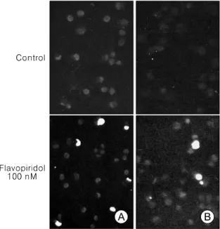
Fig. 2. Ovarian cancer cell lines, TOV-21G (A) and TOV-112D (B) labelled by the TUNEL method. Cells were treated with 100 nM flavopiridol for 24 hours.
Nuclear morphology was examined under a fluoresce- nce microscopy. Nuclei of apoptotic cells were green.
Live cells were totally blue without green spot. Original magnification, ×200.
서는 24시간 및 48시간 처치군 모두 세포 생존도의 유의한 차이는 없었으나, 100 nM 이상의 농도에서는 48시간 처치 한 경우에 더욱 생존도가 감소하는 양상을 보였다(Fig.
1A). 24시간 처치군에서는 대조군과 비교하여 300 nM의 처치 농도부터 세포의 생존도가 67.3±5.9%로 통계적으로 유의하게 감소하기 시작하여 처치 용량에 따라 통계적으 로 유의하게 생존도가 감소함을 알 수 있었다(p<0.05).
TOV-112D 난소암 세포주에서도, 50 nM의 처치 농도 에서는 세포 생존도의 유의한 차이는 없었다. 24시간 처 치군에서는 300 nM의 처치 농도부터, 48시간 처치군에 서는 100 nM의 처치 농도부터 대조군과 비교하여 세포 의 생존도가 통계적으로 유의하게 감소하기 시작하여 TOV-21G 난소암 세포주와 마찬가지로 처치 용량 및 처 치 시간에 따라 유의하게 생존도가 감소함을 알 수 있었 다(p<0.05). 한편, TOV-21G 난소암 세포주에서 세포의 생존도가 50% 이상 감소한 것은 500 nM 이상의 농도로 24시간 처치하거나 300 nM 이상의 농도로 48시간 처치 한 경우이었는데, TOV-112D 난소암 세포주에서는 300 nM 이상의 농도이면 24시간이나 48시간 동안 처치하는 경우 모두 50% 이상의 세포 생존도 감소가 나타났고, 같 은 처치 농도에서의 생존도 감소가 TOV-21G보다 더욱 큰 양상을 보여 flavopiridol에 대하여 더 높은 감수성을 가진 것으로 나타났다(Fig. 1B).
2. Flavopiridol에 의한 난소암 세포주의 세포자멸사 유도 Flavopiridol에 의한 난소암 세포주 TOV-21G와 TOV- 112D의 세포자멸사 유도를 알아보기 위하여 flavopiridol 을 100 nM, 300 nM, 500 nM의 농도로 24시간과 48시간
Fig. 3. Induction of apoptosis by flavopiridol in ovarian cancer cell lines TOV-21G. Cells were treated with the indicated concentrations of flavopiridol (100, 300, and 500 nM) for 24 and 48 hours, respectively. (A) for apoptosis, the externalization of phosphatidylserine was assessed by measuring annexin-V-Fluos binding using propidium iodide as a counterstain. Quadrant rectangular dot grams from a representative of 3 independent experiments is shown. (B) DNA fragmentation were determined using terminal deoxynucleotidyl transferase for incorporation of fluorescein- 12-dUTP at free 3'-OH DNA ends (TUNEL assay). The percentage of TUNEL-positive cells was counted for each condition at 40× magnification in five separate fields of at least 100 cells each. Data were expressed as the
Fig. 3. Continued.
동안 각각 처치 한 후 Annexin-V 염색과 TUNEL 방법으 로 조사하였다.
Annexin V 염색은 세포자멸사의 초기에 세포막 내측 에 위치하였다가 세포막 외표면으로 전위되는 phospha- tidylserine에 높은 친화력을 가지고 결합하는 annexin V-Flous의 성질을 이용한 세포자멸사 판정 방법이다. 초 기 단계의 세포자멸사가 진행 중인 세포는 annexin-V- Flous에만 염색되고 propidium iodide로는 염색되지 않는 반면, 후기단계의 세포자멸사가 진행 중인 세포 또는 괴 사(necrosis)가 진행 중인 세포들은 annexin-V-Flous와 propidium iodide로 동시에 염색되며, 살아 있는 세포는 어느 것에도 염색되지 않는 양상을 나타낸다.
TUNEL 방법에서 nuclear fragmentation을 가지는 apoptotic body는 형광 현미경 하에서 녹색으로, 대조 염 색에 의하여 apoptotic body가 없는 다른 세포의 핵은 청 색으로 관찰된다(Fig. 2). 세포자멸사의 percentage (%)는 서로 다른 위치의 다섯 곳의 현미경하 시야에서 100개 이상의 세포당 발견된 총 apoptotic nuclei의 숫자로 표현 한다.
TOV-21G 난소암 세포주에서 아무런 처치를 하지 않 은 대조군에서 발생한 세포자멸사의 변화는 annexin V 염색에서 생존세포/초기 세포자멸사 세포/후기 세포자 멸사 세포 및 괴사세포의 구성이 24시간과 48시간 배양 후 99.0/0.2/0.8%, 98.5/1.2/0.2%로 각각 나타났고, TUNEL 방법의 결과에서도 24시간과 48시간 배양 후 0.9±0.2%, 1.4±0.7%로 세포자멸사가 각각 관찰되어 대부분이 생존 세포로 구성되어 있었으나, 100 nM의 flavopiridol 처치
24시간 후부터 세포자멸사가 발생하기 시작하여 500 nM의 flavopiridol 처치 48시간 후에는 annexin V 염색에 서 36.5/44.3/17.3%, TUNEL 방법으로 조사된 세포자멸 사의 %는 53.6±5.9%로 50%가 넘는 세포자멸사의 발생 을 관찰할 수 있었다(Fig. 3). 이러한 결과는 flavopiridol 처치 용량과 배양시간에 의존적으로 세포자멸사가 진행 되고 생존 세포가 줄어드는 양상을 나타내는 것이었다.
TOV-112D 난소암 세포주에서도 아무런 처치를 하지 않은 대조군에서 발생한 세포자멸사의 변화는 annexin V 염색에서 생존세포/초기 세포자멸사 세포/후기 세포 자멸사 세포 및 괴사세포의 구성이 24시간과 48시간 배 양 후 97.3/1.7/0.9%, 97.1/2.2/0.6%로 각각 나타났고, TUNEL 방법의 결과에서도 24시간과 48시간 배양 후 2.4±0.7%, 2.1±1.0%로 세포자멸사가 각각 관찰되어 역 시 대부분이 생존세포로 구성되어 있었으나, 100 nM의 flavopiridol 처치 24시간 후부터 세포자멸사가 발생하기 시작하여, 500 nM의 flavopiridol 처치 48시간 후에는 annexin V 염색에서 19.7/35.2/42.7%로 각각 변화하였으 며, TUNEL 방법으로 조사된 세포자멸사의 %는 70.0±
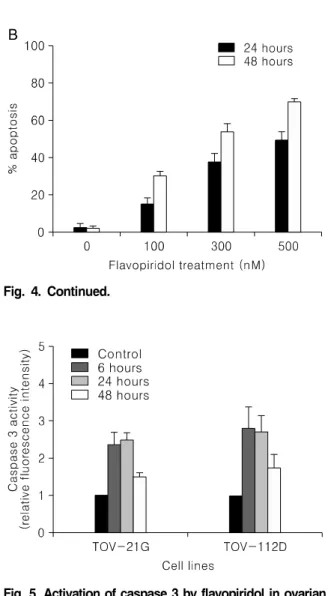
1.5%로 50%가 넘는 세포자멸사의 발생을 관찰할 수 있 었다(Fig. 4). 이러한 결과는 TOV-21G 난소암 세포주와 마찬가지로 flavopiridol 처치 용량과 배양시간에 의존적 으로 세포자멸사가 유도되는 것인데, 같은 처치 농도 및 배양시간에서의 TOV-21 G와 비교하면 세포자멸사가 더 많이 유발되는 양상을 나타내었다.
3. Flavopiridol 처치 후 caspase 3 활성도의 증가 세포자멸사에서 가장 공통적으로 활성화 되는 것으로 알려진 caspase 3가 flavopiridol에 의한 세포자멸사에서 도 활성이 증가하는지 fluorogenic substrate인 Ac-DEVD- AMC의 절단을 이용하여 측정하였다.
500 nM의 flavopiridol로 처치한 경우 TOV-21G 난소 암 세포주에서 caspase 3의 활성은 대조군에 비하여 6 시간 후에 2.4배 증가하였고, 24시간 후에는 2.5배로 활 성의 증가를 보였지만, 48시간 후에는 1.5배로 나타나 활 성 증가가 지속되지 않았다. TOV-112D 난소암 세포주 에서도 caspase 3의 활성은 대조군에 비하여 6시간 후에 2.8배로 증가하였고 24시간 후에는 2.7배로 활성의 증가 가 유지되었지만, 48시간 후에는 역시 1.7배로 활성 증가 가 유지되지는 않았다(Fig. 5). 이러한 결과는 flavopiridol
Fig. 4. Induction of apoptosis by flavopiridol in ovarian cancer cell lines TOV-112D. Cells were treated with the indicated concentrations of flavopiridol (100, 300, and 500 nM) for 24 and 48 hours, respectively. (A) For apoptosis, the externalization of phosphatidylserine was assessed by measuring annexin V-Fluos binding using propidium iodide as a counterstain. Quadrant rectangular dot grams from a representative of 3 independent experiments is shown. (B) DNA fragmentation were determined using terminal deoxynucleotidyl transferase for incorporation of fluorescein- 12-dUTP at free 3'-OH DNA ends (TUNEL assay). The percentage of TUNEL-positive cells was counted for each condition at 40× magnification in five separate fields of at least 100 cells each. Data were expressed as the
Fig. 4. Continued.
Fig. 6. The effect of flavopiridol on the expression of apoptosis related proteins (AIF, XIAP, p53, Mcl-1, Bcl-XL, Bcl-2, and Bax) in ovarian cancer cell lines TOV-21G and TOV-112D. Cells were treated with flavopiridol (100, 300, and 500 nM) for 24 hours, respectively. Aliquots of cells were transferred and protein extracts were assayed for western blot analysis.
The expression of actin were used as the loading control.
Fig. 5. Activation of caspase 3 by flavopiridol in ovarian cancer cell lines TOV-21G and TOV-112D. Cells were treated with 500 nM flavopiridol for the times indicated.
Protein extracts were obtained from aliquots of cells and assayed for caspase 3 activity using the fluorogenic substrate DEVD-AMC.
의 처치에 따라서 TOV-21G와 TOV-112D 난소암 세포주 모두에서 caspase 3의 발현 증가가 6시간 이내에 상당한 수준에 도달한다는 사실을 의미한다.
4. Flavopiridol에 의한 세포자멸사(apoptosis) 관련 단 백질의 발현 변화
Flavopiridol 처치 후에 발생하는 세포자멸사에서 관련 단백질들인 Bax, Bcl-XL, Bcl-2, p53, Mcl-1, AIF, XIAP, caspase 9 등이 어떻게 관여하는지를 알아보기 위하여 이 들의 변화를 western blot 분석으로 확인하였다 (Fig. 6).
TOV-21G와 TOV-112D 난소암 세포주 모두에서
anti-apoptosis 단백질인 Bcl-2와 pro-apoptosis 단백질인 Bax의 발현은 대조군과 flavopiridol을 처치한 실험군에 서 발현의 차이를 나타내지 않았다. 또한, caspase에 비의 존성으로 세포자멸사를 유도하는 apoptosis-inducing factor (AIF) 단백질의 발현도 두 세포주의 대조군과 처 치군 모두에서 변화가 나타나지 않았다.
Anti-apoptosis 단백질인 Mcl-1은 TOV-21G와 TOV- 112D 난소암 세포주 모두에서 300 nM의 처치 농도부터 단백질 발현이 감소하였으며, 500 nM의 처치 농도에서 는 단백질 발현을 관찰할 수 없었다.
또한 anti-apoptosis 단백질인 Bcl-XL의 발현도 Mcl-1과 비슷한 양상을 보였는데, TOV-21G와 TOV-112D 난소암 세포주 모두에서 300 nM의 처치 농도부터 단백질 발현 이 감소하여, 500 nM의 처치 농도에서는 단백질 발현이 더욱 감소하였다.
Anti-apoptosis 단백질인 inhibitors of apoptosis proteins (IAP) family의 하나인 X-linked inhibitors of apoptosis proteins (XIAP) 단백질의 발현은 TOV-21G와 TOV- 112D 난소암 세포주에서 flavopiridol을 100 nM로 처치 하였을 때부터 감소하여, 500 nM의 처치 농도에서는 발 현의 감소가 더욱 뚜렷하게 나타났다.
p53 단백질의 발현은 TOV-21G와 TOV-112D 난소암 세포주 모두에서 flavopiridol을 처치한 후 대조군보다 그 발현이 증가되었지만 처치 농도가 증가함에 따른 p53 단
Fig. 8. Flavopiridol-induced suppression of the activity of ERK, MAP kinase in ovarian cancer cell lines TOV-21G and TOV-112D. Cells were treated with flavopiridol (100, and 500 nM) for 24 hours. Cell lysates were immunoprecipitated with anti-ERK antibodies before a further incubation with protein A-Sepharose beads. These immune complexes were reacted with myelin basic protein (MBP) as a substrate and then the phosphorylated substrate was analyzed by immunoblot analysis, probing with a monoclonal phospho-MBP antibody.
Fig. 7. Activation and processing of caspase 9 during flavopiridol induced apoptosis in ovarian cancer cell lines TOV-21G and TOV-112D. Cells were treated with flavopiridol (100, 300, and 500 nM) for 24 hours.
Aliquots of cells were transferred and lysed in SDS sample buffer and lysates were subjected to western blot analysis with specific antibody, which recognized the pro-form and the active cleaved form of caspase 9, Mr 46 kDa and 34 kDa, respectively.
백질 발현의 증가는 거의 관찰되지 않았다(Fig. 6).
세포자멸사 과정에서 중요한 단백질인 caspase 중에서 upstream initiator인 caspase 9의 발현을 조사하였는데, flavopiridol 처치 전에는 TOV-21G와 TOV-112D 난소암 세포주 모두에서 비활성 상태인 pro-caspase 9의 발현 (46 kDa)은 관찰되었지만 pro-caspase 9의 앞 부위가 절 단된 활성 형태인 caspase 9의 발현(34 kDa)은 거의 나타 나지 않았다. Flavopiridol의 처치 후에는 TOV-21G에서 는 처치 농도가 증가함에 따라 pro-caspase 9의 발현은 감소하고 caspase 9의 활성은 증가하는 양상을 나타내었 고, TOV-112D에서는 발현을 관찰할 수 없었던 caspase 9의 발현이 나타났지만 처치 농도에 따른 발현 증가를 보이지는 않았으며, pro-caspase 9의 발현도 감소하지 않 았다(Fig. 7).
5. Flavopiridol 처치 후 MAP kinase 활성도의 감소 MAP kinase 중에서 성장과 분화에 대한 신호전달을 담당하는 extracellular signal-regulated protein (ERK) kinase에 대한 flavopiridol의 영향을 조사하였다. TOV- 21G와 TOV-112D 난소암 세포주 모두에서 100, 500 nM 의 flavopiridol을 24시간 처치한 경우 ERK의 substrate인 MBP의 발현이 대조군과 비교할 때, 처치 농도에 비례하 여 감소한 것으로 나타났다. 이러한 결과는 flavopiridol 이 ERK kinase의 활성을 억제한다는 사실을 보여주는 것이었다(Fig. 8).
고 찰
1991년 flavopiridol의 항종양 작용이 보고된 이후에 많
은 연구자들에 의하여 그 기전이 조사되었고, 세포주기 가 진행되는 세포에서 cdk들의 작용을 억제하여 강력한 항증식 작용을 하는 것이 중요한 기전으로 알려졌다. 그 러나 flavopiridol이 휴지기 세포(resting cell)에서도 세포 독성을 가진다는 사실이 보고되면서 flavopiridol의 작용 기전은 좀 더 복잡하고 다양할 것으로 추정되었으며, 이 후 세포의 생존과 관련된 p53 혹은 Rb 및 Bcl-2 등의 발 현을 연구한 보고들에서 flavopiridol의 세포독성 기전은 선택된 세포주에 따라 조금씩 다른 양상을 나타내는 것 으로 밝혀졌다.1,6
본 연구의 MTT assay에서 flavopiridol은 정상 P53을 발현하는 난소암 세포주 TOV-21G와 변이형 P53을 발현 하는 TOV-112D에서 모두 처치 용량과 시간에 의존적으 로 세포독성을 나타내었는데, TOV-112D 난소암 세포주 가 세포독성에 감수성이 더 높은 양상을 보였다. 이러한 결과는 정상 P53을 발현하는 A549 비소세포폐암 (non-small cell lung cancer) 세포주가 P53의 이상을 가진 Calu-6 및 NCI-H520 비소세포폐암 세포주보다 flavopiri- dol의 세포 독성에 민감하게 반응한다는 Shapiro 등의 보 고를17 비롯한 변이형 P53을 발현하는 세포주가 약제의 세포독성에 저항을 보인다는 다른 연구자들의 보고와는 상이한 결과였다. 그러나 Samouelian 등은 TOV-21G와 TOV-112D 난소암 세포주에서 시행한 약제 민감성 연구 에서 TOV-21G가 TOV-112D보다 cisplatin의 세포독성에 는 더 높은 감수성을 보이지만, camptothecin이나 paclitaxel에 대하여는 두 가지 세포주 사이에 약제 민감
성의 차이가 없다고 보고하였다.18 따라서 TOV-21G와 TOV-112D 난소암 세포주의 약제 민감성은 두 난소암 세포주가 가지는 다른 특성에서 기인할 가능성이 있으 며, TOV-112D와 비교하여 TOV-21G에서 특이하게 나타 나는 유전자의 이상은 TGF-b RII (transforming growth factor beta receptor II)와 KRAS2의 돌연변이인데,18 이것 이 flavopiridol의 세포 독성에 대한 감수성에 영향을 주 었는지는 향후의 추가적인 연구가 더 필요할 것으로 생 각된다.
본 연구에서는 flavopiridol에 의하여 유도되는 TOV- 21G와 TOV-112D 난소암 세포주의 세포자멸사에 Bcl-2, Mcl-1, Bax, Bcl-X 단백질이 관여하는지 알아보기 위해 western blot 분석을 시행하였고, 두 난소암 세포주 모두 에서 세포자멸사의 유도는 Bcl-2 및 Bax와는 관련이 없 고 Mcl-1 및 Bcl-XL의 발현감소와 밀접한 관련이 있을 것으로 판단되었다. Parker 등과 Kitada 등은 백혈병 세포 주들에서 flavopiridol 처치 후의 세포자멸사 유도가 Bcl-2 단백질의 발현 감소와 관련이 있을 가능성을 보고 하였지만,10,12 대부분의 다른 연구자들은 flavopiridol의 세포자멸사 유도는 Bcl-2 및 Bax와는 관련이 없다고 하 였으며,6 또한 최근에는 MB-468과 SKBR-3 유방암 세포 주에서 flavopiridol 투여 후 Mcl-1과 Bcl-XL 단백질의 발 현이 감소하였다는 보고와 다발성 골수종 및 폐암 세포 주에서 Mcl-1 단백질이 flavopiridol의 투여로 나타나는 세포자멸사 유도에서 중요한 역할을 한다는 보고도 있
어,19-21 본 연구의 결과는 다른 연구자들과 일치하는 것
으로 나타났다.
AIF는 Bcl-2 family를 경유한 세포자멸사 신호에 의하 여 유리되는 미토콘드리아 단백질이지만 cytochrome c 와는 달리 nuclear targeting sequence를 가지고 있어 핵 내로 전위되고, caspase에 비의존적인 양상으로 DNA 분 절 및 염색질 농축화에 관여하는 것으로 알려져 있는
데,22,23 본 연구에서는 두 난소암 세포주 모두에서 발현
의 변화가 없었다. IAP family 중 하나로서, caspase 3, 7 의 활성을 직접적으로 억제하여 세포자멸사를 방해하는 것으로 알려진 XIAP의 발현은24 TOV-21G와 TOV- 112D 난소암 세포주에서 flavopiridol 투여 후 모두 감소 하여 세포자멸사가 유도되는 기전의 하나로 판단되었 다.
Caspase 3는 세포자멸사 신호과정에서 세포내외의 경
로를 실행하는 실행자로서 활성화된 후에 poly (ADP- ribose) polymerase (PARP)를 포함한 중요한 기질을 분해 하고 기능적 활성화 혹은 불활성화를 유도하여 세포 사 망을 실행한다고 알려져 왔다.25 본 연구에서는 caspase 3의 활성이 flavopiridol 투여 후 6시간에 이미 증가되고 24시간에는 2.5-2.7배까지 나타났으며, 또한 AIF의 발현 이 flavopiridol 투여 후에 변화를 보이지 않았던 사실에 미루어 적어도 TOV-21G와 TOV-112D 난소암 세포주에 서는 flavopiridol의 세포자멸사 유도에 caspase 의존적인 경로가 중요하다는 것을 알 수 있었다. Flavopiridol의 처 치 후 caspase의 활성이 증가하는 것이 caspase 활성에 억제작용을 하는 XIAP의 발현이 감소하고, Mcl-1 및 Bcl-XL의 발현감소에 따라 나타나는 2차적인 결과로 나 타날 수 있지만, 본 연구에서 caspase 3의 활성 증가가 처치 6시간 후부터 발생한다는 사실은 flavopiridol이 caspase 3의 활성화에 직접적으로 작용하는 다른 기전이 있을 가능성을 나타내는 것이었고, caspase 3를 활성화 시키는 upstream initiator인 caspase 9의 발현도 flavopiri- dol로 처치하기 전에는 비활성 상태인 pro-caspase 9의 발 현만 나타나다가, 처치 이후에는 활성 형태인 caspase 9 의 발현이 나타나는 양상을 보여 caspase 3의 활성 증가 는 flavopiridol에 의한 caspase 9의 활성 증가에 따라 일 어나는 것으로 추정되었다. 그러나 flavopiridol이 caspase family에 직접적으로 작용하는지를 규명하기 위해서는 caspase 9과 caspase 3에 특이한 억제제를 이용한 연구가 향후 더 필요할 것이다.
이상의 결과들은 TOV-21G와 TOV-112D 난소암 세포 주에서 flavopiridol에 의하여 유도되는 세포자멸사는 XIAP와 Mcl-1 및 Bcl-XL의 발현 감소와 caspase 9의 활 성화에 의존적인 세포내 경로를 거쳐 caspase 3를 활성화 하여 발생한다는 것을 시사하였다. 하지만 본 연구에서 death ligand들에 의해 caspase 8을 활성화하는 세포외 경 로가 flavopiridol의 처치에 따라 변화를 보이지 않은 사 실을 확인하지 않았기 때문에 추가적인 연구가 시행되 어야 한다.
P53의 돌연변이 및 과발현(overexpression)은 악성 난 소종양의 50% 이상에서 흔하게 발견되고 있고, 대부분 의 항암화학제제들의 세포독성 작용은 P53에 의존적인 세 포자멸사의 유도를 통하여 일어나는 것으로 알려져 있으 며, P53의 이상이 발견되는 종양세포에서는 cisplatin과 같
은 항암화학제제들의 세포독성에 내성을 나타낸다.26,27 본 연구에서는 TOV-21G와 TOV-112D에서 p53 단백질 이 flavopiridol을 처치한 이후에 모두 발현이 증가 되었 는데, p53 돌연변이를 가진 TOV-112D에서도 TOV- 21G 에서와 같이 flavopiridol 투여한 후에 세포자멸사가 처치 농도에 따라 증가하였던 사실에 미루어 난소암 세포주 에서 flavopiridol에 의하여 유도되는 세포자멸사는 p53 에 비의존적인 것으로 추정되었다. 그러나 본 연구에서 TOV-21G 난소암 세포주에 p53 단백질의 기능을 소실시 키는 human papilloma virus (HPV)16 E6를 발현하여 TOV-21G와 세포자멸사 발생을 직접 비교하지 않았기 때문에 결론을 내릴 수는 없었다.
본 연구에서는 flavopiridol 처치 후 여러 가지 외부자 극에 의한 세포내 신호전달 경로의 하나로서 세포의 성 장과 분화 및 세포자멸사 등에 관여하는 중요한 조절인 자인 MAP kinase 가운데에서 ERK의 활성이 측정되었는 데,28 두 난소암 세포주 모두에서 감소하는 것으로 나타 나 flavopiridol의 세포자멸사 유도 과정에 ERK 활성 감 소가 직접적인 연관이 있으리라 추정되었다. 향후에, 다 른 MAP kinase로서 세포자멸사에서 활성이 증가될 수 있는 p38 및 c-Jun N-terminal kinase (JNK)의 활성을 조사 하는 연구가 더 필요하다.29
결론적으로 본 연구에서 flavopiridol은 TOV-21G와 TOV-112D 난소암 세포주에서 세포자멸사를 유발하였 는데, 세포자멸사의 양상은 TUNEL 및 annexin-V 염색 방법에서 처치 용량 및 시간에 의존적이었다. Flavopiri- dol에 의한 세포자멸사 발생기전은 Bcl-2와 Bax와는 관 계가 없었고, XIAP와 Mcl-1 및 Bcl-XL의 발현 감소와 더 불어 활성형 caspase 9의 발현 및 caspase 3의 활성 증가 가 연관되어, caspase 의존적인 세포내 경로를 거침을 알 수 있었다. 또한, flavopiridol에 의한 세포자멸사 유도에 따라 ERK의 활성 감소가 나타나 flavopiridol이 특이한 세포내 신호전달경로에 영향을 끼쳐 세포내의 생존과 사망 신호사이의 균형에 영향을 줄 것이라는 사실을 추 정할 수 있었다. Flavopiridol에 의한 세포자멸사는 p53에 비의존적이고 Bcl-2 단백질의 억제와 관련 없이 발생하 였고, 이러한 사실로 P53 유전자의 돌연변이가 흔한 난 소암에서 향후 flavopiridol이 항암화학약제로 약제내성 을 극복하고 작용하는 좋은 후보가 될 수 있을 것으로 제시된다.
참고문헌
1. Kaur G, Stetler-Stevenson M, Sebers S, Worland P, Sedlacek H, Myers C, et al. Growth inhibition with rever- sible cell cycle arrest of carcinoma cells by flavone L86-8275. J Natl Cancer Inst 1992; 84: 1736-40.
2. Zhai S, Senderowicz AM, Sausville EA, Figg WD.
Flavopiridol, a novel cyclin-dependent kinase inhibitor, in clinical development. Ann Pharmacother 2002; 36:
905-11.
3. Worland PJ, Kaur G, Stetler-Stevenson M, Sebers S, Sartor O, Sausville EA. Alteration of the phosphorylation state of p34cdc2 kinase by the flavone L86-8275 in breast carcinoma cells. Correlation with decreased H1 kinase activity. Biochem Pharmacol 1993; 46: 1831-40.
4. De Azevedo WF Jr, Mueller-Dieckmann HJ, Schulze- Gahmen U, Worland PJ, Sausville E, Kim SH. Structural basis for specificity and potency of a flavonoid inhibitor of human CDK2, a cell cycle kinase. Proc Natl Acad Sci USA 1996; 93: 2735-40.
5. Carlson B, Lahusen T, Singh S, Loaiza-Perez A, Worland PJ, Pestell R, et al. Down-regulation of cyclin D1 by tran- scriptional repression in MCF-7 human breast carcinoma cells induced by flavopiridol. Cancer Res 1999; 59:
4634-41.
6. Sedlacek HH. Mechanisms of action of flavopiridol. Crit Rev Oncol Hematol 2001; 38: 139-70.
7. Melillo G, Sausville EA, Cloud K, Lahusen T, Varesio L, SenderowAM. Flavopiridol, a protein kinase inhibitor, down-regulates hypoxic induction of vascular endothelial growth factor expression in human monocytes. Cancer Res 1999; 59: 5433-7.
8. Arguello F, Alexander M, Sterry JA, Tudor G, Smith EM, Kalavar NT, et al. Flavopiridol induces apoptosis of nor- mal lymphoid cells, causes immunosuppression, and has potent antitumor activity In vivo against human leukemia and lymphoma xenografts. Blood 1998; 91: 2482-90.
9. Patel V, Senderowicz AM, Pinto D Jr, Igishi T, Raffeld M, Quintanilla-Martinez L, et al. Flavopiridol, a novel cy- clin-dependent kinase inhibitor, suppresses the growth of head and neck squamous cell carcinomas by inducing apoptosis. J Clin Invest 1998; 102: 1674-81.
10. Parker BW, Kaur G, Nieves-Neira W, Taimi M, Kohlhagen G, Shimizu T, et al. Early induction of apop- tosis in hematopoietic cell lines after exposure to flavopiridol. Blood 1998; 91: 458-65.
11. Achenbach TV, Muller R, Slater EP. Bcl-2 independence of flavopiridol-induced apoptosis. Mitochondrial depolari- zation in the absence of cytochrome c release. J Biol Chem 2000; 275: 32089-97.
12. Kitada S, Zapata JM, Andreeff M, Reed JC. Protein kin- ase inhibitors flavopiridol and 7-hydroxy-staurosporine down-regulate antiapoptosis proteins in B-cell chronic lymphocytic leukemia. Blood 2000; 96: 393-7.
13. Raju U, Nakata E, Mason KA, Ang KK, Milas L.
Flavopiridol, a cyclin-dependent kinase inhibitor, enhan-
ces radiosensitivity of ovarian carcinoma cells. Cancer Res 2003; 63: 3263-7.
14. Motwani M, Delohery TM, Schwartz GK. Sequential de- pendent enhancement of caspase activation and apoptosis by flavopiridol on paclitaxel-treated human gastric and breast cancer cells. Clin Cancer Res 1999; 5: 1876-83.
15. Shapiro GI. Preclinical and clinical development of the cyclin-dependent kinase inhibitor flavopiridol. Clin Cancer Res 2004; 10: 4270s-5s.
16. Provencher DM, Lounis H, Champoux L, Tetrault M, Manderson EN, Wang JC, et al. Characterization of four novel epithelial ovarian cancer cell lines. In Vitro Cell Dev Biol Anim 2000; 36: 357-61.
17. Shapiro GI, Koestner DA, Matranga CB, Rollins BJ.
Flavopiridol induces cell cycle arrest and p53-in- dependent apoptosis in non-small cell lung cancer cell lines. Clin Cancer Res 1999; 5: 2925-38.
18. Samouelian V, Maugard CM, Jolicoeur M, Bertrand R, Arcand SL, Tonin PN, et al. Chemosensitivity and radio- sensitivity profiles of four new human epithelial ovarian cancer cell lines exhibiting genetic alterations in BRCA2, TGFbeta-RII, KRAS2, TP53 and/or CDNK2A. Cancer Chemother Pharmacol 2004; 54: 377-83.
19. Wittmann S, Bali P, Donapaty S, Nimmanapalli R, Guo F, Yamaguchi H, et al. Flavopiridol down-regulates anti- apoptotic proteins and sensitizes human breast cancer cells to epothilone B-induced apoptosis. Cancer Res 2003; 63: 93-9.
20. Gojo I, Zhang B, Fenton RG. The cyclin-dependent kin- ase inhibitor flavopiridol induces apoptosis in multiple myeloma cells through transcriptional repression and
down-regulation of Mcl-1. Clin Cancer Res 2002; 8:
3527-38.
21. Ma Y, Cress WD, Haura EB. Flavopiridol-induced apop- tosis is mediated through up-regulation of E2F1 and re- pression of Mcl-1. Mol Cancer Ther 2003; 2: 73-81.
22. Susin SA, Lorenzo HK, Zamzami N, Marzo I, Snow BE, Brothers GM, et al. Molecular characterization of mi- tochondrial apoptosis-inducing factor. Nature 1999; 397:
441-6.
23. Cande C, Cecconi F, Dessen P, Kroemer G. Apoptosis-in- ducing factor (AIF): Key to the conserved caspase-in- dependent pathways of cell death? J Cell Sci 2002; 115:
4727-34.
24. Deveraux QL, Takahashi R, Salvesen GS, Reed JC.
X-linked IAP is a direct inhibitor of cell-death proteases.
Nature 1997; 388: 300-4.
25. Woo M, Hakem R, Soengas MS, Duncan GS, Shahinian A, Kagi D, et al. Essential contribution of caspase 3/CPP32 to apoptosis and its associated nuclear changes.
Genes Dev 1998; 12: 806-19.
26. Laframboise S, Chapman W, McLaughlin J, Andrulis IL.
p53 mutations in epithelial ovarian cancers: possible role in predicting chemoresistance. Cancer J 2000; 6: 302-8.
27. Schuijer M, Berns EM. TP53 and ovarian cancer. Hum Mutat 2003; 21: 285-91.
28. Robinson MJ, Cobb MH. Mitogen-activated protein kin- ase pathways. Curr Opin Cell Biol 1997; 9: 180-6.
29. Xia Z, Dickens M, Raingeaud J, Davis RJ, Greenberg ME. Opposing effects of ERK and JNK-p38 MAP kin- ases on apoptosis. Science 1995; 270: 1326-31.
=ABSTRACT=
Induction of apoptosis by the kinase inhibitor flavopiridol in human ovarian cancer cell lines
Soo-Young Hur, Joon-Mo Lee
Department of Obstetrics and Gynecology, College of Medicine, The Catholic University of Korea, Seoul, Korea
Objective:Flavopiridol that inhibits cyclin-dependent kinase, can cause cell cycle arrest, induce apoptosis in human tumor cell lines. In the present study, we investigated apoptotic effects of flavopiridol and the underlying molecular mechanisms in human ovarian cancer cell lines.
Methods:We used TOV-21G and TOV-112D cell lines. The cell viability was tested by MTT assay and apoptosis was assessed by TUNEL assay and annexin-V binding. Western blot was used to examine apoptosis related protein levels.
MAP kinase activity was analyzed by non-radioactive MAP kinase assay kit.
Results:Treatment of TOV-21G and TOV-112D cells with flavopiridol (50 nM to 1000 nM) led to a dose- and time-dependent inhibition of cell growth and survival. Dose-related induction of apoptosis was also observed in these cell lines. Flavopiridol (500 nM) induced striking decreases in the levels of the antiapoptic proteins Mcl-1, Bcl-XL, and XIAP in both cell lines. In contrast, expression of Bax, Bcl-2, and AIF was not significantly influenced by flavopiridol.
Although flavopiridol resulted in accumulation of p53 in both cells, flavopiridol mediated apoptosis was p53 independent because it occurred to the same degree in TOV-112D cells in which p53 was inactivated by mutation. Flavopiridol treatment resulted in enhanced cleavage of pro-caspase 9 and activation of caspase 3. Apoptosis was associated with suppression of ERK activity.
Conclusion:Although the precise mechanisms of flavopiridol mediated cytotoxicity have not been fully defined, these data suggest that flavopiridol has activity against ovarian cancers in vitro and is worthy of continued clinical development in the treatment of ovarian cancer.
Key Words:Flavopiridol, Ovarian cancer cell lines, Apoptosis
Address reprint requests to Joon-Mo Lee
Department of Obstetrics and Gynecology, College of Medicine, The Catholic University of Korea 50, Banpo-dong, Seocho-gu, Seoul 137-701, Korea
Tel:82-2-590-1376, Fax:82-2-595-1549, E-mail:[email protected]