통한 Arsenic Trioxide와 Sulindac 병합요법의 세포고사효과
원광대학교 의과대학 내과학교실 원광의과학연구소 김학렬, 양세훈, 정은택
Combination Treatment with Arsenic Trioxide and Sulindac Induces Apoptosis of NCI-H157 Human Lung Carcinoma Cells via ROS Generation with Mitochondrial Dysfunction
Hak-Ryul Kim, M.D., Sei-Hoon Yang, M.D. and Eun-Taik Jeong, M.D.
Department of Internal Medicine, College of Medicine Wonkwang University, Iksan, Korea
Background : Arsenic trioxide (As2O3) has been used to treat acute promyelocytic leukemia, and it induces apop tosis in a variety of solid tumor cell lines including non-small cell lung cancer cells. However, nonsteroidal anti- inflammatory drugs (NSAID) can enhance tumor response to chemotherapeutic drugs or radiation. It was previously demonstrated that a combination treatment with As2O3 and sulindac induces the apoptosis of NCI-H157 human lung carcinoma cells by activating the caspase cascade. This study aimed to determine if a combination treatment augmented its apoptotic potential through other pathways except for the activation of the caspase cascade.
Material and Methods : The NCI-H157 cells were treated with As2O3, sulindac and antioxidants such as glutathione (GSH) and N-acetylcysteine (NAC). The cell viability was measured by a MTT assay, and the level of intracellular hydrogen peroxide (H2O2) generation was monitored fluorimetrically using a scopoletin-horse radish peroxidase (HRP) assay. Western blotting and mitochondrial membrane potential transition analysis were performed in order to define the mechanical basis of apoptosis.
Results : The viability of the cells was decreased by a combination treatment of As2O3 and sulindac, and the cells were protected using antioxidants in a dose-dependent manner. The increased H2O2 generation by the combination treatment was inhibited by antioxidants. The combination treatment induced changes in the mitochondrial tran smembrane potential as well as the expression of the Bcl-2 family proteins, and increased cytochrome c release into the cytosol. However, the antioxidants inhibited the effects of the combination treatment.
Conclusion : Combination treatment with As2O3 and sulindac induces apoptosis in NCI-H157 human lung carcinoma cells via ROS generation with a mitochondrial dysfunction. (Tuberc Respir Dis 2005; 59: 30-38)
Key words : Arsenic trioxide, Sulindac, Apoptosis
‡본 연구는 2004년도 원광대학교 교내 연구비 지원에
의해 이루어짐
Address for correspondence : Hak Ryul Kim, M.D.
Department of Internal Medicine, Wonkwang University Hospital, 344-2 Shinyong-dong, Iksan, Jeonbuk, 570-711, Korea
Phone : 063-850-1328 Fax : 063-855-2025 E-mail : [email protected]
Received : Feb. 21. 2005 Accepted : Jun. 9. 2005
서 론
폐암은 국내에서 위암을 제치고 암의 사망원인 중 1위를 차지하고 있다1. 폐암을 정복하기 위한 여러 노 력에도 불구하고 전체적으로 5년 생존율이 14%에 불
과할 정도로 폐암의 예후는 매우 불량하여 기존의 수 술, 방사선요법, 항암화학요법과 이들을 근간으로 한 병용요법으로는 치료의 한계를 보여주고 있어 새로 운 제재들을 이용한 다양한 치료적 방법이 요구되고 있다.
Arsenic trioxide(As2O3, 비소 삼산화물)는 재발성 또는 불응성 급성전골수성백혈병의 매우 효과적인 치 료제로 이용되고 있고2,3, 비소세포폐암을 비롯한 여러 고형종양세포에서 세포고사를 유도하나 백혈병에 비 해 상대적으로 높은 농도가 필요하여 실제적인 치료 에 제한이 있어왔다4-7.
Sulindac은 indomethacin과 구조식이 비슷한 NSAID 로 ρ-methyl sulphinyl group의 전구물질로서 sulphide
로 대사되어 prostaglandin 합성을 억제시킨다. 가족 성 선종성 용종증의 발암 예방 효과를 가지고 있는데8, 항암기능으로 설명되는 COX-2 억제기능9,10과 암세포 성장의 억제, 그리고 세포고사 유도가 기전으로 보고 되고 있다11-13. 최근 화학요법이나 방사선치료에 병합 치료시 종양의 반응성이 증가함이 알려졌으나 정확한 기전은 밝혀지지 않은 실정이다14-16.
저자들은 NCI-H157 폐암세포주에 As2O3와 suli
ndac의 병합요법시 Fas/FasL 신호전달계의 활성화와 caspase 단백질 활성화에 의해 세포고사가 유도되었 음을 보고한 바 있다17. 이 연구의 목적은 As2O3와 sulindac의 병합요법이 이러한 경로 이외에 다른 어떤 경로를 통해 세포고사능을 배가 시키는지 여부를 알 아보는데 있다.
재료 및 방법 1. 재 료
사람의 편평상피암 세포에서 유래한 NCI-H157 세 포는 한국 세포주 은행(Korean cell line bank, 서울대 학교)로부터 분양 받아 계대배양하면서 실험을 실시 하였다. 실험에 필요한 RPMI 1640, 항생제, trypsin 및 우태아 혈청(fetal bovine serum: FBS)은 GIBCO BRL사(Grand Island, NY, USA)에서 구입하였다.
Sulindac, arsenic trioxide(As2O3), Methylthiazol-2- yl-2,5-diphenyl, tetrazolium bromide(MTT)는 Sigma 사(St. Louis, USA) 제품을 사용하였고, JC-1은 Mo
lecular probes(Pitchford Avenue, OR)제품을 이용하 였다. Caspase-3 protease의 기질인 Ac-DEVD-7- amino-4-methylcoumarin(AMC)은 Calbiochem사(San Diego, USA)로부터 구입하였고, PARP, Bax, Bid, Bcl-xL에 대한 항체는 Santa Cruz사(San Diego, USA) 제품을 이용하였으며, anti-cytochrome c항체는 Pha
rmigen사(San Diego, USA)로부터 구입하였다. 또한 anti-rabbit IgG conjugated horse-radish peroxidase 와 enhanced chemiluminescence kit(ECL kit)는 Ame
rsham사(Buckinghamshine, England)에서 구입하여 사용하였다.
2. 방 법
1) NCI-H157 세포배양 및 시약처리
NCI-H157세포는 37℃, 5% CO2 배양기에서 10%
우태아 혈청이 포함된 RPMI 1640(GIBCO BRL, En
gland) 배양액으로 배양하였으며, 24시간 간격으로 배 양액을 교체하여 log phase에 있는 세포에 As2O3, suli
ndac을 단독 또는 병합처리 하였고, GSH, NAC의 항 산화제를 시약처리 30분전에 전처리한 군을 포함하였 다. 48시간 후에 세포성장 억제, 세포고사 현상 및 이에 연관된 생화학 및 분자생물학적 실험을 수행하였다.
2) 세포 생존율 측정
세포(5X10⁴ 세포수/mL)는 세포 배양판(24-well plate) 에 1mL씩 분주하여 12시간이상 CO2 세포배양기 안에 서 안정시킨 후, 실험에 필요한 시약을 처리한 다음, MTT 용액(5mg/ml in PBS)을 배양액 최종부피의 1/10이 되게 첨가하였다. 4시간 후 살아있는 세포에 의해 생성된 보라색 formazan의 검출은 MTT가 들어 있는 배양액을 모두 버리고 DMSO을 500ml을 넣고 충분히 녹인 후 96 well plate에 100ml을 넣고 분광광 도계(ELISA reader, Molecular Devices Co., Sunny
vale, USA)을 이용하여 595nm파장에서 흡광도를 측 정하였다.
3) 활성산소종(reactive oxygen species, ROS) 생성측정
세포외 H2O2의 생성은 horse radish peroxidase (HRP)에 의한 산화작용 동안에 scopoletin의 형광의 감소정도를 측정하였다. 시약처리 한 세포(5X10⁴ 세 포수/mL)의 상층액을 얻어 1U/ml의 HRP을 첨가한 후, 암실에서 scopoletin 5μM을 10분간 반응시킨 후, fluorometer(Molecular Devices Co, USA)로 350nm (ex
citation wavelength)와 460nm(emission wavelength) 의 파장에서 측정하였다.
4) Hoechst 염색
세포핵의 형태학적 변화를 조사하기 위해 시약처리 한 세포를 4% 포름알데히드(formaldehyde) 용액에서
10분간 고정시킨 후 PBS로 2회 세척하고 Hoechst 33342(Sigma) 염색약을 생리식염수에 10μM이 되게 희석하여 1시간 염색한 후 다시 생리식염수에 희석하 여 형광현미경(Leica MPS 60, Germany)으로 관찰하 였다.
5) 미토콘드리아 막전위 측정
미토콘드리아의 막전위(membrane potential)의 변 화를 조사하기 위하여 JC-1 염색을 시행하였다. 시약 처리된 세포들은 PBS로 2회 세척하였다. JC-1(10μ g/ml)은 각각 RPMI 1640 배지에 희석하여 20분간 3 7℃, CO2 배양기에서 반응시킨 후 다시 PBS로 세척하여 형광현미경(Leica MPS 60, Germany)으로 관찰하였다.
6) Western blotting
시약 처리된 세포를 포집하여 차가운 Hank's bala
nced salt solution(HBSS, pH 7.4)으로 2회 세척하였 다. 얻어진 세포는 파쇄용액(50mM HEPES pH 7.4, 150mM Nacl, 1% deoxy-cholate, 1mM EDTA, 1mM PMSF, 1μg/ml aprotinin)과 4℃에서 30분간 반응하였 다. 세포 파쇄액은 13,000rpm에서 20분간 원심분리한 후 상층액을 BCA 용액을 이용하여 단백질을 정량하 였다. 동량의 세포 파쇄액(단백질: 200μg)은 2×sample buffer와 혼합하여 100℃에서 5분간 가열한 후 12.5%
SDS-PAGE를 시행하였다. 전기영동이 끝난 gel의 단 백질은 semi-dry 방법으로 실온에서 단위 면적당 0.8mA 전하를 2시간 걸어주어 nitrocellulose membrane상에 이동시켰다. Nitrocellulose membrane은 blocking bu
ffer(5% skim milk)와 상온에서 1시간 반응하여 비특 이적 항체결합을 예방하였다. Bax, Bid, Bcl-xL, PARP 에 대한 항체는 0.01%(v/v)의 Tween-20이 포함된 3%
skim milk/TBS에 1:1000으로 희석하여 상온에서 3 시간 반응 후 이차항체인 anti-rabbit IgG conjugated horse-radish peroxidase와 1시간 반응하였다. Nitro
cellulose membrane은 TBS로 3번 세척한 후 ECL kit 를 사용하여 ECL 필름에 노출하였다.
7) 세포질과 미토콘드리아 분획의 분리
시약 처리된 세포를 포집하여 PBS로 2회 세척하였
다. 얻어진 세포는 Buffer A(250mM sucrose, 20mM HEPES pH 7.5, 10mM KCl, 1.5mM MgCl2, 1mM EDTA, 1mM EGTA, 1mM DTT, 17μg/mL PMSF, 8 μg/ml aprotinin, 2μg/ml leupeptin)와 4℃에서 20분간 반응시킨 후 homogeniger로 10회 파쇄하여 4℃, 750g 에서 10분간 원심분리 하였다. 원심분리 한 상층액을 새 튜브에 이동시킨 후 다시 4℃, 20,000g에서 10분간 원심분리 하였으며, 이때 상층액은 세포질로, 침사물 은 미토콘드리아 분획으로 사용하였다. 이후의 과정 은 상술된 Western blotting방법을 이용하였다.
8) 결과 산출 방법
표시된 결과는 3번 이상의 독립적인 실험결과이며 이들의 평균(mean)과 표준편차(standard deviation, S.D.)를 산출하여 표시하였다. 실험결과의 통계처리는 Student's t-test에 준하여 처리하였다.
결 과
1. As2O3와 sulindac 병합요법이 폐암 세포주의 생존율에 미치는 영향
Sulindac 5μM와 As2O3 2.5μM로 단독처리군과 병 합처리군으로 나누었고, 여기에 항산화제인 N-acet
ylcysteine(NAC)과 glutathione(GSH)를 함께 전처리 한 군을 포함하여 48시간 후 세포활성도를 MTT 방 법으로 측정하였다. 생존율의 결과는 Sulindac 단독처 리시 3.7%, As2O3 단독처리시에 9.1%의 미미한 감소 를 보였던 반면 병합요법 시에는 58%로 생존율의 의 의 있는 감소를 보였다. 이러한 생존율은 NAC와 GSH를 전처리한 경우 농도 의존적으로 회복됨을 관 찰할 수 있었다(Fig. 1).
2. As2O3와 sulindac 병합요법에 의한 ROS의 생성
병합요법에 의한 의미 있는 생존율의 감소가 항산 화제에 의해 회복된 결과로서 병합요법시 ROS의 생 성유무를 알아보기 위해 HRP에 의한 scopoletin 형 광의 감소를 fluorometer를 이용하여 측정한 결과 병
Control S only A only S + A
5mM 10mM 10mM 15mM 0
20 40 60 80 100
Viability
S + A S + A S + A S + A
NAC GSH
S : Sulindac 5μM A : As2O32.5μM NAC : N-acetylcysteine GSH : glutathione
*
**
**
#
#
Figure 1. Combination treatment of arsenic trioxide and sulindac decreased the viability of NCI-H157 lung cancer cells. it’s the cancer cells were protected using antioxidants in a dose-dependent manner. The cell viability was determined by a MTT assay after 48 hours. The data represent the mean±S.D. of three independent experiments. *, p <0.05 compared with the control. #, ** compared to S+A.
Control As2O3 Sulindac
As2O3 + Sulindac+ GSH As2O3+ Sulindac
Procaspase-3 (35kDa) PARP (116kDa) Cleaved PARP (85kDa)
As2O3 - + - + + +
Sulindac - - + + + +
GSH - - - - + -
NAC - - - - - +
A.
B.
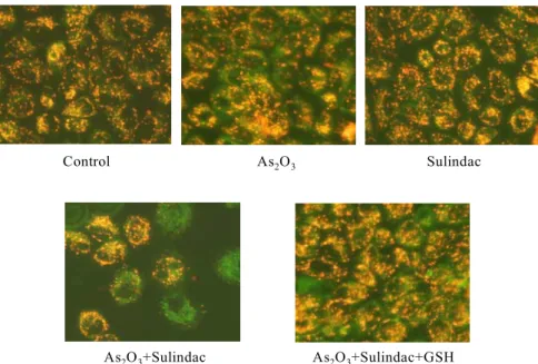
Figure 3. (A) Nuclear morphological investigation of the combination treated NCI-H157 cells. The nuclear morphology was investigated by Hoechst 33342 dye staining under fluorescence microscopy. (B) The com
bination treatment of As2O3 and sulindac induced the cleavage of procaspase-3 and PARP in NCI-H157 cells.
This effect was inhibited by antioxidants.
A : As2O32.5μM S : Sulindac 5μM N : N-acetylcysteine G : glutathione
Cont Aonly Sonly A+S A+S+N A+S+G 60
80 100 120 140 160 180 200 220
ROS production (% of control)
*
** **
Figure 2. Combination treatment of arsenic trioxide and sulindac increased the level of ROS generation in the NCI-H157 lung cancer cells. Their generation was inhibited by antioxidants. The intracellular hydrogen peroxide (H2O2) level was determined fluorimetrically using a scopoletin-horse radish peroxidase (HRP) assay.
The data represents the mean±S.D. of triplicate exper
iments. *, p <0.05 compared with the control. **, p
<0.05 compared to S+A.
합요법시에 대조군에 비해 2.1배로 의의 있게 증가하 였고, 이러한 현상은 NAC, GSH 전처리군에서 각각 1.3배, 1.4배로 감소된 소견을 보였다(Fig. 2).
3. As2O3와 sulindac 병합요법에 의한 세포고사
병합요법에 의한 세포사가 세포고사 기전에 의해 매개되었는지 여부와 이러한 세포고사에 ROS가 관여 하였는지 여부를 알아보기 위해 세포고사의 형태학적 인 특징과 관련된 단백질의 발현을 확인하였다. 먼저 세포의 핵을 Hoechst 33342 염색시 대조군은 세포핵 의 모양이 타원형의 균일한 형광염색을 나타내었고, 단독요법시에도 미미한 변화만을 보였으나, 병합요법 시에는 세포수가 적어지고 핵이 여러 조각으로 분절 된 형광절편이 관찰되었다. 이러한 현상은 GSH 전처 리군에서 회복되고 있음을 보여주었다(Fig. 3A). 세포 고사와 관련된 단백질의 발현을 알아보기 위해 하위 단계인 caspase-3 protease와 이것의 기질단백질인 PARP활성을 western blotting을 통해 확인하였다. 병 합처리군에서 35kDa의 procaspase-3가 절단되어 감 소하였고, PARP는 caspase-3 protease에 의해 116kDa 크기의 단백질이 절단되어 85kDa의 단편으로의 절단 이 관찰되었다. 이러한 현상은 NAC와 GSH를 함께 전처리한 군에서 소실됨을 확인하였다(Fig. 3B).
Control As2O3 Sulindac
As2O3+Sulindac As2O3+Sulindac+GSH
Figure 4. Combination treatment of As2O3 and sulindac induced a mitochondrial membrane potential transition (MPT) in NCI-H157 cells. This effect was inhibited by antioxidants. The cells were stained with 10 μg/ml of JC-1 and observed using fluorescent microscopy and analyzed by flow cytometry. The data is the representative of three independentexperiments.
Anti-cytochrome c blot 0 12 24 36 48
Cytosolic Mitochondria A.
B. As2O3 - + - + + +
Sulindac - - + + + +
GSH - - - - + -
NAC - - - - - +
Cytosolic
Mitochondria
Figure 5. (A) The lysate was fractionated into cyto
solic and mitochondrial portions. Two fractions were separated on 12.5% SDS-PAGE and immunoblotted for cytochrome c. The combination treatment of As2O3 and sulindac increased the cytochrome c release to cyto
solic part in a time-dependent manner. (B) This effect was inhibited by antioxidants.
4. As2O3와 sulindac 병합요법에 의한 미토콘드리 아 막전위차에 미치는 영향
병합요법에 의한 세포고사가 미토콘드리아의 기능 변화와 관련되었는지의 여부를 알아보고자 미토콘드 리아의 막전위차 변화(membrane potential transition;
MPT)를 확인하기 위하여 JC-1 염색 후에 형광현미 경을 통해 관찰하였다. 처리 후 24시간 후의 결과를 보면, 대조군에서는 JC-1의 정상색인 오렌지색 형 광이 나타났고, 단독처리군에서 이러한 변화는 미미 하였으나 병합처리군에서 녹색형광을 보였고, 이러 한 현상은 GSH 전처리군에서 상쇄됨을 확인하였다 (Fig. 4).
5. As2O3와 sulindac 병합요법이 anti-cytochrome c의 발현에 미치는 영향
미토콘드리아에서 세포질내로 방출되어 caspase-9 protease의 활성화를 유도하는 cytochrome c의 세포 질 내의 이동을 western blotting으로 관찰하였다. 이
때 세포질과 미토콘드리아 분획은 미토콘드리아에만 존재하는 단백질인 VDAC을 통해 온전히 분리됨을 확인하였다. 먼저 병합처리시 시간의존적으로 세포질 내의 cytochrome c의 양이 증가하고 미토콘드리아 내
Bax
β-actin Bid Bcl-xL
As2O3 - + - + + +
Sulindac - - + + + +
GSH - - - - + -
NAC - - - - - +
B.
Time (hr)0 12 24 30 36 42 48 As2O3 (2.5μM) + Sulindac (5μM)
Bax (21kDa)
β-actin Bid (22kDa) Bcl-xL (30kDa) A.
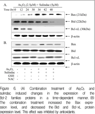
Figure 6. (A) Combination treatment of As2O3 and sulindac induced changes in the expression of the Bcl-2 families proteins in a time-dependent manner. (B) The combination treatment increased the Bax expre
ssion level, and decreased the Bid and Bcl-xL protein expression level. This effect was inhibited by antioxidants.
의 양은 현저히 감소되어 미토콘드리아로부터 세포질 내로 cytochrome c가 방출됨을 확인하였다(Fig. 5A).
48시간 후의 결과를 처리방법에 따라 다시 확인한 결 과 병합요법시의 cytochrome c의 세포질내의 증가와 미토콘드리아내의 감소 소견은 GSH와 NAC를 전처리 한 군에서 이러한 소견은 상쇄됨을 보였다(Fig. 5B).
6. As2O3와 sulindac 병합요법이 Bax, Bid 및 Bcl-xL의 발현에 미치는 영향
미토콘드리아에서 cytochrome c의 방출 및 미토콘 드리아의 막전위차를 조절한다고 알려져 있는 Bcl-2 family 단백질들의 발현양상을 확인하였다. 먼저 병합 처리시 시간의존성변화를 보면 세포고사 촉진 기능을 갖는 Bax는 증가양상을 보였고, caspase-8에 의해 분 절되는 bid의 발현은 감소됨을 확인하였다. 또한 세포 사멸 억제 단백질인 Bcl-xL의 발현은 절단되어 감소 됨을 보였다(Fig 6A). 변화를 보였던 단백질을 다시 처리방법에 따라 확인한 결과 병합처리군에서 Bax의 증가, Bid와 Bcl-xL의 발현감소를 확인할 수 있었고 이러한 현상은 GSH, NAC를 전처리한 군에서 소실됨 을 보였다(Fig. 6B).
고 찰
항암제에 의한 암세포의 사멸이 일부는 세포고사에 의하여 매개됨이 밝혀지면서, 이에 대한 생화학적 그 리고 분자생물학적인 접근이 활발히 이루어지고 있 다. 세포고사 조절기전에 중요한 미토콘드리아에 대 한 최근의 보고에 의하면 Bcl-2 family 단백질에 의해 조절되는 cytochrome c는 미토콘드리아에서 세포질 로 방출되고, 이어서 caspase-8 protease와의 결합을 통해 그 활성이 조절되어 하부 caspase-3 protease를 활성화시켜 세포고사 현상을 유도한다18,19. Bcl-2 family 단백질 중 세포고사를 억제하는 Bcl-2나 Bcl-xL 단백 질 등은 세포고사 촉진 단백질과 heterodimer를 형성 하거나 암세포에서 항암제 처리시 인산화 되어 그 기 능을 상실하게 된다. 그러나 Bax, Bak, Bid 등의 세포 고사 촉진 단백질은 미토콘드리아로 이동하여 cytochrome c를 방출할 수 있는 세공(pore)을 형성하기도 한다. 특 히 Bid 단백질은 활성화된 caspase-8 protease에 의해 절단된 후 미토콘드리아로 이동하여 세포고사를 촉진 하는 것으로 알려져 있다18,19. 따라서 세포고사를 유도 하는 다양한 자극이나 Bcl-xL을 과발현시에 미토콘 드리아 막전위의 상실이나 cytochrome c의 방출이 방 지되고, 반대로 Bax 단백질을 과발현시에는 cytochrome c의 방출이 촉진되고 미토콘드리아의 막전위차 변화 를 유도한다고 보고 되고 있다20.
As2O3는 농도에 따라 APL세포에 두 가지 방식으로 작용하는데21, 먼저 0.1-0.5μM의 저농도에서는 분화의 유도로 인해 As2O3가 직접 PML 또는 PML/RARα를 분해하는 작용을 한다22. 또한 1-2μM의 농도에서는 세포고사의 유도로 고사유발신호에 의해 미토콘드리 아 막전위의 변화가 일어나고 이에 따라 막의 투과성 이 변화되어 cytochrome c와 다른 고사유도인자가 유 리되어 caspase의 활성화가 일어난다23,24. 이밖에도 활 성산소의 활성화, 미토콘드리아의 기능변화, JNK의 활성화, tubulin의 불활성화, p53의 유도 등25이 고사유 도의 기전으로 제시되고 있으나 구체적인 고사 기전 은 밝혀져 있지 않다. 최근에 As2O3가 미토콘드리아 소실을 유발하고 미토콘드리아 투과성 전이구멍 복합 체(permeability transition pore complex, PTPC)에
직접 영향을 미쳐 고사를 유도함이 보고 되었다26. 한 편 Chen 등27은 As2O3가 ROS의 생성을 촉진하여 간 접적으로 미토콘드리아에 손상을 입혀 고사를 유도한 다고 하였다.
NSAID가 최근 생물학적 치료, 방사선치료, 기존의 항암제들과의 병합요법시 상승효과가 있음이 보고 되 고 있다. Torrance 등28은 sulindac과 EKI-569(EGFR tyrosine kinase inhibitor)의 병합치료시 장 종양을 감 소시키는데 단독치료에 비해 상승효과가 있다고 하였 다. Duffy 등29은 폐암과 백혈병 세포주에서 NSAIDs 가 anthracycline과 vinca alkaloid계의 항암제의 세포 독성을 증가시켰다고 하였고, Soriano 등30은 sulindac 과 paclitaxel이나 cisplatin의 병합요법이 소세포와 비 소세포폐암 모두에서 종양의 성장억제효과를 배가시 켰으며, Hida 등31도 비소세포폐암에서 nimesulide와 irinotecan, taxotere, VP-16, cisplatin이나 SM5887과 의 병합치료시 상승작용이 있음을 보고함으로서 암 치료시 NSAID의 효과를 증명하였다.
본 연구에서 As2O3와 sulidac과의 병합요법이 단독 치료에 비해 생존율을 의미 있게 감소시켰고, 이것은 세포내에서 ROS 형성과 관계됨을 항산화제 전처치 결과와 직접적인 H2O2 측정으로 증명하였다. 또한 병 합요법에 의해 세포고사가 유도되었고 항산화제로 전 처치시 활성화가 뚜렷히 억제됨을 확인하여 caspase 계의 활성화를 통한 세포고사가 ROS의 영향에 의한 것임을 알 수 있었다. 또한 활성화된 caspase-8의 활 성에 의하여 Bid가 truncation되어 미토콘드리아로 이 동하였고, Bcl-xL의 발현감소와 Bax의 발현증가로 인한 미토콘드리아 막전위차의 감소현상을 초래하였 다. 이로 인해 cytochrome c가 세포질로 방출되었고 caspase-3의 활성화를 통해 세포고사 현상이 유도되 어 이러한 일련의 과정에 세포내에서 생성된 ROS가 미토콘드리아에 영향을 미쳤음을 알 수 있었다.
As2O3는 체외실험에서 림프종이나 다른 고형종양 세포에서도 용량 의존적으로 고사를 유도함이 밝혀져 치료제로서 주목을 받았으나 백혈병에서 사용되는 As2O3의 치료농도가 0.5-2.0μM인 반면 5-10μM의 고 농도가 필요하기 때문에 부작용으로 인해 실제 임상 적으로 적용하지 못하고 있는 실정이다. 본 연구에서
폐암세포주에 대해 As2O3를 sulidac과의 병합요법을 통해 백혈병치료시의 치료농도에 근접한 2.5μM의 농 도에서 그들의 세포고사를 배가시킴을 확인하여 향후 치료적인 접근을 위한 이론적 토대를 제공할 수 있을 것으로 보인다.
요 약
연구배경 :
Arsenic trioxide(As2O3, 비소 삼산화물)는 급성전 골수성백혈병의 효과적인 치료제로 이용되고 있고, 비소세포폐암을 비롯한 여러 고형종양세포에서 세포 고사를 유도하나 백혈병에 비해 상대적으로 높은 농 도가 필요하여 실제적인 치료에 제한이 있어왔다.
Sulindac은 COX-2 억제, 암세포 성장의 억제 및 세포 고사 유도를 기전으로 하는 항암효과가 있고, 다른 화 학요법이나 방사선치료의 반응성을 증가시키는 것으 로 알려져 있다. 저자들은 NCI-H157 폐암세포주에서 As2O3와 sulindac의 병합요법이 Fas/FasL 신호전달 계의 활성화와 caspase 단백질 활성화 의해 세포고사 가 유도되었음을 보고한 바 있다. 이 연구의 목적은 이러한 경로 이외에 다른 기전유무를 밝히는데 있다.
방 법 :
세포 독성은 MTT 방법으로 구하였고, HRP 방법 을 이용해 ROS를 직접 측정하였다. 세포고사의 형태 학적 특성을 보기위해 핵산염색과 관련된 단백질의 발현을 확인하였다. 또한 미토콘드리아의 기능변화를 보기 위해 미토콘드리아의 막전위차를 측정하였고, anti-cytochrome c와 Bcl-2 family 단백질들의 발현 양상을 western blotting을 통해 관찰하였다.
결 과 :
단독요법에 비해 병합요법시에 생존율의 의의 있는 감소를 보였다. 이러한 생존율은 항산화제를 전처리 한 경우 농도 의존적으로 회복됨을 관찰할 수 있었다.
ROS의 생성은 병합요법시에 대조군에 비해 의의 있 게 증가하였고, 이러한 현상은 항산화제 전처리군에 서 감소된 소견을 보였다. 또한 병합요법시 핵산염색 에 의해 여러조각의 분절된 형광절편과 caspase 3, PARP의 활성을 통해 세포고사가 유도되었음을 확인
하였고, 항산화제 전처리군에서 상쇄됨을 관찰하였다.
JC-1 염색 후 형광현미경을 통한 미토콘드리아 막전 위차에 미치는 영향은 병합처리군에서 녹색형광으로 의 변화가 보였고 항산화제 전처리군에서 소실됨을 확인하였다. 또한 병합요법시의 cytochrome c의 세포 질내의 증가와 미토콘드리아내의 감소가 관찰되었고, Bax의 증가, Bid와 Bcl-xL의 발현감소를 확인할 수 있었으며 이러한 현상은 항산화제 전처리군에서 소실 됨을 보였다.
결 론 :
NCI-H157 폐암세포주에 As2O3와 sulindac의 병합 요법은 ROS생성과 미토콘드리아 기능변화를 통해 세 포고사가 유도되었다.
참 고 문 헌
1. Bae JM, Won YJ, Jung KW, Suh KA, Yun YH, Shin MH, et al. Survival of Korean cancer patients dia
gnosed in 1995. Cancer Res Treat 2002;34:319-25.
2. Sun HD, Ma L, Hu XC, Zhang TD. Treatment of acute promyelocytic leukemia by Ailing-1 therapy with use of syndrome differentiation of traditional Chinese medicine. Chin J Comb Trad Chin Med West Med 1992;12:170-1.
3. Soignet SL, Frankel SR, Douer D, Tallman MS, Kantarjian H, Calleja E, et al. United States multi
center study of arsenic trioxide in relapsed acute pro
myelocytic leukemia. J Clin Oncol 2001;19:3852-60.
4. Shen ZY, Tan LJ, Cai WJ, Shen J, Chen C, Tang XM, et al. Arsenic trioxide induces apoptosis of oeso
phageal carcinoma in vitro. Int J Mol Med 1999;4:
33-7.
5. Akao Y, Nakagawa Y, Akiyama K. Arsenic trioxide induces apoptosis in neuroblastoma cell lines th
rough the activation of caspase 3 in vitro. FEBS Lett 1999;455:59-62.
6. Zheng J, Deng YP, Lin C, Fu M, Xiao PG, Wu M.
Arsenic trioxide induces apoptosis of HPV16 DNA- immortalized human cervical epithelial cells and selectively inhibits viral gene expression. Int J Can
cer 1999;82:286-92.
7. Zhang TC, Cao EH, Li JF, Ma W, Qin JF. Induction of apoptosis and inhibition of human gastric cancer MGC-803 cell growth by arsenic trioxide. Eur J Can
cer 1999;35:1258-63.
8. Giardiello FM, Spannhake EW, DuBois RN, Hylind LM, Robinson CR, Hubbard WC, et al. Prostagla
ndin levels in human colorectal mucosa: effects suli
ndac in patients with familial adenomatous poly
posis. Dig Dis Sci 1998;43:311-6.
9. Smalley WE, DuBois RN. Colorectal cancer and non- steroidal anti-inflammatory drugs. Adv Pharmacol 1997;39:1-20.
10. Dannenberg AJ, Zakim D. Chemoprevention of colon- rectal cancer through inhibition of cyclooxygenase-2.
Semin Oncol 1999;26:499-504.
11. Shim H, Yang SH, Bak SM, Jeong ET. Sodium salicylate(NaSaL) induces poptosis of NCI-H1299 lung carcinoma cells via activation caspase-3 pro
tease. Tuberc Respir Dis 2002:53:485-96.
12. Shiff SJ, Qiao L, Tsai LL, Rigas B. Sulindac sulfide, an aspirin-like compound, inhibits proliferation, ca
uses cell cycle quiescence, and induces apoptosis in HT-29 colon adenocarcinoma cells. J Clin Invest 1995;96:491-503.
13. Chan TA, Morin PJ, Vogelstein B, Kinzler KW. Me
chanism underlying nonsteroidal antiinflammatory drug-mediated apoptosis. Proc Natl Acad Sci U S A 1998;95:681-6.
14. Milas L. Cyclooxygenase-2 (COX-2) enzyme inhibitors as potential enhancers of tumor radioresponse. Semin Radiat Oncol 2001;11:290-9.
15. Koki AT, Leahy KM, Masferrer JL. Potential utility of COX-2 inhibitors in chemoprevention and chemo
therapy. Expert Opin Investig Drugs 1999;8:1623-38.
16. Milas L, Kishi K, Hunter N, Mason K, Masferrer JL, Tofilon PJ. Enhancement of tumor response to gamma-radiation by an inhibitor of cyclooxygenase-2 enzyme. J Natl Cancer Inst 1999;91:1501-4.
17. Kim HR, Yang SH, Jeong ET. Induces apoptosis of NCI-H157 human lung carcinoma cells via activation of caspase cascade by combination treatment with arsenic trioxide and sulindac. Tuberc Respir Dis 2004:56:381-92.
18. Adams JM, Cory S. The Bcl-2 protein family: ar
biters of cell survival. Science 1998:281:1322-6.
19. Antonsson B, Martinou JC. The Bcl-2 protein family.
Exp Cell Res 2000:256:50-7.
20. Vander Heiden MG, Chandel NS, Williamson EK, Schumacker PT, Thompson CB. Bcl-xL regulates the membrane potential and volume homeostasis of mitochondria. Cell 1997:91:627-37.
21. Chen GQ, Shi XG, Tang W, Xiong SM, Zhu J, Cai X, et al. Use of arsenic trioxide(As2O3) in the treatment of acute promyelcytic leukemia(APL): I. As2O3 exerts dose-dependent dual effects on APL cells. Blood 1997;
89:3345-53.
22. Chen GQ, Zhu J, Shi XG, Ni JH, Zhong HJ, Si GY, et al. In vitro studies on cellular and molecular
mechanisms of arsenic trioxide in the treatment of acute promyelocytic leukemia: arsenic trioxide induces NB4 cell apoptosis with downregualation of Bcl-2 expression and modulation of PML-RARα/PML pro
teins. Blood 1996;88:1052-61.
23. Jung BH, Park C, Kim HR, Park MR. Arsenic tri
oxide induces apoptosis of HL-60 cells via activation of intrinsic caspase protease with mitochondrial dysfunction. Cancer Res Treat 2002;34:308-15.
24. Uslu R, Sanli UA, Sezgin C, Karabulut B, Terzioglu E, Omay SB, et al. Arsenic trioxide-mediated cyto
toxicity and apoptosis in prostate and ovarian ca
rcinoma cell lines. Clin Cancer Res 2000;6:4957-64.
25. Park WH, Seol JG, Kim ES, Hyun JM, Jung CW, Lee CC, et al. Arsenic trioxide-mediated growth inhibition in MC/CAR myeloma cells via cell cycle arrest in association with induction of cyclin-de
pendent kinase inhibitor, p21, and apoptosis. Cancer Res 2000;60:3065-71.
26. Larochette N, Decaudin D, Jacotot E, Brenner C, Marzo I, Susin SA, et al. Arsenite induces apoptosis via a direct effect on the mitochondrial permeability transition pore. Exp Cell Res 1999;249:413-21.
27. Chen YC, Lin-Shiau SY, Lin JK. Involvement of re
active oxygen species and caspase 3 activation in arsenite-induced apoptosis. J Cell Physiol 1998;177:
324-33.
28. Torrance CJ, Jackson PE, Montgomery E, Kinzler KW, Vogelstein B, Wissner A, et al. Combinational chemoprevention of intestinal neoplasia. Nat Med 2000;6:1024-8.
29. Duffy CP, Elliott CJ, O'Connor RA, Heenan MM, Coyle S, Cleary IM, et al. Enhancement of chemotherapeutic drug toxicity to human tumour cells in vitro by a subset of NSAIDs. Eur J Cancer 1998;34:1250-9.
30. Soriano AF, Helfrich B, Chan DC, Heasley LE, Bunn PA Jr, Chou TC. Synergistic effects of new chemo
preventive agents and conventional cytotoxic agents against human lung cancer cell lines. Cancer Res 1999;59:6178-84.
31. Hida T, Kozaki K, Muramatsu H, Masuda A, Shimizu S, Mitsudomi T, et al. Cyclooxygenase-2 inhibitor induces apoptosis and enhances cytotoxicity of var
ious anticancer agents in non-small cell lung cancer cell lines. Clin Cancer Res 2000;6:2006-11.