INTRODUCTION
Lung cancer became the leading cause of cancer death in the world; however, even the early stage does not have a sat- isfactory long-term survival rate after complete resection. Dur- ing follow-up after surgical resection in our center, we noted a possible difference in recurrence patterns between squamous cell carcinoma and non-squamous cell carcinoma. After resec- tion of non-small cell lung cancer, histologic type is noted as a prognostic factor in some reports, but not in others. Recently, some studies have reported on the prognostic significance of several biological factors in the early stage of non-small cell lung cancer (1, 2). Histologic characterization offers a starting point for the analysis of biological prognostic factors. The aim of the present study was to evaluate the pattern of recurrence after curative resection of pathological stage I and II non-small cell lung cancers, with special attention to the cell type.
MATERIALS AND METHODS
We reviewed the clinical records of 525 patients operated on for pathologic stage I and II lung cancer between January 1995 and December 2000. Four hundred and forty-seven lobectomies, 75 pneumonectomies, and 3 wedge resections were performed. Eighteen patients with in-hospital mortality, including operative mortality, were excluded. Another 25
patients were also excluded because they had carcinoid tumor, or mucoepidermoid carcinoma. Clinicopathologic character- istics of the remaining 482 patients are described in Table 1.
We obtained follow-up data by chest CT at 3 month intervals for the first 2 yr and at 6 month intervals after 2 yr. Recurrence was evaluated with our hospital records and information from other hospitals. Recurrence pattern was classified into two categories: locoregional and distant metastasis. Locoregional recurrence included cancer development to supraclavicular nodes, mediastinal nodes, pleural effusion or seeding, bronchial stump and ipsilateral lung. Distant metastasis categorized metastasis to contralateral lung, brain, bone, liver, adrenal and other organs. Simultaneous locoregional and distant metas- tasis was considered distant metastasis group.
Sex, pathological stage, tumor differentiation (well, mod- erate, poor), extent of resection (wedge resection, lobectomy including sleeve resection, and pneumonectomy), and histo- logic type (squamous cell carcinoma, nonsquamous carcinoma including adenocarcinoma and large cell carcinoma) were eval- uated by univariate and multivariate analysis. Survival rates were calculated by the actuarial method and compared by the log-rank test with SPSS software.
RESULTS
Median follow-up period was 40 months (2-99 months) Yong Soo Choi, Young Mog Shim, Kwhanmien Kim, Jhingook Kim*
Department of Thoracic and Cardiovascular Surgery, Samsung Medical Center, Sungkyunkwan University School of Medicine, Seoul, Korea
Address for correspondence Jhingook Kim, M.D.
Department of Thoracic and Cardiovascular Surgery, Samsung Medical Center, Sungkyunkwan University School of Medicine, 50 Ilwon-dong, Gangnam-gu, Seoul 135-710, Korea
Tel : +82.2-3410-3483, Fax : +82.2-3410-1680 E-mail : [email protected]
674 J Korean Med Sci 2004; 19: 674-6
ISSN 1011-8934
Copyright � The Korean Academy of Medical Sciences
Pattern of Recurrence after Curative Resection of Local (Stage I and II) Non-Small Cell Lung Cancer: Difference According to the Histologic Type
The aim of the present study was to evaluate the pattern of recurrence after com- plete resection of pathological stage I, II non-small cell lung cancer, especially accord- ing to the cell type. We reviewed the clinical records of 525 patients operated on for pathologic stage I and II lung cancer. The histologic type was found to be squamous in 253 and non-squamous in 229 patients. Median follow-up period was 40 months.
Recurrences were identified in 173 (36%) of 482 enrolled patients; distant metasta- sis in 70%, distant and local recurrence in 11%, and local recurrence in 19%. Dis- tant metastasis was more common in non-squamous than in squamous cell carci- noma (p=0.044). Brain metastasis was more frequently identified in non-squamous than in squamous cell carcinoma (24.2% vs. 7.3%. p=0.005). Multivariate analyses showed that cell type is the significant risk factor for recurrence-free survival in stage I and stage II non-small cell lung cancer. Recurrence-free survival curves showed that non-squamous cell carcinoma had similar risks during early periods of follow-up and more risks after 2 yr from the operation compared to squamous cell carcinoma.
Pathological stage and histologic type significantly influence recurrence-free survival.
Key Words :Carcinoma, Non-Small-Cell Lung; Pneumonectomy; Neoplasm Recurrence, Local
Received : 23 March 2004 Accepted : 7 May 2004
Pattern of Recurrence in Local NSCLC 675
after operation. Recurrences were identified in 173 (36%) of 482 patients during follow-up; distant metastasis 70%, simul- taneous distant metastasis and local recurrence 11%, and local recurrence 19%. Distant metastasis was more common in non- squamous cell carcinoma (adenocarcinoma and large cell car- cinoma) than in squamous cell carcinoma (p=0.044). Brain metastasis was found more frequently in non-squamous cell carcinoma than in squamous carcinoma (24.2% vs. 7.3%. p=
0.005). There were no significant differences between local and distant recurrences, according to either stage I or stage II (p=0.382). Overall actuarial 5-yr survival rates for stage I and stage II were 74% and 42% (p<0.001), respectively. Over-
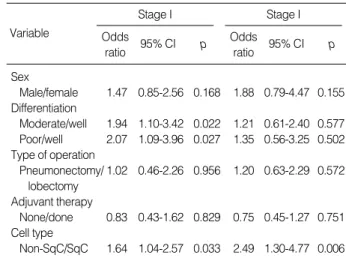
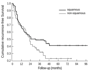
all 5-yr recurrence-free survival rates were also statistically dif- ferent (stage I, 57% and stage II, 36%, p<0.001). Univariate analyses of recurrence-free survival in stage I and stage II are described in Table 2. Multivariate analyses by the Cox regres- sion test showed that cell type is the significant risk factor for recurrence-free survival in both stage I and stage II non-small cell lung cancer (Table 3). Recurrence-free survival curves show- ed similar risks during early periods of follow-up for non-squamous cell carcinoma, and more risks after 2 yr from the operation when compared to squamous cell carcinoma (Fig. 1, 2).
DISCUSSION
We were fortunate to obtain nearly complete follow-up of patients after surgical resection for local non-small cell lung
Variable Stage I
SqC Non-SqC p
Stage II SqC Non-SqC p Age (yr)
≤60 61 85 0.069 40 19 0.725
>60 109 100 43 25
Sex
Male 160 96 0.001 81 25 0.001
Female 10 89 2 19
Differentiation
Well 27 38 0.003 14 10 0.161
Moderate 96 49 47 13
Poor 29 24 14 7
Extent of resection
Lobectomy 150 175 0.010 52 38 0.007
Pneumonectomy 20 8 30 6
Adjuvant therapy
None 154 177 0.056 46 32 0.057
Done 16 8 37 12
Table 1.Clinicopathological characteristics of 355 stage I and 127 stage II patients
SqC, squamous cell carcinoma.
Stage I
5-yr survival p 5-yr RFS p
Variable Stage II
5-yr survival p 5-yr RFS p
Sex
Male 69% 0.0078 55% 0.0959 39% 0.1900 36% 0.8626
Female 84% 65% 58% 33%
Differentiation
Well 79% 0.1843 64% 0.0285 52% 0.3020 34% 0.6317
Moderate 68% 52% 42% 39%
Poor 72% 52% 34% 38%
Extent of resection
Lobectomy 76% 0.0233 58% 0.9178 44% 0.5610 35% 0.7481
Pneumonectomy 53% 54% 39% 40%
Adjuvant therapy
None 75% 0.0822 57% 0.7061 46% 0.5907 39% 0.4472
Done 53% 57% 36% 30%
Cell type
Squamous 72% 0.6226 60% 0.2189 43% 0.5326 42% 0.1191
Non-Squamous 76% 55% 41% 23%
Table 2.Univariate analyses of recurrence-free survival
RFS, recurrence-free survival.
Variable
Stage I Odds
ratio 95% CI p
Stage I Odds
ratio 95% CI p Sex
Male/female 1.47 0.85-2.56 0.168 1.88 0.79-4.47 0.155 Differentiation
Moderate/well 1.94 1.10-3.42 0.022 1.21 0.61-2.40 0.577 Poor/well 2.07 1.09-3.96 0.027 1.35 0.56-3.25 0.502 Type of operation
Pneumonectomy/ 1.02 0.46-2.26 0.956 1.20 0.63-2.29 0.572 lobectomy
Adjuvant therapy
None/done 0.83 0.43-1.62 0.829 0.75 0.45-1.27 0.751 Cell type
Non-SqC/SqC 1.64 1.04-2.57 0.033 2.49 1.30-4.77 0.006 Table 3.Multivariate correlates of recurrence-free survival
CI, confidence interval; SqC, squamous cell carcinoma.
676 Y.S. Choi, Y.M. Shim, K. Kim, et al.
cancer. Our study was initiated based on our initial observa- tions that recurrence patterns appeared to be different, accord- ing to the histology at outpatient follow-up. According to previous reports, the histologic type of the tumor is one of the determinants of survival in patients with resected local non-small cell lung cancer. In the Lung Cancer Study Group, cancer recurrences and cancer-related death were more fre- quent and recurrence rates were higher in patients with non- squamous histology (3). However, Martini et al. reported that there was no difference in overall survival between squamous and non-squamous cancers (4). Ramacciato et al. also reported that histologic type did not play a statistically significant role in the total incidence of recurrence (5). Rena et al. reported better 5-yr survival rates for adenocarcinoma than for squa- mous cell carcinoma (6). Our data demonstrated similar results to those of the Lung Cancer Study Group. Recent data report- ed by Okada et al. demonstrated that advanced stage, high involvement of lymph nodes, male gender, and non-squamous cancer were independent, unfavorable prognostic factors in completely-resected lung cancer patients (7). We confirmed that the poor prognosis of non-squamous cell carcinoma be- comes evident after 2 yr. This finding could be explained by the fact that distant metastases were more common in non- squamous cell tumors than in the squamous type.
Our data also demonstrated that brain metastases were more common in non-squamous carcinoma than in squamous cell carcinoma, and some reports and preceding report by our insti- tute showed corresponding results to ours (8-10). Whether the patients benefit from the therapy for single brain metas- tases remains another issue for study.
REFERENCES
1. Cagini L, Monacelli M, Giustozzi G, Moggi L, Bellezza G, Sidoni A, Bucciarelli E, Darwish S, Ludovini V, Pistola L, Gregorc V, Tonato
M. Biological prognostic factors for early stage completely resected non-small cell lung cancer. J Surg Oncol 2000; 74: 53-60.
2. D’Amico TA, Aloia TA, Moore MB, Herndon JE 2nd, Brooks KR, Lau CL, Harpole DH Jr. Molecular biologic substaging of stage I lung cancer according to gender and histology. Ann Thorac Surg 2000;
69: 882-6.
3. Thomas PA Jr, Rubinstein L. Malignant disease appearing late after operation for T1 N0 non-small cell lung cancer. The Lung Cancer Study Group. J Thorac Cardiovasc Surg 1993; 106: 1053-8.
4. Martini N, Bains MS, Burt ME, Zakowski MF, McCormack P, Rusch VW, Ginsberg RJ. Incidence of local recurrence and second primary tumors in resected stage I lung cancer. J Thorac Cardiovasc Surg 1995; 109: 120-9.
5. Ramacciato G, Paolini A, Volpino P, Aurello P, Balesh AM, D’Andrea N, Del Grande E, Passaro U, Tosato F, Fegiz G. Modality of failure following resection of stage I and stage II non-small cell lung cancer.
Int Surg 1995; 80: 156-61.
6. Rena O, Oliaro A, Cavallo A, Filosso PL, Donati G, Di Marzio P, Maggi G, Ruffini E. Stage I non-small cell lung carcinoma: really an early stage? Eur J Cardiothorac Surg 2002; 21: 514-9.
7. Okada M, Nishio W, Sakamoto T, Harada H, Uchino K, Tsubota N.
Long-term survival and prognostic factors of five-year survivors with complete resection of non-small cell lung carcinoma. J Thorac Car- diovasc Surg 2003; 126: 558-62.
8. Mussi A, Pistolesi M, Lucchi M, Janni A, Chella A, Parenti G, Rossi G, Angeletti CA. Resection of single brain metastasis in non-small-cell lung cancer: prognostic factors. J Thorac Cardiovasc Surg 1996; 112:
146-53.
9. Ohta Y, Oda M, Tsunezuka Y, Uchiyama N, Nishijima H, Takanaka T, Ohnishi H, Kohda Y, Yamashita J, Watanabe G. Results of recent therapy for non-small-cell lung cancer with brain metastasis as the initial relapse. Am J Clin Oncol 2002; 25: 476-9.
10. Ham HS, Kang SJ, An CH, Ahn JW, Kim HC, Lim SY, Suh GY, Kim KM, Chung MP, Kim HJ, Kim JG, Kwon OJ, Shim YM, Rhee CH.
Clinical characteristics of recurred patients with stage I, II non-small cell lung cancer. Tuberc Respir Dis 2000; 48: 428-37.
Cumulative recurrence-free Survival
1.0 0.9 0.8 0.7 0.6 0.5 0.4 0.3 0.2
0 12 24 36 48 60 72 84 96
Follow-up (months)
Fig. 1.Recurrence-free survival according to the cell type in stage I patients.
squamous non-squamous
Cumulative recurrence-free Survival
1.0 0.9 0.8 0.7 0.6 0.5 0.4 0.3 0.2
0 12 24 36 48 60 72 84 96
Follow-up (months)
Fig. 2.Recurrence-free survival according to the cell type in stage II patients.
squamous non-squamous