INTRODUCTION
Primary cranial chondrosarcoma is not an uncommon neo- plasm and usually arises at the synchondroses of the skull base, particularly the sphenoid bone and the clival basioc- ciput (1-3). Primary intracranial extraosseous chondrosarco- ma is rare and most are attached to the dura, the presumed site of origin of these tumors (4-7). However, seven cases of primary intracranial chondrosarcomas unrelated to the cranium or the meninges have been reported and all of these tumors were histologically typed as mesenchymal variants (3, 5).
Recently, the authors experienced a case of primary intra- parenchymal myxoid chondrosarcoma of the brain without any attachment to the cranium or the meninges. We believe it worthwhile to document the clinical manifestations and radiological findings of this case, because to the best of our knowledge, this condition has not been previously reported.
The authors discuss pathologic differential diagnosis between histologically similar tumors, such as chordoma, parachor- doma, chordoid meningioma and chordoid glioma for the meticulous histologic differentiation of tumors, although cords of epithelioid cells reminiscent of typical chordomas are not the significant histologic finding in present case.
CASE REPORT
A 43-yr-old man was admitted to the neurosurgery depart-
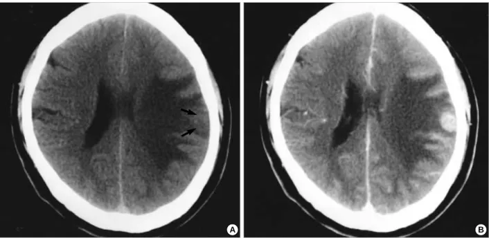
ment due to a history of occipital headache, nausea, and vom- iting of two months’ duration. He complained of general weakness, myalgia, tinnitus in the left ear, and an intermit- tent tingling sensation in the distribution of the bilateral mandibular division of the V cranial nerve. He had been man- aged with an insulin pump due to longstanding diabetes mellitus, and the generalized vague symptoms, mentioned previously, were considered to be complications of the diabetes mellitus. Neurological and physical examinations revealed no abnormalities. A pre-contrast computed tomography (CT) scan showed a 2 cm, non-calcified, slightly low density mass with severe peritumoral edema in the left parietal lobe.
A post-contrast CT scan revealed strong homogeneous en- hancement of the mass (Fig. 1), which was of low signal intensity on T1-weighted magnetic resonance (MR) imag- ing and high signal intensity on T2-weighted MR imag- ing. Post-contrast MR imaging revealed a homogeneously enhancing mass in the left parietal lobe (Fig. 2). A bone scan showed no abnormal uptake. A serum tumor marker study, performed under the impression of metastatic brain tumor, was negative for AFP, CEA, CA125, CA19-9, PAP, and PSA.
Whole body 18F-FDG-PET (positron emission tomography) showed a focal hypermetabolic lesion in the parietal area, however, there was no abnormality outside the brain. The patient underwent a left parietal osteoplastic craniotomy under the impression of atypical malignant glioma, lym- phoma, or metastatic tumor. Skull bone and dural surface looked normal and a cruciate dural incision was made. There
So-Hyang Im, Dong Gyu Kim, In Ae Park*, Je G. Chi*
Departments of Neurosurgery and Pathology*, Seoul National University College of Medicine, Clinical Research Institute, Seoul National University Hospital, Seoul, Korea
Address for correspondence Dong Gyu Kim, M.D.
Associate Professor, Department of Neurosurgery Seoul National University Hospital , 28 Yongon- dong, Chongno-gu, Seoul 110-744, Korea Tel : +82.2-760-2874, FAX : +82.2-744-8459 E-mail : gknife @ plaza.snu.ac.kr
301 J Korean Med Sci 2003; 18: 301-7
ISSN 1011-8934
Copyright � The Korean Academy of Medical Sciences
Primary Intracranial Myxoid Chondrosarcoma
: Report of a Case and Review of the Literature
The authors present a case of primary intracranial extraosseous myxoid chon- drosarcoma without any attachment to the cranium or the meninges. The clinical and radiological findings of the primary intraparenchymal tumor are described with a review of the literature concerning cranial and intracranial myxoid chon- drosarcoma.
Key Words : Chondrosarcoma; Central Nervous System Neoplasms
Received : 7 December 2001 Accepted : 14 May 2002
was no adhesion or attachment between the dura and the cerebral cortex and the cortical surface also showed a normal architecture. An incision was made on the surface of the parietal lobe and a well encapsulated mass was found just beneath the cortex. The tumor was readily dissected from the surrounding gliotic plane and an en-bloc removal was performed. The immediate postoperative course was unevent- ful. The patient received postoperative radiotherapy of 5,940 cGy in 33 fractions. He remains in good medical condition and follow-up MR images taken three years after the opera- tion showed no recurrence.
Pathological Findings
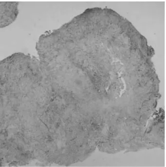
Macroscopically, the tumor had a nodular and lobulated appearance and measured 1.2×1.0×1.0 cm (Fig. 3). It had a grayish surface and showed a yellowish gray gelatinous area on serial section. It was firm to slightly hard in consistency.
Microscopically, the periphery of the tumor was surrounded partly by a thin fibrous tissue and partly by a somewhat com- pressed brain parenchyma. On low power cross-sectional view, the tumor had paucicellular areas interspersed with areas con- taining moderate numbers of cellular foci. The tumor showed an associated basophilic myxoid matrix, separated by thin
Fig. 1.(A) Pre-contrast computed tomography (CT) showing an ill-defined isodense mass (arrows), with severe surrounding edema in the left parietal cortex. (B) Strong homogeneous enhancement of the mass as seen in postcontrast CT.
A B
Fig. 2.(A) Axial T2-weighted magnetic resonance (MR) image showing slightly high signal intensity mass with severe surrounding edema in the left parietal cortex (arrow). (B) Sagittal T1-weighted MR image revealing an ill-defined low signal intensity mass (arrow). (C) Strong homogeneous enhancement of the mass as seen on the axial post-contrast T1-weighted MR image. The mass is located just beneath the dura, however, there was no connection to the dura.
A B C
Primary Intracranial Myxoid Chondrosarcoma 303
connective tissue bands, and was composed of round or slightly elongated cells separated by abundant myxoid stroma. The individual cells possessed small hyperchromatic, slightly irregular nuclei and a narrow rim of deeply eosinophilic cyto- plasm, and were arranged in short anastomosing cords and strands in myxoid matrix (Fig. 4, 5). The histology of this
tumor resembled that of an extraskeletal myxoid chondrosar- coma of other soft tissues. Microscopically, neither hemor- rhage nor necrosis was observed. Mitoses were extremely rare.
Portions of the tumor contained numerous thin-walled vessels.
The following antibodies were used for immunohistochemi- cal analyses; a polyclonal antibody to S100 (1:400; Dako A/S, Glostrup, Denmark) and monoclonal antisera to glial fibril- lary acidic protein (GFAP) (Dako, 1:500), vimentin (clone V9;
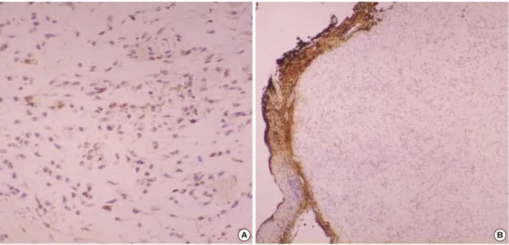
1:100; Dako A/S, Glostrup, Denmark), epithelial membrane antigen (EMA) (clone E29; 1:50; Dako A/S, Glostrup, Den- mark), cytokeratin 19 (clone RCK 108; 1:50; Dako A/S, Glostrup, Denmark), and cytokeratin, high molecular weight (clone 34 E12; 1:50; Dako A/S, Glostrup, Denmark). Immu- nohistochemical studies were performed using the avidin- biotin-peroxidase complex method. Diaminobenzidine was used as a substrate. On immunohistochemical studies, the neoplastic cells showed a weak positive staining for S100, positive staining for vimentin, and a lack of staining for EMA, cytokeratin 19, GFAP, and high molecular weight cytoker- atin, which confirmed the diagnosis of myxoid chondrosar- coma (Fig. 6).
DISCUSSION
Primary cranial chondrosarcomas normally arise from the skull base and are usually located extradurally (1-3). Prima- ry intracranial extraosseous chondrosarcomas have been less commonly reported than cranial chondrosarcomas and their locations include the cerebral convexity, falx, fourth ventricle,
Fig. 3.Photomicrograph showing a nodular and lobulated appear- ance of tumor (H&E, ×10).
Fig. 4.High power photomicrograph of the tumor shows epithe- lioid to spindle shaped cells dispersed in a myxochondroid matrix (H&E, ×400).
Fig. 5.Photomicrograph of the tumor cells, which are arranged in strands and cords within a large amount of stroma. Round or slight- ly elongated cells are separated by abundant myxoid stroma (H&E,
×200).
cerebellum, and thalamus (3, 6-10). Theoretically, a chon- drosarcoma should originate from the mesenchymal tissues, like cartilage, therefore, those arising from the skull base are quite natural. Intracranial chondrosarcomas are also thought to arise from the mesenchymal elements of the central ner- vous system, such as, the primitive multipotential mesenchy- mal cells or their mature descendents (fibroblasts, meningeal cells, and pial cells) located within the leptomeninges, the pia-arachnoid surrounding blood vessels or in the vessel walls, the stroma of the choroid plexus and aberrant embryonal cartilagenous rests (4, 7, 9-16). In the case of primary intra- parenchymal chondrosarcoma, misplaced embryonal carti- lagenous rests or primitive multipotential mesenchymal cells in leptomeningeal sheaths around vessels or the vessel walls have been suggested to be origins without definitive evidence (3, 5, 7, 8, 10, 17-21). The tumor in present case was locat- ed just beneath the cortex so that the leptomeningeal tissue or a vessel in the depths of the sulcus might be a possible origin. This hypothesis was suggested by some authors (13, 22) and our case might be an example.
Histologically, three subtypes of chondrosarcomas have been described; classic chondrosarcoma, mesenchymal chon- drosarcoma, and myxoid chondrosarcoma (5, 17). Most of the primary intracranial extraosseous chondrosarcomas show a dural involvement. However, those within the brain paren- chyme without any attachment to the cranium or the meninges are very rare with only seven cases reported. These include a thalamic, three cerebellar, two frontal, and one parietal tumors (3, 5, 8, 11, 12, 17). All of these primary intraparenchymal chondrosarcomas were of a mesenchymal histological sub- type. A case of radiation-induced classic chondrosarcoma of
the cerebellum occurred 16 yr after radiation therapy for a cerebellar astrocytoma (23). However, primary intraparenchy- mal myxoid chondrosarcoma without any dural involvement has not been previously reported.
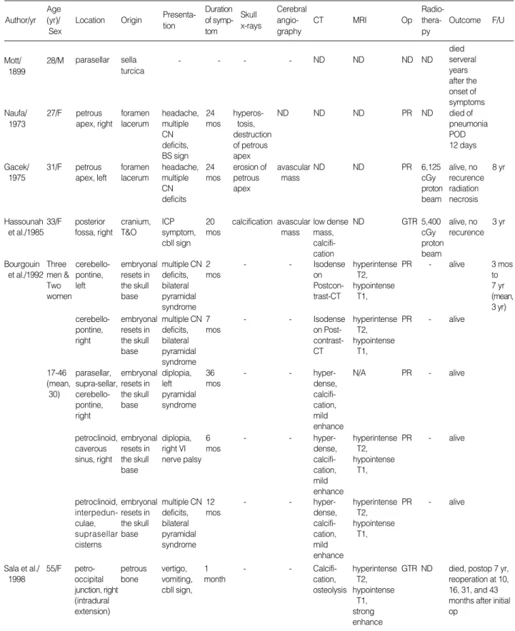
The classic cranial and intracranial chondrosarcomas usu- ally arise at the skull base and most frequently affect adults (3, 7, 18). The classic subtype has a better prognosis than the mesenchymal subtype (5, 7, 18). Intracranial extraosseous mesenchymal chondrosarcoma usually occurs in the frontopari- etal region and is highly vascular (5, 7). It is the most aggres- sive subtype with a tendency for recurrence and metastasis (3, 5, 17). The 5-yr survival rate is about 40%, and only occa- sional long-term survivals have been reported (7). Cranial or intracranial myxoid chondrosarcoma is a rare variant and only 10 cranial cases and four intracranial cases have been reported (2, 3, 7, 9, 10, 13, 14, 19-21) (Table 1, 2).
Calcification was more common in cranial myxoid chon- drosarcoma. The masses were found to be of low density or isodense on CT scan. MR imaging showed the tumors to be of low signal intensity on T1-weighted images and of high signal intensity on T2-weighted images. Tumors showed modest to strong enhancements on CT and/or MRI. Severe peritumoral brain edema was seen only in the present case.
Three patients who had undergone a gross total removal with or without postoperative radiation therapy were in good medi- cal condition during the follow-up period of 13 months to three years. Two patients who had undergone a gross total removal experienced tumor recurrences. Five patients who underwent a partial resection with or without postoperative radiation therapy were alive during the follow-up period of three months to eight years (mean 5.5 yr).
Fig. 6.On immunohistochemical studies, the myxoid chondrosarcoma shows a weak positive staining for S100 (A, ×200) and a lack of staining for GFAP (B, ×40).
A B
Primary Intracranial Myxoid Chondrosarcoma 305
Author/yr Age (yr)/
Sex
Presenta- tion
Duration of symp- tom
Op Radio- thera- py
Outcome F/U Skull
x-rays Origin
Location
Cerebral angio- graphy
CT MRI
Table 1.Clinical features of 10 patients with cranial myxoid chondrosarcoma
Mott/
1899
28/M parasellar sella turcica
- - - - ND ND ND ND
died serveral years after the onset of symptoms Naufa/
1973
27/F petrous apex, right
foramen lacerum
headache, multiple CN deficits, BS sign
24 mos
hyperos- tosis, destruction of petrous apex
ND ND ND PR ND died of
pneumonia POD 12 days
Gacek/
1975
31/F petrous apex, left
foramen lacerum
headache, multiple CN deficits
24 mos
erosion of petrous apex
avascular mass
ND ND PR 6,125
cGy proton beam
8 yr alive, no recurence radiation necrosis
Hassounah et al./1985
33/F posterior fossa, right
cranium, T&O
ICP symptom, cbll sign
20 mos
calcification avascular mass
low dense mass, calcifi- cation
ND GTR 5,400
cGy proton beam
3 yr alive, no recurence
Bourgouin et al./1992
Three men &
Two women
cerebello- pontine, left
embryonal resets in the skull base
multiple CN deficits, bilateral pyramidal syndrome
2 mos
- - Isodense
on Postcon- trast-CT
hyperintense T2, hypointense
T1,
PR - 3 mos
to 7 yr (mean, 3 yr) alive
17-46 (mean,
30)
parasellar, supra-sellar, cerebello- pontine, right
embryonal resets in the skull base
diplopia, left pyramidal syndrome
36 mos
- - hyper-
dense, calcifi- cation, mild enhance
N/A PR - alive
petroclinoid, caverous sinus, right
embryonal resets in the skull base
diplopia, right VI nerve palsy
6 mos
- - hyper-
dense, calcifi- cation, mild enhance
hyperintense T2, hypointense
T1,
PR - alive
petroclinoid, interpedun- culae, suprasellar cisterns
embryonal resets in the skull base
multiple CN deficits, bilateral pyramidal syndrome
12 mos
- - hyper-
dense, calcifi- cation, mild enhance
hyperintense T2, hypointense
T1,
PR - alive
cerebello- pontine, right
embryonal resets in the skull base
multiple CN deficits, bilateral pyramidal syndrome
7 mos
- - Isodense
on Post- contrast- CT
hyperintense T2, hypointense
T1,
PR - alive
‐ : information is not available, ND: not done, T&O: temporo-occipital, yr: year, CN:cranial nerve, BS: brain stem, ICP: intracranial pressure, cbll: cerebellar, mos: months, CT: computed tomography, MRI: magnetic resonance imaging, T2: T2-weighted image, T1: T1-weighted image, enhance:enhancement, Op: operation, PR: partial resection, GTR: gross total resection, POD: postopeation day, F/U: follow-up.
55/F Sala et al./
1998
petro- occipital junction, right (intradural extension)
petrous bone
vertigo, vomiting, cbll sign,
1 month
- - Calcifi-
cation, osteolysis
hyperintense T2, hypointense
T1, strong enhance
GTR ND died, postop 7 yr, reoperation at 10, 16, 31, and 43 months after initial op
Histologically, chordoma consists of epithelial cells with abundant, foamy cytoplasm, resembling the cells in adeno- carcinoma (24). Chondroid chordomas have morphologic features similar to those of typical chordomas except the additional features of cartilagenous foci resembling those of chondroma or conventional chondrosarcoma (25). Parachor- doma resembles chordoma histologically with nests of uni- form-appearing, vacuolated epithelioid cells deposited in a myxochondroid matrix, however, with a wider range of appear- ance (26, 27). Chordoid meningioma presents as encapsulated mass. Histologically, chordoid meningiomas were composed of meningothelial cells, mimicking the features of chordoma with nests and cords of epithelioid and spindle cells with abun- dant myxoid matrix, and often associated with peritumoral lymphoplasmacellular infiltration (28). Chordoid glioma occurs preferentially in the third ventricle with a discrete margin (29). Histologically, the tumor also reminiscents of
chordoma with cords and lobules of oval-to-polygonal epithe- lioid cells in a mucoid matrix (29).
Immunohistochemically, chordoma and parachordoma coexpress S-100 protein, cytokeratin and EMA, and the two neoplasms differ in their detailed cytokeratin immunophe- notype, whereas myxoid chondrosarcoma consistently lacked cytokeratin (26). Chordoid meningiomas were immunohis- tochemically consistently negative for S-100 protein, cyto- keratin and EMA (30). The cells of chordoid glioma showed diffuse and intense immunoreactivity for GFAP and vimentin, whereas immunoreactivity for EMA and cytokeratin was nonreactive to focally reactive (29, 31, 32). Immunohisto- chemical finding of present case, showing slight and focal reactivity for S-100 protein, whereas negative immunoreac- tivity for cytokeratin, GFAP and EMA, is not compatible with neither chordoma and related tumors, chordoid menin- gioma nor chordoid glioma but is compatible with myxoid
Davidson/
1981
hemisphere, left (dural attach- ment)
symptom, cbll sign
days mass rhage,
mild enhance, hydro- cephalus
mos no recur- rence
Salcman et al./1992
28/F falx, left sheath of blood vessels
headache, hemipare- sis, dys- phasia
2 mos
ND ND isodense
mass, minimal enhance
hyperintense T2, hypointense
T1, modest enhance
GTR ND 22
mos alive,
recurred at 10 mos 2nd Op &15,000 cGy 125I-bra- chytherapy died of local Sato et al./
1993
43/F pineal region
mesenchy- mal cells in the tentorium
blurred vision, gait disturbance
2 mos
- - multi-
lobulated, cystic mass, moderate enhance
- PR 6,000
cGy chemo- therapy
recurrence with meningeal dissemi- nation, POD 3 yr Present
case
43/M parietal cortex, left
? ICP
symptom 2 mos
normal ND Isodense mass, severe edema, strong enhance
hyperintense T2, hypointense
T1, strong enhance, severe edema
GTR 5,940 cGy photon beam
3 yr alive,
no recur- rence
‐ : information is not available, ND: not done, yr: year, ICP: intracranial pressure, mos: months, CT: computed tomography, MRI: magnetic resonance imaging, T2: T2-weighted image, T1: T1-weighted image, enhance: enhancement, Op: operation, PR: partial resection, GTR: gross total resection, POD: postopeation day, F/U: follow-up, 2nd: second.
Primary Intracranial Myxoid Chondrosarcoma 307
chondrosarcoma.
Primary intracranial myxoid chondrosarcoma is so uncom- mon that no definitive statement can be made about the opti- mal treatment and prognosis. However, most patients showed a relatively benign clinical course during the follow-up period.
Radical excision might play an important role in the man- agement. Postoperative radiotherapy should be considered to prevent recurrence and progression of the tumor.
REFERENCES
1. Bahr AL, Gayler BW. Cranial chondrosarcomas. Report of four cases and review of literature. Radiology 1977; 124: 151-6.
2. Gacek RR. Diagnosis and management of primary tumors of the petrous apex. Ann Otol Rhinol Laryngol 1975; 84: 1-20.
3. Hassounah M, Al-Mefty O, Akhtar M, Jinkins JR, Fox JL. Primary cranial and intracranial chondrosarcoma. A survey. Acta Neu- rochir (Wien) 1985; 78: 123-32.
4. Alpers BJ. Cerebral osteochondroma of dural origin. Ann Surg 1935;
101: 27-37.
5. Bingaman KD, Alleyne CH, Olson JJ. Intracranial extraskeletal mesenchymal chondrosarcoma: case report. Neurosurgery 2000;
46: 207-12.
6. Gerszten PC, Pollack IF, Hamilton RL. Primary parafalcine chon- drosarcoma in a child. Acta Neuropathol 1998; 95: 111-4.
7. Salcman M, Scholtz H, Kristt D, Numaguchi Y. Extraskeletal myx- oid chondrosarcoma of the falx. Neurosurgery 1992; 31: 344-8.
8. Heros RC, Martinez AJ, Ahn HS. Intracranial mesenchymal chon- drosarcoma. Surg Neurol 1980; 14: 311-7.
9. Scott RM, Dickersin R, Wolpert SM, Twitchell T. Myxochondrosar- coma of the fourth ventricle. Case report. J Neurosurg 1976; 44:
386-9.
10. Smith TW, Davidson RI. Primary meningeal myxochondrosarcoma presenting as a cerebellar mass: case report. Neurosurgery 1981;
8: 577-81.
11. Parker JR, Zarabi MC, Parker JC Jr. Intracerebral mesenchymal chondrosarcoma. Ann Clin Lab Sci 1989; 19: 401-7.
12. Raskind R, Grant S. Primary mesenchymal chondrosarcoma of the cerebrum: report of a case. J Neurosurg 1966; 24: 676-8.
13. Naufal PM. Primary sarcomas of the temporal bone. Arch Otolaryn- gol 1973; 98: 44-50.
14. Sato K, Kubota T, Yoshida K, Murata H. Intracranial extraskeletal myxoid chondrosarcoma with special reference to lamellar inclu- sions in the rough endoplasmic reticulum. Acta Neuropathol 1993;
86: 525-8.
15. Korten AG, ter Berg HJ, Spincemaille GH, van der Laan RT, Van de Wel AM. Intracranial chondrosarcoma: review of the literature and report of 15 cases. J Neurol Neurosurg Psychiatry 1998; 65:
88-92.
16. Scheithauer BW, Rubinstein LJ. Meningeal mesenchymal chondrosar- coma: Report of 8 cases with review of the literature. Cancer 1978;
42: 2744-52.
17. Harsh GR IV, Wilson CB. Central nervous system mesenchymal chondrosarcoma: Case report. J Neurosurg 1984; 61: 375-81.
18. Rubinstein LJ. Sarcomas of the nervous system. In : Minckler J (ed) Pathology of the nervous system, vol 2. McGraw-Hill, New York, 1971: 2144-64.
19. Bourgouin PM, Tampieri D, Robitaille Y, Robert F, Bergeron D, del Carpio R, Melancon D, Ethier R. Low-grade myxoid chondrosar- coma of the base of the skull: CT, MR, and histopathology. J Com- put Assist Tomogr 1992; 16: 268-73.
20. Mott FW. Chondrosarcoma springing from the sella turcica. Arch Neurol Psychiat 1899; 1: 432-3.
21. Sala F, Talacchi A, Beltramello A, Iuzzolino P, Bricolo A. Intracra- nial myxoid chondrosarcoma with early intradural growth. J Neu- rosurg 1998; 42: 159-63.
22. Lynch PG, Uriburu E. An intracranial cartilage-containing meningeal tumor. Case report. J Neurosurg 1973; 39: 261-4.
23. Bernstein M, Perrin RG, Platts ME. Radiation-induced cerebellar chondrosarcoma. Case report. J Neurosurg 1984; 61: 174-7.
24. Povysil C, Matejovsky Z. A comparative ultrastructural study of chondrosarcoma, chordoid sarcoma, chordoma and chordoma periphericum. Pathol Res Pract 1985; 179: 546-59.
25. Rossiello R, Ferrara G, Varricchio A, Baldi A, Motta S, Motta G.
Chondroid chordoma of the lateral skull base. ORL J Otorhinolaryn- gol Relat Spec 2001; 63: 114-8.
26. Folpe AL, Agoff SN, Willis J, Weiss SW. Parachordoma is immuno- histochemically and cytogenetically distinct from axial chordoma and extraskeletal myxoid chondrosarcoma. Am J Surg Pathol 1999;
23: 1059-67.
27. Fisher C. Parachordoma exists--but what is it?. Adv Anat Pathol 2000; 7: 141-8.
28. Couce ME, Aker FV, Scheithauer BW. Chordoid meningioma: a clinicopathologic study of 42 cases. Am J Surg Pathol 2000; 24:
899-905.
29. Brat DJ, Scheithauer BW, Staugaitis SM, Cortez SC, Brecher K, Burger PC. Third ventricular chordoid glioma: a distinct clinico- pathologic entity. J Neuropathol Exp Neurol 1998; 57: 283-90.
30. Mori S, Oka K, Hakozaki H, Soga Y, Hayano M, Oka T, Nakazato Y, Mori N. Chordoid meningioma. A case report. Pathol Res Pract 2001; 197: 515-8.
31. Ricoy JR, Lobato RD, Baez B, Cabello A, Martinez MA, Rodriguez G. Suprasellar chordoid glioma. Acta Neuropathol 2000; 99: 699- 703.
32. Cenacchi G, Roncaroli F, Cerasoli S, Ficarra G, Merli GA, Gian- gaspero F. Chordoid glioma of the third ventricle: an ultrastructural study of three cases with a histogenetic hypothesis. Am J Surg Pathol 2001; 25: 401-5.