D I A B E T E S & M E T A B O L I S M J O U R N A L
This is an Open Access article distributed under the terms of the Creative Commons At- tribution Non-Commercial License (http://creativecommons.org/licenses/by-nc/3.0/) which permits unrestricted non-commercial use, distribution, and reproduction in any medium, provided the original work is properly cited.
The Causes and Consequences of Low Levels of High Density Lipoproteins in Patients with Diabetes
Philip J. Barter
The Heart Research Institute, Sydney, Australia
Type 2 diabetes is commonly accompanied by a low level of high density lipoprotein cholesterol (HDL-C) that contributes to the increased cardiovascular risk associated with this condition. Given that HDLs have the ability to improve increase the uptake of glucose by skeletal muscle and to stimulate the secretion of insulin from pancreatic beta cells the possibility arises that a low HDL concentration in type 2 diabetes may also contribute to a worsening of diabetic control. Thus, there is a double case for raising the level of HDL–C in patients with type 2 diabetes: to reduce cardiovascular risk and to improve glycemic control. Approaches to raising HDL–C include lifestyle factors such as weight reduction, increased physical activity and stopping smoking. Of currently available drugs, the most effective is niacin. Newer formulations of niacin are reasonably well tolerated and have the ability to in- crease HDL–C by up to 30%. The effect of niacin on cardiovascular events in type 2 diabetes is currently being tested in a large- scale clinical outcome trial.
Keywords: Diabetes mellitus, type 2; High density lipoprotein; Insulin secretion; Insulin resistance; Lifestyle
Corresponding author: Philip J. Barter
The Heart Research Institute, 7 Eliza Street, Newtown, Sydney 2042, Australia E-mail: [email protected]
INTRODUCTION
Many patients with type 2 diabetes have a low concentration of high density lipoprotein cholesterol (HDL–C). The fact that a low level of HDL-C is a predictor of cardiovascular disease, it follows that a low level of HDL–C in patients with type 2 diabe- tes may contribute to the increased cardiovascular risk associ- ated with this condition. The precise cause of the low HDL–C in type 2 diabetes is not known but may be the consequence of insulin resistance, augmented very low density lipoprotein production and increased activities of cholesteryl ester trans- fer protein (CETP) and endothelial lipase. Recent studies showing that HDLs have the ability to improve increase the uptake of glucose by skeletal muscle [1] and to stimulate the secretion of insulin from pancreatic beta cells [2] raise the possibility that the low HDL concentration in type 2 diabetes may also contribute to a worsening of diabetic control or, in-
deed, to the progression of prediabetes to the full diabetic state. Therefore, a low concentration of HDLs may not only be a consequence of the diabetes and a contributor to the increased cardiovascular risk in diabetics but may also result in a wors- ening of glycemic control in people with type 2 diabetes.
POSSIBLE MECHANISMS RESPONSIBLE FOR A LOW HDL-C IN DIABETES
The typical dyslipidemia in type 2 diabetes includes an elevat- ed level of plasma triglyceride, a low level of HDL–C and a low density lipoprotein (LDL) fraction that is characterized by small dense particles [3]. The low level of HDL–C in these patients is associated with the presence of HDL particles are also smaller and denser than normal. Since it is known that the particle size of HDLs correlates inversely with the concentration of plasma triglyceride, the low level of HDL–C associated with diabetes pISSN 2233-6079 · eISSN 2233-6087
may be secondary to an elevated level of plasma triglyceride.
This inverse relationship between the concentrations of HDL–
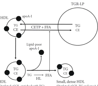
C and plasma triglyceride is the consequence of an increased rate of HDL catabolism that may be secondary to triglyceride enrichment of HDL particles in a mechanism dependent on the activities of CETP and hepatic lipase [4] as outlined in Fig. 1.
CARDIO-PROTECTIVE PROPERTIES OF HDLS
HDLs have a number of properties with the potential to protect against cardiovascular disease (Table 1) [5]. The best known of these relates to the ability of HDLs to promote the efflux of cholesterol from macrophages in the artery wall. However,
HDLs have several additional properties with the potential to protect against the consequences of atherosclerotic vascular disease. These include anti-oxidant, anti-thrombotic and anti- inflammatory effects and the ability to promote both the func- tion and repair of the endothelium. Some of the potentially protective functions of HDLs may be compromised in patients with diabetes. There is also evidence that HDLs have the capac- ity to improve diabetic control and possibly to protect against the development of type 2 diabetes [2].
HDL FUNCTION IS COMPROMISED IN DIABETES Apolipoprotein (apo) A-I (the major HDL apolipoprotein) may become glycated in people with diabetes. Such glycation has been reported to impair both the ability of HDLs to pro- mote cholesterol efflux from macrophages [6] and their ability to inhibit vascular inflammation [7]. This finding is of poten- tial patho-physiological significance given that subjects with type 2 diabetes, especially those with micro- and macro-vas- cular complications, tend to be in a pro-inflammatory state.
This highlights the importance of maintaining good glycemic control in people with diabetes and raises the possibility that therapeutic intervention with cross-link breakers, which re- portedly prevent protein modifications, may have the poten- tial to decrease the risk of the microvascular, and possibly the macrovascular complications that accompany diabetes. It is of interest to note that the endothelial vasoprotective effects of HDLs are impaired in patients with type 2 diabetes [8]. How- ever, when diabetic subjects are treated with niacin, the endo- thelial protective properties of HDLs are restored [8].
EFFECTS OF HDLS ON GLUCOSE HOMEOSTASIS There is a growing body of experimental evidence that HDLs have beneficial effects on glucose homeostasis by both increas- ing pancreatic beta cell function and by enhancing plasma glucose disposal.
Table 1. Potential protective properties of high density lipo- proteins
• Promote cholesterol efflux from macrophages in artery wall
• Anti-oxidant properties
• Anti-thrombotic properties
• Inhibit vascular inflammation
• Improve endothelial function
• Promote endothelial repair
• Improve diabetic control HDL
TGR-LP
TGCE
TGCE TG
CE CETG
TG FFA
HL CETP + FFA
HDL(Depleted of CE, enriched with TG) Small, dense HDL (Depleted of CE, TG and apoA-I) apoA-I
Lipid-poor apoA-I
Fig. 1. Cholesteryl ester transfer protein (CETP) promotes the transfer of cholesteryl esters (CE) from high density lipopro- teins (HDLs) to triglyceride-rich lipoproteins (TGR-LPs) in exchange for triglyceride (TG) to generate HDLs that are de- pleted of CE and enriched in TG. This TG enrichment pro- vides HDLs with the preferred substrate for hepatic lipase.
Subsequent hydrolysis of the newly acquired HDL TG by he- patic lipase leads to a reduction in volume of the particle core, a consequent decrease in particle size and a dissociation of lip- id-free/lipid-poor apolipoprotein (apo) A-I from the HDL particle surface. The dissociation of lipid-poor apoA-I from HDLs provides an explanation for why the concentrations of both HDL–C and apoA-I tend to be low in states of hypertri- glyceridemia such as occur in type 2 diabetes. Given that free fatty acids (FFAs) enhance the CETP-mediated remodelling of HDLs, such remodelling (and the associated low levels of HDL–C and apoA-I) may be exaggerated in patients with type 2 diabetes, a condition in which the concentration of FFAs in plasma is elevated as a consequence of the associated insulin resistance.
HDLs and pancreatic beta cell function
The ATP binding cassette transporter A1 (ABCA1) promotes efflux of cholesterol from cells to lipid-free/lipid-poor apoA-I in the extracellular space. It has been suggested that ABCA1 deletion results in cholesterol accumulation in the cell mem- brane of beta cells, with a subsequent inhibition of the exocy- tosis of insulin from secretory granules, and inhibition of in- sulin secretion [9]. It has also been reported that HDLs have beneficial effects on beta cells by inhibiting apoptosis [10].
And more recently, it has been shown in studies conducted in vitro using both Min6 cells and primary islets that HDLs iso- lated from human plasma and also the major HDL proteins, apoA-I and apoA-II, increase insulin synthesis and secretion up to 5-fold [2].
HDL and muscle glucose uptake
The observation that HDLs increase cellular glucose uptake in cultures of primary human skeletal muscle cells isolated from patients with type 2 diabetes mellitus [1] adds further support to the view that HDLs have the capacity to improve diabetic control (and possibly delay the development of new onset dia- betes) by several mechanisms.
STRATEGIES FOR RAISING HDL–C IN PATIENTS WITH TYPE 2 DIABETES
Lifestyle
Weight reduction
Most overweight people have a low level of HDL–C. Further- more, weight reduction is usually accompanied by an increase in the HDL–C level, although to be effective, the weight loss needs to be substantial and sustained. The mechanism under- lying a relationship between body weight and HDL-C concen- tration is uncertain. However, the fact that most patients with type 2 diabetes are overweight provides a strong basis for rec- ommending weight reduction as a strategy to raise the level of HDL–C in such patients.
Physical activity
High levels of aerobic activity are associated with high levels of HDL–C. Furthermore, increasing the level of physical activity in people with low levels of HDL–C, especially in those who are overweight, increases the HDL–C concentration. An exer- cise-induced increase in the level of HDL–C may be second- ary to an increased activity of lipoprotein lipase and the conse-
quent reduction in concentration of plasma triglyceride. It has been argued that the single most important preventable cause of low HDL–C in the modern world is a low level of physical activity.
A recent meta-analysis has confirmed the benefit of regular aerobic exercise on raising HDL–C levels and provided some insights into how much exercise is required [11]. The analysis included 25 randomized controlled studies that were designed to evaluate the effect of exercise training on HDL–C levels.
Overall, the mean exercise-induced increase in HDL–C was 2.53 mg/dL (P<0.001). Importantly, an increase in HDL–C concentration was apparent only in people who expended at least 900 kcal or exercised for at least 120 minutes each week.
In these people, every 10 minutes prolongation of exercise per session was associated with a 1.4 mg/dL increase in HDL–C.
In further analyses it was found that the increase in HDL–C was greatest in people whose body mass index was <28 kg/m2. These findings re-enforce recommendations for increasing levels of activity as a cardio-protective strategy in people with type 2 diabetes.
Alcohol consumption
Alcohol consumption increases the level of HDL–C, possibly secondary to an inhibition of CETP. However, it should be emphasized that it is not known whether the HDL–C eleva- tion associated with alcohol consumption is cardio-protective.
Smoking cessation
Smoking reduces the concentration of HDL-C and smoking cessation is associated with an up to 10% increase in HDL-C level. The mechanism by which smoking reduces the level of HDL–C is not known.
Pharmacological management
Levels of HDL–C are increased by treatment with several classes of currently available lipid-modifying agents. These in- clude fibrates, statins, and (especially) niacin, with evidence accumulating that such increases do translate into a reduced risk of cardiovascular disease.
Fibrates
The ability of fibrates to increase in concentration of HDL–C in patients with type 2 diabetes is rather modest, with increas- es of only 2% to 3% reported in two large clinical trials [12,13].
But treatment with fibrates (including gemfibrozil, bezafibrate,
and fenofibrate) is highly effective in reducing coronary risk in people with the combination of low HDL–C and elevated tri- glyceride [14]. In contrast, people with normal levels of HDL–
C and triglyceride levels appear to derive little macro-vascular benefit from treatment with fibrates. These results have been observed in both diabetic and non-diabetic subjects. However, the cardiovascular benefits of fibrates in people with low HDL–
C and high triglyceride are largely unrelated to fibrate-induced changes in either HDL–C or triglyceride. This suggests that a dyslipidemia characterized by a low HDL–C and high triglyc- eride identifies a group of people who derive substantial bene- fit from treatment with a fibrate but that the benefit is by a mechanism other than by changes in lipid concentrations.
Statins
Statins raise the level of HDL–C by 3% to 15%. The increase is greatest in those in whom the baseline level of HDL–C is low [15]. Given that the level of HDL–C tends to be low in patients with type 2 diabetes, it might be expected that the HDL–C in- crease induced by a statin would be greater in diabetics than in non-diabetics. The reality is the opposite, with evidence that the increase in level of HDL–C following treatment with a statin is much less in diabetic than in non-diabetic patients [15]. The explanation and the clinical implications of a reduced HDL–C response to statins in people with diabetes is not known.
Statins have been reported to have an adverse effect on glu- cose homeostasis, including a possible increase in develop- ment of new onset diabetes [16]. The mechanism is not known. The cholesterol content of pancreatic beta islet cells does impact of insulin synthesis and secretion with evidence that an increase in cell cholesterol content decreases insulin secretion [17]. However, given that statin treatment will, if anything, decrease cell cholesterol levels, this is an unlikely ex- planation for the observed increase in new onset diabetes in people treated with statins. It is possible that statins decrease insulin sensitivity in the liver or muscle, although there is no direct experimental evidence to support this. Thus, it is cur- rently not known why statins impact adversely on glucose ho- meostasis. However, given the strong evidence that statins markedly reduce cardiovascular risk patients with type 2 dia- betes [18], the clinical impact of a small increase in new onset diabetes is probably very small.
Niacin
Niacin increases HDL–C by up to 35% [19]. In addition, treat-
ment with niacin is associated with a change in the subpopula- tion distribution of HDLs towards larger particles [20], a change predictive of both a slowing of coronary disease progression and a reduction in cardiovascular events.
The precise mechanism by which niacin increases the con- centration of HDL-C is not known, although there is evidence that it delays the catabolism of HDL particles, possibly by de- creasing activity of CETP. There is also evidence that niacin increases the synthesis of apoA-I.
In addition to increasing the plasma concentration of HDL–
C and apoA-I, niacin has the capacity to modify HDLs in such a way that their function is enhanced. In one study HDLs were isolated from patients with type 2 diabetes and compared with HDLs isolated from healthy subjects [8]. Effects of the isolated HDLs on endothelium-dependent vasodilation and early en- dothelial progenitor cell-mediated endothelial repair were measured. Whereas the HDLs from healthy subjects stimulat- ed endothelial nitric oxide production, reduced endothelial oxidant stress and improved both endothelium-dependent va- sodilation and early endothelial progenitor cell-mediated en- dothelial repair, these beneficial endothelial effects of HDL were not observed in the HDLs isolated from diabetic patients.
However, after the diabetic patients had been treated for 3 months with niacin, the ability of the isolated HDLs to stimu- late endothelial nitric oxide, to reduce superoxide production and to promote endothelial progenitor cell-mediated endothe- lial repair were greatly improved [8].
Niacin has also been reported both to enhance the anti-in- flammatory effects of HDLs [21] and to inhibit vascular in- flammation by a mechanism that appears to be unrelated to changes in plasma lipids [22].
There is mounting evidence that treatment with niacin re- duces cardiovascular events and promotes regression of ath- erosclerosis [23] as revealed by imaging studies in human, with emerging evidence that the benefit is related (at least in part) to the magnitude of the increase in HDL–C.
If niacin is so effective, why is it not more widely used? The original, immediate release forms of niacin caused severe flushing that resulted in many people stopping the medication.
And those who did take it were often unable to achieve the recommended therapeutic dose of 2 g per day. The flushing problem has been substantially reduced by the development of newer extended release formulations of niacin. Not only is the extended release form better tolerated but it may be taken as a single daily dose rather than in three divided doses as was the
case with the immediate release form of the drug [24]. Flush- ing is reduced even more when an extended release form of niacin is combined with the anti-flushing agent, laropiprant, allowing more patients to take the drug at its recommended dose of 2 g per day [25]. The mechanism by which niacin causes flushing is understood. It binds to a receptor in the skin that leads to the production of PGD2. PGD2 then binds to the DP1 receptor causing dilation of the blood vessels in the skin leading to flushing. Laropiprant blocks the DP1 receptor and thus inhibits the flushing caused by niacin
Like statins, niacin is also known to have adverse effects on glucose homeostasis by decreasing insulin sensitivity and pos- sibly worsening diabetic control. Whether the effects of niacin on glucose homeostasis are of clinical importance is uncertain but will be determined by the results of ongoing cardiovascu- lar clinical outcome trials with niacin.
CONCLUSION
Many patients with type 2 diabetes have a low plasma concen- tration of HDL–C that may contribute to an increased risk of developing cardiovascular disease. The observation that HDLs have beneficial effects on pancreatic beta cell function and glucose uptake by skeletal muscle, adds support to the propo- sition that HDL-raising in people with type 2 diabetes may be anti-atherogenic as a consequence of both direct effects on the artery wall and also by improving diabetic control. It will be of great interest to see whether studies with HDL raising agents such as CETP inhibitors currently under investigation in clini- cal trials have beneficial effects on diabetic control.
REFERENCES
1. Drew BG, Duffy SJ, Formosa MF, Natoli AK, Henstridge DC, Penfold SA, Thomas WG, Mukhamedova N, de Courten B, Forbes JM, Yap FY, Kaye DM, van Hall G, Febbraio MA, Kemp BE, Sviridov D, Steinberg GR, Kingwell BA. High-density li- poprotein modulates glucose metabolism in patients with type 2 diabetes mellitus. Circulation 2009;119:2103-11.
2. Fryirs MA, Barter PJ, Appavoo M, Tuch BE, Tabet F, Heather AK, Rye KA. Effects of high-density lipoproteins on pancreatic beta-cell insulin secretion. Arterioscler Thromb Vasc Biol 2010;
30:1642-8.
3. Mooradian AD. Dyslipidemia in type 2 diabetes mellitus. Nat Clin Pract Endocrinol Metab 2009;5:150-9.
4. Barter PJ. Hugh sinclair lecture: the regulation and remodel- ling of HDL by plasma factors. Atheroscler Suppl 2002;3:39-47.
5. Barter PJ, Nicholls S, Rye KA, Anantharamaiah GM, Navab M, Fogelman AM. Antiinflammatory properties of HDL. Circ Res 2004;95:764-72.
6. Hoang A, Murphy AJ, Coughlan MT, Thomas MC, Forbes JM, O’Brien R, Cooper ME, Chin-Dusting JP, Sviridov D. Advanced glycation of apolipoprotein A-I impairs its anti-atherogenic properties. Diabetologia 2007;50:1770-9.
7. Nobecourt E, Tabet F, Lambert G, Puranik R, Bao S, Yan L, Davies MJ, Brown BE, Jenkins AJ, Dusting GJ, Bonnet DJ, Curtiss LK, Barter PJ, Rye KA. Nonenzymatic glycation im- pairs the antiinflammatory properties of apolipoprotein A-I.
Arterioscler Thromb Vasc Biol 2010;30:766-72.
8. Sorrentino SA, Besler C, Rohrer L, Meyer M, Heinrich K, Bahl- mann FH, Mueller M, Horvath T, Doerries C, Heinemann M, Flemmer S, Markowski A, Manes C, Bahr MJ, Haller H, von Eckardstein A, Drexler H, Landmesser U. Endothelial-vaso- protective effects of high-density lipoprotein are impaired in patients with type 2 diabetes mellitus but are improved after extended-release niacin therapy. Circulation 2010;121:110-22.
9. Kruit JK, Kremer PH, Dai L, Tang R, Ruddle P, de Haan W, Brunham LR, Verchere CB, Hayden MR. Cholesterol efflux via ATP-binding cassette transporter A1 (ABCA1) and cholester- ol uptake via the LDL receptor influences cholesterol-induced impairment of beta cell function in mice. Diabetologia 2010;
53:1110-9.
10. Kruit JK, Brunham LR, Verchere CB, Hayden MR. HDL and LDL cholesterol significantly influence beta-cell function in type 2 diabetes mellitus. Curr Opin Lipidol 2010;21:178-85.
11. Kodama S, Tanaka S, Saito K, Shu M, Sone Y, Onitake F, Suzu- ki E, Shimano H, Yamamoto S, Kondo K, Ohashi Y, Yamada N, Sone H. Effect of aerobic exercise training on serum levels of high-density lipoprotein cholesterol: a meta-analysis. Arch In- tern Med 2007;167:999-1008.
12. Keech A, Simes RJ, Barter P, Best J, Scott R, Taskinen MR, Forder P, Pillai A, Davis T, Glasziou P, Drury P, Kesaniemi YA, Sullivan D, Hunt D, Colman P, d’Emden M, Whiting M, Ehnholm C, Laakso M; FIELD study investigators. Effects of long-term fenofibrate therapy on cardiovascular events in 9795 people with type 2 diabetes mellitus (the FIELD study):
randomised controlled trial. Lancet 2005;366:1849-61.
13. ACCORD Study Group, Ginsberg HN, Elam MB, Lovato LC, Crouse JR 3rd, Leiter LA, Linz P, Friedewald WT, Buse JB, Gerstein HC, Probstfield J, Grimm RH, Ismail-Beigi F, Bigger
JT, Goff DC Jr, Cushman WC, Simons-Morton DG, Byington RP. Effects of combination lipid therapy in type 2 diabetes mel- litus. N Engl J Med 2010;362:1563-74.
14. Barter PJ, Rye KA. Is there a role for fibrates in the manage- ment of dyslipidemia in the metabolic syndrome? Arterioscler Thromb Vasc Biol 2008;28:39-46.
15. Barter PJ, Brandrup-Wognsen G, Palmer MK, Nicholls SJ. Ef- fect of statins on HDL-C: a complex process unrelated to changes in LDL-C: analysis of the VOYAGER Database. J Lip- id Res 2010;51:1546-53.
16. Sattar N, Preiss D, Murray HM, Welsh P, Buckley BM, de Craen AJ, Seshasai SR, McMurray JJ, Freeman DJ, Jukema JW, Mac- farlane PW, Packard CJ, Stott DJ, Westendorp RG, Shepherd J, Davis BR, Pressel SL, Marchioli R, Marfisi RM, Maggioni AP, Tavazzi L, Tognoni G, Kjekshus J, Pedersen TR, Cook TJ, Got- to AM, Clearfield MB, Downs JR, Nakamura H, Ohashi Y, Mizuno K, Ray KK, Ford I. Statins and risk of incident diabe- tes: a collaborative meta-analysis of randomised statin trials.
Lancet 2010;375:735-42.
17. Hao M, Bogan JS. Cholesterol regulates glucose-stimulated in- sulin secretion through phosphatidylinositol 4,5-bisphosphate.
J Biol Chem 2009;284:29489-98.
18. Cholesterol Treatment Trialists’ (CTT) Collaborators, Kearney PM, Blackwell L, Collins R, Keech A, Simes J, Peto R, Armitage J, Baigent C. Efficacy of cholesterol-lowering therapy in 18,686 people with diabetes in 14 randomised trials of statins: a meta- analysis. Lancet 2008;371:117-25.
19. Duggal JK, Singh M, Attri N, Singh PP, Ahmed N, Pahwa S,
Molnar J, Singh S, Khosla S, Arora R. Effect of niacin therapy on cardiovascular outcomes in patients with coronary artery disease. J Cardiovasc Pharmacol Ther 2010;15:158-66.
20. Kuvin JT, Dave DM, Sliney KA, Mooney P, Patel AR, Kimmel- stiel CD, Karas RH. Effects of extended-release niacin on lipo- protein particle size, distribution, and inflammatory markers in patients with coronary artery disease. Am J Cardiol 2006;98:
743-5.
21. Yvan-Charvet L, Kling J, Pagler T, Li H, Hubbard B, Fisher T, Sparrow CP, Taggart AK, Tall AR. Cholesterol efflux potential and antiinflammatory properties of high-density lipoprotein after treatment with niacin or anacetrapib. Arterioscler Thromb Vasc Biol 2010;30:1430-8.
22. Wu BJ, Yan L, Charlton F, Witting P, Barter PJ, Rye KA. Evi- dence that niacin inhibits acute vascular inflammation and improves endothelial dysfunction independent of changes in plasma lipids. Arterioscler Thromb Vasc Biol 2010;30:968-75.
23. Bruckert E, Labreuche J, Amarenco P. Meta-analysis of the ef- fect of nicotinic acid alone or in combination on cardiovascu- lar events and atherosclerosis. Atherosclerosis 2010;210:353-61.
24. Guyton JR. Extended-release niacin for modifying the lipo- protein profile. Expert Opin Pharmacother 2004;5:1385-98.
25. Maccubbin D, Koren MJ, Davidson M, Gavish D, Pasternak RC, Macdonell G, Mallick M, Sisk CM, Paolini JF, Mitchel Y.
Flushing profile of extended-release niacin/laropiprant versus gradually titrated niacin extended-release in patients with dys- lipidemia with and without ischemic cardiovascular disease.
Am J Cardiol 2009;104:74-81.