Primary squamous cell carcinoma of the stomach is a rare epithelial tumor with an incidence ranging from 0.04% to 0.4% of all diagnosed gastric cancers (1). To date, only 80 cases have been reported in the English lit- erature (2). Moreover, the multidetector CT (MDCT) findings associated with primary squamous cell carcino- ma of the stomach have not yet been reported. We re- port a case of primary squamous cell carcinoma of the stomach, which was manifested as a large unlcerofun- gating mass by a MDCT.
Case Report
A 40-year-old man diagnosed with a gastric mass via an endoscopic examination from a local clinic, was ad- mitted at the Guro Hospital of Korea University, for fur- ther evaluation and treatment. The patient had been in
good health prior to his illness, but began suffering from epigastric soreness and nausea for six months. The onset of pain began intermittently, and gradually became more constant. A series of laboratory examinations per- formed after being admitted did not reveal any abnor- malities except for anemia (hemoglobin 9.8 g/dL).
Moreover, the CEA and CA 19-9 serum level was with- in the normal range.
The CT images of the abdomen were acquired using a 16-slice MDCT scanner (Sensation 16; Siemens, Germany). A contrast agent was injected intravenously at 3 cc/sec, followed by image acquisition 75 seconds lat- er. The MDCT study was performed at the following pa- rameters: 120 kV, 140 mA, and a 0.75-mm beam colli- mation at a 10-mm/rotation table speed. The CT images were obtained at a supine position, from just above the diaphragm, to immediately below the pelvis. All the ob- tained images were stored in DICOM (Digital Imaging and Communications in Medicine) format. Digital data were sent to a PACS server (πview; Infinitt, Seoul, Korea), and were distributed to workstations (πview;
Infinitt). The 3D reconstruction was achieved by down- loading all images onto a local hard drive of a display
J Korean Radiol Soc 2008;59:37-40
─ 37 ─
CT Findings of Primary Squamous Cell Carcinoma of the Stomach: A Case Report1
Kyoung Min Kim, M.D., Chang Hee Lee, M.D., Kyeong Ah Kim, M.D., Cheol Min Park, M.D.
1Department of Diagnostic Radiology, Guro Hospital of Korea University, Korea
Received December 7, 2006 ; Accepted October 15, 2007
Address reprint requests to : Chang Hee Lee, M.D., Department of Diagnostic Radiology, Guro Hospital of Korea University, 80 Guro-dong, Guro-gu, Seoul 152-703, South Korea
Tel. 82-2-2626-1338 Fax. 82-2-6280-9076 E-mail: [email protected]
Primary squamous cell carcinoma is a rare tumor of the stomach with an incidence ranging from 0.04% to 0.4% of all diagnosed gastric cancers. We report a case of squa- mous cell carcinoma in the stomach associated with hypertrophic gastropathy and ob- served as a huge mass and wall thickening on the greater curvature site by a multide- tector CT.
Index words :Stomach
Stomach neoplasms Carcinoma
Squamous cell Tomography
workstation.
An axial CT scan revealed a large ulcerofungating mass (greater than 8 cm) involving the gastric body and antrum, with a particular presence on the posterior wall and greater curvature side, as well as thickening of the adjacent mucosal folds (Fig. 1). The invasion of the transverse colon was suspected from the interpretation of the coronal reformatted images (Fig. 2). In addition, thickened gastric mucosal folds also appeared near this
mass (Fig. 2). A CT gastrography and virtual gastroscopy images revealed a large ulcerofungating mass at the pos- terior wall and the greater curvature side of the gastric body and antrum (Fig. 3, 4). However, no distant metas- tasis was evident on the preoperative imaging studies.
A total gastrectomy, esophagojejunostomy, and seg- mental resection of the transverse colon was performed, and the resultant surgical specimen showed an ulcero- fungating mass measuring 6 cm by 8 cm at the greater curvature site of the gastric body and antrum (Fig. 5). In addition, the gastric mucosa located adjacent to the tu- mor was thickened with prominent rugal folds. The pathologic diagnosis yielded a well-differentiated squa- mous cell carcinoma with an invasion of the perigastric
Kyoung Min Kim, et al: CT Findings of Primary Squamous Cell Carcinoma of the Stomach
─ 38 ─
Fig. 3. A CT gastrography image with a 3D surface rendering technique shows a large ulcerfungating mass on the greater curvature side of the gastric body and antrum (arrows).
Fig. 4. A CT virtual gastroscopy image shows a large ulcero- fungating mass (arrow) observed at the posterior wall of the gastric body and antrum. See markedly thickened gastric mu- cosal fold (arrowhead) at the lesser curvature side of the gastric body.
Fig. 2. A coronal reformatted image reveals a large ulcerfungat- ing mass on the greater curvature of the body and antrum (ar- rows). The invasion of the transverse colon is suspected (ar- rowhead). Thickened gastric mucosal folds (double arrow) are also seen near the mass.
Fig. 1. Axial CT scan showing a large ulcerofungating mass (ar- row) involving the gastric body and antrum and is particularly present on the posterior wall, greater curvature side, and thickening of the adjacent mucocal folds (arrowhead).
fat and extension to the transverse colonic wall (Fig. 6).
No metastasis was found in all the 61 resected lymph nodes. The prominent mucosa of the gastric body was confirmed by hypertrophic gastropathy. In addition, no adenocarcinomatous component was identified, even after examining all the sections in the specimen under a microscope. The patient did not show postoperative complications, and a gastrograffin swallow obtained 14 days after the operation revealed no anastomotic leak.
Lastly, the patient was treated with 5 cycles of 5-FU and cisplan.
Discussion
Primary squamous cell carcinoma is an extremely rare tumor of the stomach, with an incidence of 0.04-0.4%
of all cases of gastric cancer (1). To date, only 80 cases of this illness have been published. The diagnostic prereq- uisite for primary squamous cell carcinoma of the stom- ach, as defined by Parks (3), is as follows: the tumor must not be located in the cardia, the tumor must not extend into the esophagus, and there must be no evi- dence of squamous cell carcinoma in any other organ.
The pathologic diagnosis is based on the following crite- ria: well-defined cell masses with whorl and pearl for- mation, no evidence of glandular differentiation within the tumor, intense cytoplasmic eosinophilia, and im- munohistochemical staining revealing the presence of high-molecular-weight cytokeratin (4). However, the pathogenesis of pure gastric squamous cell carcinoma
remains obscure. Some pathologists have implicated squamous metaplasia of the gastric mucosa as the pre- cursor of the transformation to squamous cell carcino- ma.
The data pertaining to the patient’s prognosis are also contradictory. While chemotherapy combined with sur- gical resection is reported to improve the prognosis, the consensus is that gastric squamous cell carcinomas are aggressive tumors.
Of the 80 published cases of this disease, gastric squa- mous cell carcinoma tends to occur in the proximal third of the stomach, most frequently along the lesser curvature, and it affects a slightly younger age group with a greater male preponderance compared to adeno- carcinoma (2).
According to the literature, the CT findings of squa- mous cell carcinomas of the stomach were huge masses within the gastric wall and were different from adeno- carcinomas of the stomach (1, 5). However, the MDCT findings have not yet been reported. In recent years, with the development of MDCT technology, three-di- mensional images such as MPR images, CT gastrogra- phy, and virtual gastroscopy have been introduced and provide more information on diagnosing gastric dis- eases. Particularly, these techniques accentuate the stomach wall and folds such that a CT gastrography and virtual gastroscopy can visualize mucosal lesions of the stomach, similar to a barium study and endoscopy (6).
Based on our study, a squamous cell carcinoma of the stomach is presented as a huge ulcerofungating mass on the posterior wall and the greater curvature side, as well as the thickening of mucosal folds due to associated hy- pertrophic gastropathy on MDCT images. The invasion of the transverse colon was preoperatively diagnosed on
J Korean Radiol Soc 2008;59:37-40
─ 39 ─ Fig. 5. Photograph of the resected stomach shows an ulcero- fungating mass (arrow). Note the thickened gastric mucosal folds (arrowheads) on the greater curvature site, which turned out to be hypertrophic gastropathy microscopically.
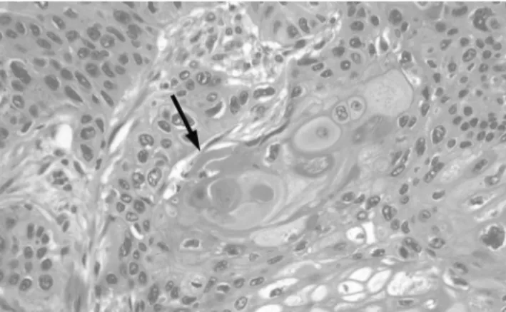
Fig. 6. Photomicrograph shows the feature of a well-differenti- ated squamous cell carcinoma. Keratin material is seen in the center as a pink-colored material (arrow). (H & E stain, ×150).
the coronal reformatted images. A virtual gastroscopy also showed an ulcerofungating mass at the posterior wall of the gastric body and antrum. There was, howev- er, no specific finding suggesting the presence of squa- mous cell carcinoma, which was different from the common malignant gastric tumors such as adenocarci- nomas and lymphomas.
In conclusion, we experienced a case of primary squa- mous cell carcinoma of the stomach which manifested as a huge ulcerofungating mass on the posterior wall and greater curvature side of the body and antrum.
However, it was difficult to differentiate a case of squa- mous cell carcinoma from other malignant gastric tu- mors, including adenocarcinomas and lymphomas on CT. Nevertheless, when a huge ulcerofungating gastric mass is found at CT, primary squamous cell carcinoma should be considered.
References
1. Schmidt C, Schmid A, Luttges JE, Kremer B, Henne-Bruns D.
Primary squamous cell carcinoma of the stomach. Report of a case and review of literature. Hepatogastroenterology 2001;48:1033-1036 2. Dursun M, Yaldiz M, Isikdogan A, Yilmaz G, Canoruc F, Ormeci
N, et al. Primary squamous cell carcinoma of the stomach: a case report and review of the literature. Eur J Gastroenterol Hepatol 2003;15:329-330
3. Parks RE. Squamous neoplasm of the stomach. Am J Roentgenol Radium Ther Nucl Med 1967;101:447-449
4. Mori M, Iwashita A, Enjoji M. Squamous cell carcinoma of the stomach: report of three cases. Am J Gastroenterol 1986;81:339-342 5. Hara J, Masuda H, Ishii Y, Aoki N, Nakayama H, Komura K, et al.
Exophytic primary squamous cell carcinoma of the stomach. J Gastroenterol 2004;39:299-300
6. Kim JH, Park SH, Hong HS, Auh YH. CT gastrography. Abdom Imaging 2005;30:509-517
Kyoung Min Kim, et al: CT Findings of Primary Squamous Cell Carcinoma of the Stomach
─ 40 ─
대한영상의학회지 2008;59:37-40
위에서 발생한 원발성 편평상피세포암의 CT 소견: 증례 보고1
1고려대학교 구로병원 영상의학과
김경민・이창희・김경아・박철민
원발성 편평상피세포암(primary squamous cell carcinoma)은 위에서 매우 드물게 발생하는 종양으로 위에서 생 기는 종양 중 0.04-0.4%를 차지한다. 저자들은 multidetector CT에서 위의 대만곡(greaster curvature)에서 벽비 후를 동반한 커다란 종괴로 나타난 비후성 위병(hypertrophic gastropathy)을 동반한 원발성 편평상피세포암의 증 례를 경험하였기에 그 CT 소견을 보고하고자 한다.