457
Copyrights © 2013 The Korean Society of Radiology
INTRODUCTION
Undifferentiated embryonal sarcoma of the liver (UES) is a rare and highly malignant hepatic neoplasm of mesenchymal origin that occurs predominantly in children, usually those 6-10 years old (1). Adult UES are extremely rare, especially in the over 60 years (2). To our knowledge, no prior study has clarified how fast a tumor mass grows, even though it is known as a rap- id-growing tumor. We report the unique case of adult UES that provide verification of the high growth rate of the tumor.
CASE REPORT
A 64-year female was admitted to emergency room of our hospital for acute right upper quadrant abdominal pain. Physi- cal examination revealed a tender mass in the right upper quad- rant of abdomen without fever. Laboratory data of liver func- tion, tumor marker such as carcinoembryonic antigen (CEA), carbohydrate antigen (CA) 125, CA19-9, and alpha-fetoprotein (AFP) were within normal range, except leukocytosis (white blood cell 14.070/uL, neutrophils 80.5%). He had a history of
colon cancer (TxN0M0 by CT staging) with curative endoscopic mucosal resection 4 years ago. There had been no tumor recur- rence during the follow-up period of about 4 years by abdominal CT. On the last follow-up CT before visiting to emergency room, there was a 0.4 cm hypodense lesion in segment 5 of liver (Fig.
1A). At that time, it was presumed to be a hepatic cyst.
Initial abdominal ultrasonography (US) showed the huge mass that was predominantly solid, heterogeneous in echo- genicity, and hypovascular with cystic component in the periph- ery (Fig. 1B). Abdominal CT showed a 11.0 cm-sized, well-cir- cumscribed hypoattenuating mass in the right lobe of the liver.
On iodinated contrast enhanced CT, this cystic was compart- mentalized by septations and showed no solid portion or calcifi- cation. Hemoperitoneum was seen mainly in the perihepatic space (Fig. 1C).
T2-weighted MR images showed a well-circumscribed large mass which was markedly hyperintense with multiple thick sep- ta and cavities of variable sizes in the right lobe of liver as well.
Coronal T2-weighted MR images showed clearly the ruptured portion of the tumor with intraperitoneal fluid (Fig. 1D). T1- weighted MR images showed the mass was predominantly hy-
Case Report
pISSN 1738-2637 / eISSN 2288-2928 J Korean Soc Radiol 2013;69(6):457-460 http://dx.doi.org/10.3348/jksr.2013.69.6.457
Received May 15, 2013; Accepted October 18, 2013 Corresponding author: O-Nyoung Kwon, MD Department of Radiology, Daegu Fatima Hospital, 576-31 Sinam-dong, Dong-gu, Daegu 701-600, Korea.
Tel. 82-53-940-7177 Fax. 82-53-954-7414 E-mail: [email protected]
This is an Open Access article distributed under the terms of the Creative Commons Attribution Non-Commercial License (http://creativecommons.org/licenses/by-nc/3.0) which permits unrestricted non-commercial use, distri- bution, and reproduction in any medium, provided the original work is properly cited.
We report the radiologic findings of a rare case of undifferentiated embryonal sar- coma of liver (UES) in a 64-year-old female. To our knowledge, this is the unique case of adult UES that provide verification of the high growth rate of the tumor.
Index terms
Undifferentiated Embryonal Sarcoma of the Liver Adult
Verification CT MR
Undifferentiated Embryonal Sarcoma of the Liver in an Adult:
The Verification of the High Growth Rate in the Tumor
성인에서 발견된 간의 미분화 배아육아종증: 증례 보고O-Nyoung Kwon, MD, Seong Hoon Kim, MD, Ji-Youl Shin, MD, Hyun Woong Shin, MD
Department of Radiology, Daegu Fatima Hospital, Daegu, Korea
Undifferentiated Embryonal Sarcoma of the Liver in an Adult: The Verification of the High Growth Rate in the Tumor
458
J Korean Soc Radiol 2013;69(6):457-460 jksronline.orggel-form embolization and right hepatic lobectomy were per- formed successfully. The solitary, large cystic mass of the liver was confirmed as primary UES (Fig. 1H).
During the retrospective evaluation of CT scans, we conclud- ed that the hypodense lesion was early UES in that the center of UES corresponds with the location of it. UES had grown from 0.4 cm to 11.0 cm in a maximal diameter for 7 months.
DISCUSSION
UES is a rare, highly malignant neoplasm of the liver predom- inantly in children (1). UES in the old, especially over 40 s is ex- tremely rare (2).
Patients frequently present with abdominal pain, fever, and weight loss. According to the literature as our case, laboratory findings of liver function and tumor marker such as AFP, CEA and CA19-9 are normal in most cases (2).
pointense with inner the foci of high signal intensity, represent- ing hemorrhage (Fig. 1E). Gadolinium contrast enhanced T1- weighted MR images showed heterogeneous enhancement of thick septa and the periphery in the mass (Fig. 1F). The dense peripheral rim was hypointense on T1- and T2-weighted MR images corresponding to the fibrous pseudocapsule. Therefore, initial diagnosis was ruptured cystic tumor such as cystadenoma or cystadenocarcinoma.
Positron emission tomography-CT showed diffuse mild fluo- rodeoxyglucose uptake in the peripheral portion of the mass, but not in the inner portion of the mass (Fig. 1G). On US-guid- ed biopsy, pathologic finding was not distinct. Consequently, surgical resection was considered.
Because of aggravated abdominal pain and increased size of the tumor on the follow-up CT, persistent bleeding from the cystic tumor was suspected. Angiography showed hypovascular mass with abnormal feeding vessels inside a tumor, and then
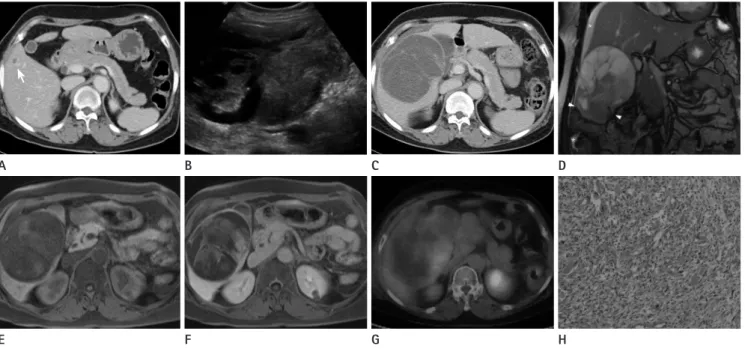
Fig. 1. A 64-year female with undifferentiated embryonal sarcoma of the liver.
A. 7 months ago, abdominal CT showed a 0.4 cm hypodense lesion (arrow) in the segment 5 of the liver.
B. Transverse US images show the huge and predominantly solid mass in the liver with peripheral cystic components.
C. Contrast enhanced CT shows the 11.0 cm-sized hypoattenuating mass in the right lobe of liver with multiple thick septa.
D. Coronal T2-weighted MR images show the large heterogeneous mass in the right lobe of the liver. Upper portion of the tumor is hyperintense with septation and lower portion is hypointense. In the lowest part of the mass, rupture (arrowheads) with asscociated intraperitoneal fluid (as- terisk) is shown.
E, F. Unenhanced T1-weighted and gadolinium contrast enhanced T1-weighted MR images show progressive enhancement of the periphery and septa.
G. PET-CT shows diffuse mild FDG uptake in the peripheral portion of the mass.
H. Photomicrograph (original magnification, × 200; H&E stain) shows undifferentiated spindle-shaped and large stellate cells in abundant myx- oid stroma.
Note.-FDG = fluorodeoxyglucose, PET = positron emission tomography, US = ultrasonography E
A
F B
G C
H D
O-Nyoung Kwon, et al
459
jksronline.org J Korean Soc Radiol 2013;69(6):457-460
eral abdominal CT scans had been taken regularly. We could notice that the maximal diameter was increased from 0.4 cm to 11.0 cm just in 7 months on CT. As our knowledge, this is a first and valuable report in the way that it is possible to express the growth rate numerically.
REFERENCES
1. Stocker JT, Ishak KG. Undifferentiated (embryonal) sarcoma of the liver: report of 31 cases. Cancer 1978;42:336-348 2. Li XW, Gong SJ, Song WH, Zhu JJ, Pan CH, Wu MC, et al.
Undifferentiated liver embryonal sarcoma in adults: a re- port of four cases and literature review. World J Gastroen- terol 2010;16:4725-4732
3. Buetow PC, Buck JL, Pantongrag-Brown L, Marshall WH, Ros PR, Levine MS, et al. Undifferentiated (embryonal) sarcoma of the liver: pathologic basis of imaging findings in 28 cases. Radiology 1997;203:779-783
4. Ros PR, Olmsted WW, Dachman AH, Goodman ZD, Ishak KG, Hartman DS. Undifferentiated (embryonal) sarcoma of the liver: radiologic-pathologic correlation. Radiology 1986;
161:141-145
5. Joshi SW, Merchant NH, Jambhekar NA. Primary multiloc- ular cystic undifferentiated (embryonal) sarcoma of the liver in childhood resembling hydatid cyst of the liver. Br J Radiol 1997;70:314-316
6. Martí-Bonmatí L, Ferrer D, Menor F, Galant J. Hepatic mesenchymal sarcoma: MRI findings. Abdom Imaging 1993;18:176-179
7. Psatha EA, Semelka RC, Fordham L, Firat Z, Woosley JT.
Undifferentiated (embryonal) sarcoma of the liver (USL):
MRI findings including dynamic gadolinium enhancement.
Magn Reson Imaging 2004;22:897-900
8. Vermess M, Collier NA, Mutum SS, Crofton ME. Misleading appearance of a rare malignant liver tumour on computed tomography. Br J Radiol 1984;57:262-265
9. Baron PW, Majlessipour F, Bedros AA, Zuppan CW, Ben- Youssef R, Yanni G, et al. Undifferentiated embryonal sar- coma of the liver successfully treated with chemotherapy and liver resection. J Gastrointest Surg 2007;11:73-75 The UES has unique characteristic radiologic finding as in
our case, that is a huge mass predominantly solid appearance on US, but cystic appearance on CT and MRI due to the high water content of the prominent myxoid stroma (3).
At US, UES appears iso- to hyperechoic, mainly solid mass correlates well with the pathologic findings (3, 4). The anechoic, cystic lesions correlate with cystic degeneration, old hemorrhage and necrosis (3).
The CT shows typically predominantly cystic mass as water attenuation with septa of variable thickness (3). The large por- tion of the mass is water-attenuation correlates with myxoid stroma of the pathologic findings (3-5). Occasionally, a pseudo- capsule may be observed by a thick and enhancing peripheral rim (4). Acute hemorrhage may be also observed by central foci of high attenuation with fluid-debris levels (3). Internal calcifica- tions are uncommon (4). Predominantly peripheral enhance- ment, especially on delayed images, have been reported after in- travenous administration of iodinated contrast materials (3).
MR imaging characteristics also resemble those of cystic le- sion as same as CT. Therefore, large portions of the mass are hy- pointense on T1-weighted images and hyperintense on T2- weighted images. Some high signal intensity on T1- and T2- weighted images represents intratumoral hemorrhage, which are better seen with MR imaging than CT (3, 4, 6). A hypoin- tense on T1- and T2-image represents also the fibrous pseudo- capsule (4). Heterogeneous enhancement of the peripheral and solid portions of the mass have been reported after intravenous administration of gadolinium contrast materials (7).
Owing to its cystic appearance at CT and MR imaging, some- times UES can be mistaken for cystic tumor (8). Especially UES in the old or with presenting by rupture, initial correct diagnosis is not easy as our case.
Until recently, the prognosis of UES was poor, with mean sur- vival of 12 months after diagnosis, even though complete surgi- cal resection (1). But recent authors report use of multiagent, adjuvant, or neoadjuvant chemotherapy followed by resection offer the best long-term results and possibly a cure (9).
UES was known high malignant tumor and the fact that the tumor grows fast have been mentioned in several reports (2).
However, it has not been supported by strong evidence in any other studies. In our case, during a colon cancer follow-up, sev-
Undifferentiated Embryonal Sarcoma of the Liver in an Adult: The Verification of the High Growth Rate in the Tumor
460
J Korean Soc Radiol 2013;69(6):457-460 jksronline.org성인에서 발견된 간의 미분화 배아육아종증: 증례 보고
권오녕 · 김성훈 · 신지열 · 신현웅
간에 발생한 미분화성 배아육아종은 매우 드문 중간엽 기원의 악성종양이다. 주로 소아에서 발생하며 성인에서는 극히 드 문 질환이다. 미분화성 배아육아종은 매우 빠르게 성장하는 종양으로 알려져 있다. 하지만 저자들이 조사한 바로는 현재 까지 문헌에서 이 종양이 정확히 얼마나 빠른 성장을 보이는지 분명하게 말할 수 있는 보고는 찾을 수 없었다. 저자들은 64세 여자 환자에서 종양의 직경이 7개월 동안 0.4 cm에서 11.0 cm로 빠르게 커지는 것을 경험하였기에, 그 영상소견을 보고하고자 한다.
대구파티마병원 영상의학과