Characteristics of the Abdominal and Neck Flexor Muscles of Children with Cerebral Palsy
Purpose: The purpose of this study was to compare the activities of the abdominal and neck flexor muscles of children with and without cerebral palsy (CP) while lifting the head in a supine position.
Methods: The subjects were eight children with CP and eight children without the disease. The activities of the external abdominal oblique (EO), internal abdominal oblique (IO), rectus abdominis (RA), sternocleidomastoid (SCM), and RA/SCM muscles were collected by surface electromyography (EMG) when the children lifted their heads. A Mann-Whitney U test was used to compare the activity of each muscle during the head-lifting exercise. Statistical significance was accepted at p<0.05.
Results: The activities of the EO, IO, and RA, and RA/SCM muscles differed significantly between the children with and without CP, but there was no significant between-group difference in the activity of the SCM muscle.
Conclusion: These findings suggest that the abdominal muscles are not employed as much in the activities of children with CP compared to those without the disease. Additionally, those with CP were more dependent on the neck flexor muscle during the head-lifting exercise in a supine position.
Key Words:Abdominal muscle, Cerebral palsy, EMG
Sung-Jin Choi1, Dae-Hyouk Bang1, Hyun-Jung So1, Won-Seob Shin2
1Department of Physical Therapy, Graduate School of Daejeon University, 2Department of Physical Therapy, College of Natural Science, Daejeon University
The Journal of Korean Society of Physical Therapy Original articles
I. Introduction
Cerebral palsy (CP) is a motor disorder associated with movement, posture, and balance problems that is caused by nonprogressive brain damage at an early stage of development.1,2 Interference with the normal development of muscles results in abnormalities of muscle tone, reflexes, and movement patterns, all of which cause muscle weakness and imbalance. These neuromotor and musculoskeletal disorders change over time.3,4 Another problem is that deficits
Copylight ⓒ 2014 The Korea Society of Physical Therapy
This is an Open Access article distribute under the terms of the Creative Commons Attribution Non-commercial License (Http:// creativecommons.org/license/by-nc/3.0.) which permits unrestricted non-commercial use, distribution,and reproduction in any medium, provided the original work is properly cited.
Received Nov 6, 2014 Revised Dec 14, 2014 Accepted Dec 16, 2014
Corresponding author Won-Seob Shin, [email protected]
in postural control in children with CP.5 One of the primary goals of postural control is to stabilize the head in space.6 Children with CP are unable to adequately stabilize their head in space during dynamic tasks and this may be related to immature or abnormal patterns of trunk muscle activation.7 Stabilization of the head’s position in space is one of the motor strategies used to establish a stable frame of reference during movement.8
A healthy infant is able to control its neck muscles by the time it is 3 or 4 months. By the time the infant is about 5 months, the trunk muscles have developed into a coordinated postural pattern. In an 8-month-old infant who can sit independently, the neck and trunk muscles are coordinated when the infant’s body sways forwards or backwards.9,10 One example of spontaneous motor behavior of an infant that can be easily observed during the first year of life, particularly
supine position.
II. Methods
1. Subjects
Eight children with CP aged 7 to 14 who were in B hospital in Daejeon and eight children without the disease were recruited for this study. All the subjects and their guardians voluntarily signed written consent forms. The inclusion criteria were children with spastic diplegia CP who could lift their heads from the floor, children who had no fear, children who did not show an adverse reaction to the surface electromyographic equipment, and children who had no musculoskeletal diseases that could affect the outcome. In the group of children with CP could not walk without assistive devices and had limited self-mobility (GMFCS levels IV and V). The degree of disability for children with CP are described by the GMFCS (Gross Motor Function Classification System), a 5-level system in which GMFCS level I indicates minimal impairment and children at the GMFCS level V cannot sit upright or ambulate independently.22 We also excluded other impairments in cognition, attention, or sensory making them unable to follow direction and uncorrected visual impairments and recent injection of botulinium toxin, surgery, or any planned medical or surgical interventions to modify effects of CP during the period of the study.
Eight children without CP who had no neurological, developmental, or musculoskeletal diseases were also included, and the consent of their parents or guardians was obtained.
2. Experimental procedure
After taking off their shirt, the child lay down in a comfortable position, and the electrode and the ground electrode were attached to the child’s body. The electrodes were placed at the midpoint of the muscle length for the sternocleidomastoid (SCM) muscle and at the muscle belly 2 cm outward from the umbilicus for the RA muscle. The electrodes for the EO were placed halfway between the anterior superior iliac spine and the lower rib, parallel to the muscle fibers. The electrodes for the IO were placed in the middle between the anterior superior iliac spine and midline of the body, just above the inguinal between 6 and 9 months of age, is active head lifting from
supine.11 The abdominal muscles play a particularly important role in providing stability for movement and adjustment of the head by fixing the chest during head movements.12 The abdominal muscles consist of the rectus abdominis (RA), internal abdominal oblique (IO), external abdominal oblique (EO, and transverse abdominis (TA). These muscles primarily play a role in bending the trunk.12 However, they are sometimes involved in helping to stabilize the trunk while lifting the head.12 Core stability refers to the ability to control the location and movements of the trunk on the pelvis, and it is regulated by the interaction of the greater and lesser muscles.13,14 These trunk muscles provide stability for the spine by maintaining the correct alignment of the body while standing or sitting. They are an essential element in functional performance and provide a basic support mechanism for moving the limbs or maintaining balance.15
Children with CP have poor trunk stability due to the low muscle tone of the trunk and proximal muscles and muscle weakness.16 They find it difficult to maintain sufficient stability of the head when performing dynamic tasks due to the instability of the trunk muscles.17 The lack of head control and trunk stability makes it impossible for them to maintain a correct posture and leads to problems with posture. As a result, an abnormal pattern of muscle activity occurs, resulting in various problems, such as muscle imbalance, weakness, and atrophy, in addition to motor coordination disorders.18,19 In particular, the weakening of the abdominal muscles in those with CP interferes with the stability of the trunk and leads to secondary dysfunction of the arms and legs.19,20 Children with CP often struggle with head and trunk stability, even during functional tasks.21
Quantitative data on the activities of the abdominal muscles during neck exercises to stabilize the trunk in children with CP are lacking. Studies are needed that compare the patterns of muscle activation of the trunk in children with and without CP. We believe that such objective data can enhance the understanding of the physical characteristics of children with cerebral palsy. Therefore, the objective of this study was to compare the activity of the abdominal and neck flexor muscles of children with and without CP during head lifting in a
ligament. The electrodes ran parallel to the muscle fibers and were placed 2 cm apart to minimize any confusion between the muscles.23 After checking that the subject was in a stable state in a supine position, the subject was instructed to raise his/her head from the ground as quickly and as high as possible. Electromyographic signals were collected while the subject held up his/her head for 5 sec, and the mean value of two measurements was compared. Optimal stabilization has been considered to be increased muscle activation of the abdominal muscles when compared with the neck flexor.2,7,14,15 To determine the synergistic relation between the muscles in this experiment, we calculated the ratio of the RA and SCM muscle activity.
3. EMG measurement
In this study, a 16-channel electromyograph (Telemyo 2400T, Noraxon, USA) was used to measure the activities of the EO, IO, RA, and SCM muscles during the head-lifting exercise.
Each surface electromyography (EMG) electrode was attached to the muscle according to the method of Cram et al.23 The ground electrode was attached to the anterior superior iliac spine on the dominant side. Prior to the EMG measurements, dead skin was removed with sandpaper after hair removal to minimize any resistance of the skin that may occur at the point of the electrode attachment. The skin was disinfected with alcohol, and disposable Ag/AgCl electrodes (Red Dot , 3M, USA) were attached to the skin.24
The sampling rate of the surface EMG measurement was at 1024 ㎐, and the measured signals were amplified 1785 times.
An 80-250 ㎐ band-pass filter was used to remove noise caused by the surrounding environment during the surface EMG measurements. To compare of activity of different
muscles and the activity of these between individuals, EMG signal are normalized. Typically, a normalized EMG signal is expressed in relation to a reference value obtained from maximal voluntary isometric contraction (MVIC) or reference voluntary contraction (RVC). In CP, the muscle activity during rest or passive movement can be used as a reference value.25 Therefore, we measured the resting muscle activity in supine position and the muscle activity values during resting for
%RVC, and they were compared and analyzed by normalizing the data for each subject. The collected EMG data were analyzed with the MyoResearch (Master 1.07 XP, Noraxon, USA) program.26
4. Statistical analyses
The collected data were statistically processed using SPSS version 18.0. The general characteristics of the subjects were analyzed using descriptive statistics, and the means and standard deviations were calculated for each group.
The activity of each muscle during the head-lifting exercise was compared with a Mann-Whitney U test. The statistical significance level for all the analyses was set at p<0.05.
Ⅲ. Results
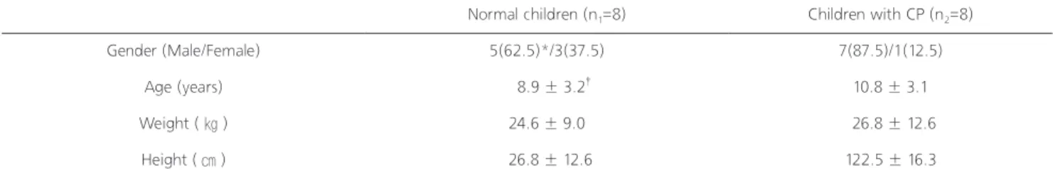
The general characteristics of the children with and without CP are presented in Table 1. There were no significant between-group differences in the ages (z=-1.33, p=0.19), weights (z=0.53, p=0.60), or heights (t=-0.58, p=0.56) of the children.
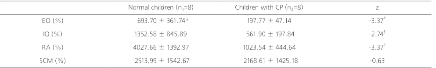
The activities of the EO, IO, and RA muscles were significantly lower in the children with CP than in those without the disease during the head-lifting exercise (p<0.05).
*Number (%), †Mean ± SD
Normal children (n1=8) Children with CP (n2=8)
Gender (Male/Female) 5(62.5)*/3(37.5) 7(87.5)/1(12.5)
Age (years) 8.9 ± 3.2† 10.8 ± 3.1
Weight ( ㎏ ) 24.6 ± 9.0 26.8 ± 12.6
Height ( ㎝ ) 26.8 ± 12.6 122.5 ± 16.3
Table 1. Ground reaction forces and distances moved by the shoulder at four chair heights.
The activity of the SCM muscle did not differ significantly between the two groups (Table 2).
The activity ratio of the RA to SCM muscle was significantly lower in the children with CP than in those without CP (p<0.05) (Table 3).
IV. Discussion
This study used surface EMG to determine whether the activities of the abdominal and neck flexor muscles of children with and without CP differed. This study found that the activities of abdominal muscles in children with CP while lifting the head in a supine position were significantly lower than in those without CP.
In the previous study, Hwang27 reported that the ability of muscle contraction was different during head lift between cerebral palsy and healthy children. In particular, muscle onset time differences between SCM and RA during head lift in supine position were significantly shorter in healthy children than in children with CP. They also demonstrated that the difference in muscle contraction capacity was expected to work hard to lift head from a supine position with cerebral palsy. Similarly, our study showed that the muscle activity of neck flexor and abdominal was different during head lift between cerebral palsy and healthy children. In the case of those without CP, the activities of the RA, EO, and IO increased up to 40.27 times, 13.52, times and 6.93 times,
respectively, when the subjects lifted their heads as compared with when they were lying down to the starting position. The large muscles located in the superficial area generated greater muscle activities, demonstrating that the superficial muscles of the abdomen worked to provide stability when performing the head-lifting exercise in a supine position. As reported earlier, the RA and other superficial abdominal muscles play a role in providing stability when the limbs are required to move.28 In contrast, the activities of the RA, EO, and IO in the children with CP increased only by 3.93 times, 3.51 times, and 2.40 times, respectively. Thus, they were substantially lower than healthy children, illustrating that the CP children experienced decreased mobilization and difficulties with co-contraction of the superficial abdominal muscles while lifting their heads.
On the other hand, there were no significant between- group differences in the activity of the SCM muscle and the neck flexor muscle. The activity ratio of the RA to the SCM muscle was 0.56 in the children with CP and 2.68 in those without CP. Thus, the muscle activity ratio of those with CP was significantly reduced (up to about five times), demonstrating that the activity of the abdominal muscles decreased compared to that of the neck flexor muscle during the head-lifting exercise in the children with CP.
The abdominal muscles play an important role in providing trunk stability, preventing the trunk from twisting or distorting and improving functional movement.29 However, in children with CP, the abdominal muscles do not perform
EO: External oblique, IO: Internal oblique, RA: Rectus abdominis, SCM: Sternocleidomastoid
*Mean ± SD, †p<0.05
RA/SCM: The activity ratio of rectus abdominis to sternocleidomastoid muscle
*Mean ± SD, †p<0.05
Normal children (n1=8) Children with CP (n2=8) z
EO (%) 693.70 ± 361.74* 197.77 ± 47.14 -3.37†
IO (%) 1352.58 ± 845.89 561.90 ± 197.84 -2.74†
RA (%) 4027.66 ± 1392.97 1023.54 ± 444.64 -3.37†
SCM (%) 2513.99 ± 1542.67 2168.61 ± 1425.18 -0.63
Normal children (n1=8) Children with CP (n2=8) z
RA/SCM 2.68 ± 2.93* 0.56 ± 0.31 -2.74†
Table 2. Comparison of abdominal muscles and neck flexor muscle
Table 3. Comparison of the activity ratio of RA to SCM muscle
properly because the co-contraction of the proximal muscles, including the trunk muscle, is insufficient during movements of the head and trunk. Thus, the development of the abdominal muscles tends to decline and be less active in children with CP than in healthy children.15
Therefore, the stability of the trunk is insufficient in children with CP. Functional movement is difficult in the absence of trunk stability.30 As a result of compensatory action, abnormal changes occur in the musculoskeletal system, thereby disturbing normal development.30 Therefore, in the therapeutic approach to children with CP, the co-activation of the abdominal muscles should be taken into account to improve the stability of the trunk during movements of the head and limbs. Children with CP are known to have deficits in trunk stability during dynamic tasks.21 Our results confirm and expand these findings by providing evidence that children with CP while walking difficult have deficits in abdominal muscles stability for head stability and deficits in trunk control during head lifting in a supine position. However, conventional treatment methods do not focus on the activity of the abdominal muscles following the head movement. Through this study, will be able to try the therapeutic approach of abdominal and neck muscles.
The present study contains a number of limitations, which affect the interpretation of the results. The small number of subjects may limit the generalization of the results. Further, the activities of the abdominal muscles were evaluated only during one motion: lifting the head. The motion of lifting the head is the basic motion to the functional movement in the development process of children with CP and one of the basic training activities performed in the supine position in general.
However, the results of muscle activity patterns in one direction (i.e., head lifting) cannot be translated to those in other directions. Future studies of a larger number of subjects are needed to evaluate the activities and the recruitment of muscles in various motions.
References
1. Swaiman KF, Ashwal S. Pediatric neurology: Principles &
practice. St. Louis, Mosby, 1999.
2. Lee HY, Kim K, Cha YJ. A survey on stress and coping style in mothers of cerebral palsied children. J Korean Soc Phys Ther.
2012;24(2):98-106.
3. Knox V, Evans AL. Evaluation of the functional effects of a course of bobath therapy in children with cerebral palsy: a preliminary study. Dev Med Child Neurol. 2002;44(7):447-60.
4. Choi HJ, Nam KW. The effect of weight-support treadmill training on the balance and activity of daily living of children with spastic diplegia. J Korean Soc Phys Ther. 2012;24(6):398- 404.
5. Bax M, Goldstein M, Rosenbaum P et al. Proposed definition and classification of cerebral palsy. Developmental Medicine &
Child Neurology. 2005;47(8):571-6.
6. Assaiante C, Amblard B. An ontogenetic model for the sensorimotor organization of balance control in humans.
Human Movement Science. 1995;14(1):13-43.
7. Saavedra S, Woollacott M, van Donkelaar P. Head stability during quiet sitting in children with cerebral palsy: Effect of vision and trunk support. Experimental brain research.
2010;201(1):13-23.
8. Wallard L, Bril B, Dietrich G et al. The role of head stabilization in locomotion in children with cerebral palsy. Annals of physical and rehabilitation medicine. 2012;55(9):590-600.
9. Harbourne RT, Giuliani C, Neela JM. A kinematic and electromyographic analysis of the development of sitting posture in infants. Developmental psychobiology. 1993;26(1):51-64.
10. Hirschfeld H, Forssberg H. Epigenetic development of postural responses for sitting during infancy. Experimental Brain Research. 1994;97(3):528-40.
11. Van Haastert IC, Groenendaal F, Van De Waarsenburg MK et al.
Active head lifting from supine in early infancy: An indicator for non-optimal cognitive outcome in late infancy. Developmental Medicine & Child Neurology. 2012;54(6):538-43.
12. Kendall FP, McCreary EK, Provance P. Muscles, testing and function: With posture and pain. 4th ed. Baltimore, Williams &
Wilkins, 1993.
13. Kibler WB, Press J, Sciascia A. The role of core stability in athletic function. Sports medicine. 2006;36(3):189-98.
14. Marshall PW, Murphy BA. Core stability exercises on and off a swiss ball. Arch Phys Med Rehabil. 2005;86(2):242-9.
15. Tecklin JS. Pediatric physical therapy. Philadelphia, Williams &
Wilkens, 1999.
16. Mayston MJ. People with cerebral palsy: effects of and perspectives for therapy. Neural Plast. 2001;8(1-2):51-69.
17. Saavedra S, Woollacott M, van Donkelaar P. Head stability during quiet sitting in children with cerebral palsy: effect of vision and trunk support. Exp Brain Res. 2010;201(1):13-23.
18. Choi MH, Lee DH, Ro HL. Effect of task-oriented training and neurodevelopmental treatment on the sitting posture in children
with cerebral palsy. J Phys Ther Sci. 2011;23(2):323-5.
19. Bobath B. Adult hemiplegia: Evaluation and treatment. Oxford, Heinemann Medical Books, 1990.
20. Gillen G, Burkhardt A. Stroke rehabilitation: A function-based approach. St. Louis, Mosby, 1998.
21. Van Der Heide JC, Fock JM, Otten B et al. Postural control during reaching in preterm children with cerebral palsy.
Developmental Medicine & Child Neurology. 2004;46(4):253-66.
22. Palisano R, Rosenbaum P, Walter S et al. Development and reliability of a system to classify gross motor function in children with cerebral palsy. Developmental Medicine & Child Neurology.
1997;39(4):214-23.
23. Cram J, Kasman G, Holtz J. Introduction to surface electromyography. Gaithersburg, Aspen Publishers, 1998.
24. Kim BK, Lee MH, Kim GC. Comparison of abdominal muscle activity during exercises using a sling and swiss-ball. J Korean Soc Phys Ther. 2013;25(3):149-54.
25. Soderberg GL, Knutson LM. A guide for use and interpretation of kinesiologic electromyographic data. Phys Ther.
2000;80(5):485-98.
26. Chang JS, Lee HY, Lee MY. The study of lumbar erector spinea and rectus abdominis activations according to the different gait velocities in young healthy adults. J Korean Soc Phys Ther.
2012;24(3):186-90.
27. Hwang SK. Comparison of the onset times of antigravity flexor muscle activity during head lift in supine position between children with cerebral palsy and healthy children. Yong-In University. Dissertation of Master's Degree. 2002.
28. O'Sullivan PB, Twomey L, Allison GT. Altered abdominal muscle recruitment in patients with chronic back pain following a specific exercise intervention. J Orthop Sports Phys Ther.
1998;27(2):114-24.
29. Vera-Garcia FJ, Grenier SG, McGill SM. Abdominal muscle response during curl-ups on both stable and labile surfaces.
Phys Ther. 2000;80(6):564-9.
30. Cho S, Oh B, Hwang B. The effects of hippotherapy on the activities of trunk muscles in preterm born children with spastic cerebral palsy. Journal of Special Education & Rehabilitation Science. 2012;51(1):349-64.