790
대한안과학회지 2009년 제 50 권 제 5 호 J Korean Ophthalmol Soc 2009;50(5):790-793 DOI : 10.3341/jkos.2009.50.5.790
www.ophthalmology.org
= 증례보고 =
접수번호 : 50-05-10-19모양체종양 형태로 발생한 과립세포종양 1예
안재환⋅서성관⋅허 준
인제대학교 의과대학 부산백병원 안과학교실
목적: 모양체종양 형태로 발생한 과립세포종양 1예를 경험하였기에 보고하고자 한다.
증례요약: 42세 여자가 내원 1달 전부터 시작된 우안 시력저하를 주소로 내원하였다. 과거력상 특이 병력 없었고, 세극등검사상 모양 체 고랑내 종괴 소견과 이와 관련된 것으로 보이는 합병성 백내장이 보였다. 모양체종양 및 합병성 백내장에 대해 경공막 모양체종양 절제 생검, 초음파수정체 유화술 및 후방 인공 수정체 삽입술을 시행하였고, 생검 결과 과립세포종양으로 판명되었다. 술 후 6개월간의 경과관찰에서 종양의 재발 소견은 보이지 않았다.
결론: 모양체종양 형태로 발생한 과립세포종양을 조직학적 소견으로 확진하였기에 보고하고자 한다.
<대한안과학회지 2009;50(5):790-793>
■ 접 수 일: 2008년 10월 28일 ■ 심사통과일: 2009년 1월 8일
■ 통 신 저 자: 서 성 관
부산시 진구 개금동 633-165 인제대학교 부산백병원 안과
Tex: 051-890-6016, Fax: 051-890-6329 E-mail: [email protected]
* 본 논문의 요지는 2008년 대한안과학회 제99회 춘계학술대회에서 포스터로 발표되었음.
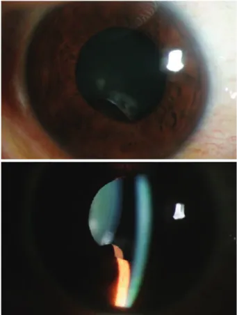
Figure 1. Slitlamp photograph shows 0.4×0.4 cm- sized mass behind inferior iris.
과립세포종양은 드물게 발생하는 연부조직의 종양으로 보통 양성소견을 보이며 눈과 그 부속기를 포함한 신체의 대부분의 기관에서 발생할 수 있다.1
안구 내에서 발생한 과립세포종양은 1966년 Cunha et al 에 의해 1예가 보고된 바 있고 국내에서는 현재까지 보고된 바 없다.2저자는 안구 내 모양체 종양의 형태로 발생한 과립 세포종양 1예를 경험하였기에 보고하고자 한다.
증례보고
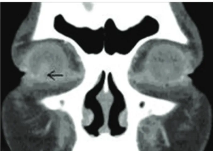
42세의 여성이 내원 한 달 전부터 시작된 우안 시력저하 를 주소로 내원하였다. 과거력상 특이 병력 없었고, 세극등 검사에서 7시 방향 홍채 뒤쪽으로 4×4 mm 크기의 종괴 병 변이 보였으며, 산동을 하면 동공연 위로 종양의 일부가 노 출되었다(Fig. 1). 종괴의 직접적인 접촉으로 인한 수정체 혼탁 소견이 보였다. 교정시력은 우안 0.32, 좌안 1.0이었 으며, 안압은 정상이었다. 다른 안과적 검사상 특이 소견은 보이지 않았다. 안와 컴퓨터전산화단층촬영상 0.5 cm 크기 의 경계가 명확하고, 균일하게 조영 증강이 되는 종괴가 보 였다(Fig. 2).
모양체 종양 및 이로 인한 합병성 백내장에 대해 경공막
모양체 종양 절제생검, 홍채절제술, 초음파수정체 유화술 및 후방 인공수정체 삽입술을 시행하였다.
조직학적 검사상 종괴는 과립질의 세포질을 나타내는 다 각형 형태의 세포로 구성되어 있고, 유사분열은 거의 보이 지 않았으며, S-100 protein에 강하게 염색되는 과립세포
791 - 안재환 외 : 모양체종양 형태의 과립세포종양 -
www.ophthalmology.org
Figure 4.The tumor cells reveal potent reaction for S- 100 protein (Immunohistochemical stain, ×100).
Figure 5. Tumor remnant was not seen, and IOL was positioned well on seven days postop.
Figure 2. Preoperative orbital CT finding. 0.5×0.5 sized well defined isodense mass lesion with intense enhancement in inferolateral aspect of right iris.
Figure 3. The tumor consist of sheet-like polygonal cells with glanular cytoplasmosis, and mitoses are rarely seen (H&E, ×100).
종양의 특징을 나타내었다(Fig. 3, 4).
술 후 6개월간의 경과관찰에서 우안 교정시력은 1.0으로 향상되었고, 종양의 재발은 없었다(Fig. 5).
고 찰
1922년 Abrikossoff가 처음 과립세포종양의 조직학적 특징을 기술한 바 있다.3 임상적으로 이 종양의 대부분은 양성이고, 주로 40~60세 사이에 나타난다. 과립세포종양은 일반적으로 하나의 병변으로 나타나고, 통증 등의 동반 증 상은 없으며, 천천히 성장하는 특징을 가지고 있다.4 주로 두경부의 피하조직에서 발생하며, 혀, 흉벽, 팔, 소화기, 후두, 그리고 다른 여러 장기에서도 발생할 수 있다.5~11 두 군데이 상의 부위에서 동시에 발생된 경우도 있으며, 선천적인 잇
몸종의 형태로도 나타난 적이 있다.12
Abrikossoff는 1926년 이 종양이 미성숙가로무늬근에서 기원하는 근육모세포종의 일종이라고 보고하였다.3 현재 과 립세포종양의 조직학적 형성에 대한 주장은 논란거리로 남 아있다. 최근의 여러 연구에서 이 종양이 면역조직화학적 검사와 전자현미경 검사에서 말초신경의 축삭과 연관되어 있음이 밝혀짐으로써 많은 학자들이 신경집세포 기원의 종 양일 가능성이 크다고 생각하고 있다.13~17
악성 과립세포종양은 연부 조직 육종의 드문 형태이다.
보통 심부 연부 조직에 위치하고 크기는 4 cm 이하이다. 이는 모든 과립세포종양의 1~3%에 해당된다.18 조직학적으로 양성과 악성의 과립세포종양을 구분하는 것이 어렵기 때문에 종종 임상적 경과만으로 판단을 해야 한다. 전형적으로 악 성 과립세포종양은 전이를 일으키기 전 보통 일 년 이내에 재발을 일으킨다.17,19본 증례의 경우 약 6개월의 경과 관찰 중 재발소견은 보이지 않았다.
과립세포종양은 안와, 눈꺼풀, 결막, 눈물낭, 상공막 등 거의 모든 안주위 조직에서 발생할 수 있다.1,20또한 경부에
792
- 대한안과학회지 2009년 제 50 권 제 5 호 -
www.ophthalmology.org
서 기원한 악성 과립세포종양이 안와 전이된 증례도 보고 된 바 있다.21안구 외 부위에서의 과립세포종양은 대개 증 상이 없이 우연히 발견되는 경우가 대부분이지만 안 주위 과립세포종양의 경우 그 발생부위에 따라 눈물흘림, 눈꺼풀 부종, 눈꺼풀 처짐, 복시, 안구돌출 등의 증상이 나타날 수 있다.1 본 증례의 경우 모양체종양의 형태로 나타나 그 후 면이 수정체와 접촉하였기 때문에 합병성 백내장이 발생하 였으며, 이로 인한 시력저하가 왔다.
Jaeger et al은 안 주위 과립세포종양을 보인 31 명의 증례 들을 보고하였는데, 거의 같은 성비(남:여 16:14)를 보였고, 흥미롭게도 75%가 왼쪽에서 나타났다. 대부분 40~70세 사 이에 발생하였고, 진단되기 전 증상의 유병기간은 2주에서 5년으로 다양하게 나타났다.1
1966년 Cunha et al은 24세 백인여성의 안구 내에서 발 생한 과립세포종양 1예를 보고한 바 있다.2이 증례는 3개월 전부터 시작된 우안의 비측 공막의 정맥울혈이 있었으며, 세극등검사상 우안의 상비측 전방 내에 경계가 명확하고, 혈관형성이 잘 되어 있는 연분홍색의 고형 종괴가 위치하고 있었다. 동공연은 종괴로 인해 이측으로 전위되어 있었으나, 각막과 수정체 등 다른 안내 조직에는 특이 소견이 없는 경우 였다. 치료로써 각막윤부 기반 공막피판을 통한 종괴 절제 술을 시행하였고 조직학적 검사상 과립세포종양으로 확진 되었다.
본 증례는 Cunha et al의 보고 이후 두 번째이며 국내에 서는 최초로 보고되는 안내 과립세포종양의 증례이다. 이전 의 증례는 전방으로 자라 들어온 홍채종양의 형태로 나타 났지만 본 증례는 홍채와 수정체 사이의 모양체종양 형태 로 나타나 이로 인해 시력을 저하시킨 합병성 백내장이 발 생하였다는 점이 다르다. 두 증례 모두 각막윤부 기반 공막 피판을 통해 홍채 일부와 함께 제거되었으며, 술 후 재발 소견은 보이지 않았다. 세극등검사상 홍채 및 모양체종양이 관찰되었을 때 홍채낭종, 홍채흑색종, 평활근종, 모양체흑 색종 등과 함께 빈도가 극히 드물긴 하지만 과립세포종양도 의심해 보아야 하겠다.
참고문헌
1) Jaeger MJ, Green WR, Miller NR, Harris GJ. Granular cell tumor of the orbit and ocular adnexae. Surv Ophthalmol 1987;31:417-23.
2) Cunha SL, Lobo FG. Granular-cell myoblastoma of the anterior uvea. Br J Ophthalmol 1966;50:99-101.
3) Abrikossoff A. Über Myome, ausgehend von der quergestreiften willkürlichen Muskulatur. Virchows Arch Path Anat Physiol 1926;260:215-33.
4) Regezi JA, Batsakis JG, Courtney RM. Granular cell tumors of the head and neck. J Oral Surg 1979;37:402-6.
5) Williams HK, Williams DM. Oral granular cell tumours: a histological and immunocytochemical study. J Oral Pathol Med 1997;26:164-9.
6) Melo CR, Melo IS, Schmitt FC, et al. Multicentric granular cell tumor of the colon: report of a patient with 52 tumors. Am J Gastroenterol 1993;88:1785-7.
7) Compagno J, Hyams VJ, Ste-Marie P. Benign granular cell tumors of the larynx: a review of 36 cases with clinicopathologic data.
Ann Otol Rhinol Laryngol 1975;84:308-14.
8) Lack EE, Worsham GF, Callihan MD, et al. Granular cell tumor:
a clinicopathologic study of 110 patients. J Surg Oncol 1980;13:
301-16.
9) Albuquerque L, Pimentel J, Costa A, Cristina L. Cerebral granular cell tumors: report of a case and a note on their nature and expected behavior. Acta Neuropathol 1992;84:680-5.
10) Markesbery WR, Duffy PE, Cowen D. Granular cell tumors of the central nervous system. J Neuropathol Exp Neurol 1973;32:
92-109.
11) Chetty R, Kalan MR. Malignant granular cell tumor of the breast.
J Surg Oncol 1992;49:135-7.
12) Singh B. Multicentric granular cell tumors. South Med J 1993;
86:144-67.
13) Sobel HJ, Marquet E. Granular cells and granular cell lesions.
Pathol Annu 1974;9:43-79.
14) Ordonez NG, Mackay B. Granular cell tumor: a review of the pathology and histogenesis. Ultrastruct Pathol 1999;23:207-22.
15) Feyrter F. Über eine eigenartige Geschwulstform des Nervenge- webes im menschlichen Verdauungsschlauch. IV. Teil der Beiträge zur Geschwulstlehre (nach Untersuchungen am menschlichen Magen und Darm). Virchows Arch (Pathol Anat) 1935;295:480-501.
16) Charles NC, Fox DM, Glasberg SS, Sawicki J. Epibulbar granular cell tumor. Report of a case and review of the literature. Ophthal- mology 1997;104:1454-6.
17) Enzinger FM, Weiss SW. Soft Tissue Tumors. St. Louis: CV Mosby, 1983;745-56.
18) Brooks JJ. Textbook of Uncommon Cancer. Sussex, England:
John Wiley & Sons, 1988;669-82.
19) Jacobiec FA, Bilyk JR, Font RL. An Atlas and Textbook, 4th ed.
Philadelphia: WB Saunders, 1996;2674-5.
20) Charles NC, Fox DM, Glasberg SS, Sawicki J. Epibulbar granular cell tumor. Report of a case and review of the literature. Oph- thalmology 1997;104:1454-6.
21) Callejo SA, Kronish JW, Decker SJ, et al. Malignant granular cell tumor metastatic to the orbit. Ophthalmology 2000;107:550-4.
793
=ABSTRACT=
A Case of Granular Cell Tumor in Form of Ciliary Body Tumor
Jae Hwan Ahn, MD, Sung Gwan Seo, MD, Jun Her, MD
Department of Ophthalmology, Inje University, Pusan Paik Hospital, Busan, Korea
Purpose: To report a case of a granular cell tumor in the form of cilliary body tumor.
Case Summary: A 42-year-old women visited our clinic with blurred vision of the right eye, which began was 1 month earlier ago. Her medical history was unremarkable. Slit-lamp examination revealed a ciliary body tumor and complicated cataract due to a mass lesion. For treatment, an excisional biopsy of the ciliary body tumor and phacoemulsification were performed. The pathologic finding confirmed the diagnosis of a granular cell tumor. There was no evidence of a recurrent lesion of the tumor at the 6-month postoperative follow-up.
Conclusions: We confirmed a granular cell tumor in the form of a cilliary body tumor based on pathologic findings.
J Korean Ophthalmol Soc 2009;50(5):790-793 Key Words: Cilliary body tumor, Granular cell tumor
Address reprint requests to Sung Gwan Seo, MD
Department of Ophthalmology, Inje University, Pusan Paik Hospital
#633-165, Gaegum-dong Busanjin-gu, Busan 614-735, Korea
Tex: 82-51-890-6016, Fax: 82-51-890-6329, E-mail: [email protected]
- 안재환 외 : 모양체종양 형태의 과립세포종양 -
www.ophthalmology.org