CASE REPORT
첫 진단 10년 후 위출구폐색 증상으로 나타난 이소성 췌장에서 기인한 위선암
정희수, 이진, 남경한1, 정수진, 오은혜, 박용은, 박종하, 김태오
인제대학교 의과대학 인제대학교 해운대백병원 내과, 병리학1
Gastric Adenocarcinoma Arising from Heterotopic Pancreas Presenting as Gastric Outlet Obstruction 10 Years after the First Diagnosis
Hee Soo Jung, Jin Lee, Kyung Han Nam1, Su Jin Jeong, Eun Hye Oh, Yong Eun Park, Jongha Park and Tae Oh Kim
Departments of Internal Medicine and Pathology1, Inje University Haeundae Paik Hospital, Inje University College of Medicine, Busan, Korea
Gastric heterotopic pancreas is a relatively uncommon incidental finding. On the other hand, the presentation of gastric ad- enocarcinoma arising from a heterotopic pancreas is rare. This paper reports a case of gastric adenocarcinoma arising from a heterotopic pancreas that presented as a gastric outlet obstruction 10 years after the initial diagnosis of a suspicious sub- mucosal tumor. Endoscopy revealed a pyloric stricture with prepyloric wall thickening and a complete gastric outlet obstruction.
Abdominal and pelvic computed tomography exposed a severely distended gastric lumen at the antrum with heterogeneously en- hancing circumferential wall thickening in the prepyloric antrum and pylorus. Because conservative treatment was ineffective and a malignancy could not be excluded, laparoscopic subtotal gastrectomy with a gastrojejunostomy was performed for histological confirmation and treatment. The histopathology diagnosis was advanced gastric carcinoma arising from heterotopic pancreatic tissue. (Korean J Gastroenterol 2020;76:37-41)
Key Words: Pancreas; Stomach neoplasm; Adenocarcinoma; Gastric outlet obstruction
Received March 27, 2020. Revised April 28, 2020. Accepted May 5, 2020.
CC This is an open access article distributed under the terms of the Creative Commons Attribution Non-Commercial License (http://creativecommons.org/licenses/
by-nc/4.0) which permits unrestricted non-commercial use, distribution, and reproduction in any medium, provided the original work is properly cited.
Copyright © 2020. Korean Society of Gastroenterology.
교신저자: 이진, 48108, 부산광역시 해운대구 해운대로 875, 인제대학교 의과대학 인제대학교 해운대백병원 내과
Correspondence to: Jin Lee, Department of Internal Medicine, Inje University Haeundae Paik Hospital, Inje University College of Medicine, 875 Haeun-daero, Haeundae-gu, Busan 48108, Korea. Tel: +82-51-797-0200, Fax: +82-51-797-1341, E-mail: [email protected], ORCID: https://orcid.org/0000-0003-2404-385X
Financial support: None. Conflict of interest: None.
INTRODUCTION
Heterotopic pancreas is a relatively uncommon abnormality that can occur in any region in the gastrointestinal tract. The most common site is the stomach, particularly the antrum and prepyloric region. Although the typical endoscopic find- ings of a heterotopic pancreas have been described as a sub- mucosal lesion with central umbilication, it is difficult to differ- entiate it from other diseases, such as gastrointestinal stro- mal tumors (GISTs), leiomyomas, or malignancies. In partic-
ular, gastric adenocarcinoma arising from a heterotopic pan- creas is rare,1-8 and the clinical course and management of gastric heterotopic pancreas are well established. There are no reports of gastric adenocarcinoma arising from a hetero- topic pancreas that was confirmed after a long-term follow-up.
This paper reports a case of gastric adenocarcinoma arising from a heterotopic pancreas presenting with a gastric outlet obstruction 10 years after the initial diagnosis of a suspicious submucosal tumor.
A B C D
Fig. 1. Endoscopic and EUS images in 2008. (A-C) Endoscopic images showing a lobulated lesion with central umbilication. (D) EUS image showing an indistinct, heterogeneous, and intermediate hypoechoic lesion involving the second and third layers of the stomach (arrow). EUS, endoscopic ultrasonography.
A B C
Fig. 2. Endoscopic images on periodic follow-up endoscopy (A) 2011, (B) 2014, and (C) 2016.
CASE REPORT
A 75-year-old man was referred to the authors’ hospital in October 2019 with dyspepsia and nausea that had lasted for more than three weeks. The patient had lost 3 kg of body weight over 1 month but had an unremarkable medical history.
The laboratory findings upon admission showed a white blood cell count of 7,000/mm3 (normal range, 4,000 to 10,000), hemoglobin 15.8 g/dL (14 to 18), amylase 37 U/L (28 to 100), lipase 31 U/L (13 to 60), CEA 5.7 ng/mL (0 to 4.3), and CA 19-9 37 U/mL (0 to 34). No specific abnormal findings were found except for increased CEA and CA 19-9 levels.
The patient was diagnosed with a suspicious submucosal tumor on the pyloric ring at another hospital in 2008 (Fig. 1).
The endoscopy findings revealed a lobulated lesion, approx- imately 2 cm in size, with central umbilication. The EUS find- ings revealed an indistinct, heterogeneous, and intermediate hypoechoic lesion involving the second and third layers. The lesion was considered to be a heterotopic pancreas on both
the endoscopic and EUS findings. No abnormal findings were seen on the periodic follow-up endoscopy until three years ago (Fig. 2).
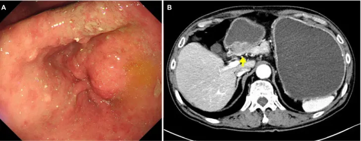
Endoscopy performed at the authors’ hospital revealed an approximately 3 cm sized submucosal lesion and pylorus nar- rowing with prepyloric wall thickening. A complete gastric out- let obstruction was noted, and the endoscope could not pass through the stricture (Fig. 3A). No lesions, such as ulcers or inflammation, were observed on the mucosal surface, and a biopsy was performed on the mucosa around the stricture.
The biopsy results were reported as chronic active gastritis with intestinal metaplasia. A follow-up endoscopic biopsy was performed five days later, and the histologic diagnosis was chronic gastritis with no evidence of a malignancy. Abdominal and pelvic CT revealed a severely distended gastric lumen at the antrum with heterogeneously enhancing circumferential wall thickening in the pre-pyloric antrum and pylorus, suggest- ing stomach cancer (T3) (Fig. 3B). A differential diagnosis of acute heterotopic pancreatitis and adenocarcinoma arising in
A B C D
Fig. 4. Gross and histopathology findings. (A) Cut sections of the whitish solid mass that invaded the subserosa. (B) The tumor showed well to poorly differentiated glands in the heterotopic pancreas. Non-neoplastic heterotopic pancreas (arrows) consisting of ducts and acini in the submucosa on the upper right side (H&E stain, ×1.25). (C) Photomicrograph of histopathology specimen shows heterotopic pancreatic tissue consisting of ducts (arrow) and acini (arrowhead). Well-differentiated adenocarcinoma is noted (on the left) (H&E stain, ×100). (D) High grade pancreatic intraepithelial neoplasia (PanIN) is characterized by papillary elements lined by cells with significant cytologic atypia (arrowhead) and low-grade PanIN (arrow) shows duct lined by flat epithelium composed of tall columnar mucin-producing cells with no cytologic atypia.
Moderately differentiated malignant glands infiltrating into the fibrotic stroma (on the left side) (H&E, ×200).
A B
Fig. 3. Endoscopic and contrast-enhanced abdominal computed tomography (CT) images at 2019. (A) Endoscopic image showing an approximately 3 cm sized submucosal lesion and pylorus narrowing. (B) CT image showing a severe distended gastric lumen at the antrum with heterogeneously enhancing circumferential wall thickening in the pre-pyloric antrum and pylorus (arrow).
a heterotopic pancreas was considered due to the increased tumor size and the resulting gastric outlet obstruction during the follow-up period. On the other hand, the patient had no symptoms of acute severe abdominal pain or elevated pancre- atic enzyme levels. Conservative treatment with an intra- venous proton pump inhibitor and fasting for eight days was ineffective, and abdominal CT did not exclude a malignancy.
Therefore, a surgical approach was considered. A laparo- scopic subtotal gastrectomy with gastrojejunostomy was per- formed for histological confirmation and treatment.
The gross findings of the subtotal gastrectomy specimen revealed a mass with erosion in the distal antrum. The cut sections revealed an ill-defined whitish and solid mass with small cystic spaces (Fig. 4A). The mass measured 3.5×1.7×1.3 cm
and involved the entire thickness of the gastric wall.
Microscopically, the tumor contained malignant glands that developed in the heterotopic pancreas (Fig. 4B). The hetero- topic pancreas was located in the submucosa and muscularis propria. The mucosa over the tumor was intact, except in the regional area of the erosion. The non-neoplastic heterotopic pancreatic tissue showed ducts and acini (Fig. 4C). Dilatation of the ducts was also observed. The tumor was characterized by a random distribution of well-to-poorly differentiated glan- dular structures. The tumor cells were medium to large with eosinophilic cytoplasm and marked nuclear pleomorphism.
The majority of the dilated ducts showed papillary features of the neoplastic epithelium as well as transitional areas be- tween the non-neoplastic epithelium and dysplasia. The high
grade pancreatic intraepithelial neoplasia (PanIN) was charac- terized by papillary elements lined with cells with significant cytologic atypia, and low-grade PanIN showed a duct lined with a flat epithelium composed of tall columnar mucin-pro- ducing cells with no cytologic atypia (Fig. 4D). Lymphovascular invasion was identified. The tumor penetrated the subserosal connective tissues without invading the visceral peritoneum.
The pathological diagnosis of an adenocarcinoma (T3 N0 stage) arising from the heterotopic pancreas was concluded.
The patient received chemotherapy with capecitabine and intravenous oxaliplatin after surgery. Follow-up CT and endos- copy were scheduled 6 months after surgery.
DISCUSSION
Heterotopic pancreas is often asymptomatic, and sub- sequent cystic formation, pancreatitis, and malignant changes can occur.9,10 On the other hand, gastric adenocarcinoma aris- ing from the heterotopic pancreas is rare, and there have been no previously reported cases of gastric adenocarcinoma arising from the heterotopic pancreas, which were confirmed after a long-term follow-up.
The typical endoscopic findings of a heterotopic pancreas are a well-circumscribed submucosal lesion with central umbilication. On the other hand, as umbilication is noted in less than half of cases, it is difficult to differentiate hetero- topic pancreas from other diseases, such as GIST, leiomyoma, or submucosal gastric carcinoma. The EUS findings of a heter- otopic pancreas are a hypoechoic or intermediate echogenic heterogeneous lesion with indistinct margins. This most com- monly arises from the third or fourth layer, or a combination of the two layers.
For a diagnosis of carcinoma arising from a heterotopic pancreas, the following three criteria have been proposed.11 First, the carcinoma must be within or close to the ectopic pancreas. Second, a transitional area between the pancreatic structures and carcinoma must be present. Third, non-neo- plastic pancreatic tissue must be comprised of well-developed acini or ductal structures. The present case meets the above three criteria (Fig. 4).
The malignant transformation of a gastric heterotopic pan- creas is rare but has been reported in the literature. In addi- tion, clinicians need to consider the possibility of a malignant transformation. The early diagnosis of malignant changes aris-
ing in the gastric heterotopic pancreas may be difficult be- cause the surface of the lesion is usually covered with normal mucosa. Although various diagnostic techniques yield results, EUS-guided fine needle aspiration (FNA) or fine needle biopsy (FNB) are useful methods for making a histologic diagnosis of submucosal lesions.12,13 EUS-FNA has been reported to be helpful in the diagnosis of heterotopic pancreas.14 A periodic follow-up by endoscopy or EUS should be considered because malignant changes from the heterotopic pancreas can occur after more than 10 years, as in this case. In addition, EUS-FNA or FNB may be useful for an early diagnosis of the malignant transformation of heterotopic pancreas if the size of the lesion increases or shows atypical changes.
The guidelines for the management of gastric heterotopic pancreas have not been well established. Gastric heterotopic pancreas is usually asymptomatic and requires no treatment.
Histologic confirmation through a resection is required in cases with a high risk, an increasing size, and symptoms, such as ulceration, bleeding, and pyloric obstruction. Unlike GIST, which sets a size of approximately 2 cm or more as high-risk criteria,15 a heterotopic pancreas does not have a clear standard for size in the high-risk criteria. On the other hand, as the size of the heterotopic pancreas was more than 2 cm in most cases reported previously as a gastric adenocarcinoma arising from a heterotopic pancreas,8 the size of the lesion may need to be considered as an important risk factor, such as in GIST.
Surgery is frequently needed to make a definitive diagnosis and plan further treatment because of the difficulty in differ- entiating a heterotopic pancreas from other diseases, such as GIST, leiomyoma, neuroendocrine tumors, or other malignancies.
Although a surgical resection is usually recommended for the treatment of symptomatic heterotopic pancreas, an endoscopic resection, such as endoscopic submucosal dissection or mu- cosal resection, can be considered after taking into account the size and location of the lesion in cases with benign features.
Periodic follow-up endoscopy or EUS is recommended if there are no symptoms.
In conclusion, although the incidence of gastric ad- enocarcinoma arising from a heterotopic pancreas is rare, a careful diagnostic and therapeutic approach should be con- sidered in patients with gastric heterotopic pancreas. In addi- tion, clinicians need to consider the possibility of a malignant transformation.
REFERENCES
1. Jeng KS, Yang KC, Kuo SH. Malignant degeneration of hetero- topic pancreas. Gastrointest Endosc 1991;37:196-198.
2. Herold G, Kraft K. Adenocarcinoma arising from ectopic gastric pancreas: two case reports with a review of the literature. Z Gastroenterol 1995;33:260-264.
3. Osanai M, Miyokawa N, Tamaki T, Yonekawa M, Kawamura A, Sawada N. Adenocarcinoma arising in gastric heterotopic pan- creas: clinicopathological and immunohistochemical study with genetic analysis of a case. Pathol Int 2001;51:549-554.
4. Jeong HY, Yang HW, Seo SW, et al. Adenocarcinoma arising from an ectopic pancreas in the stomach. Endoscopy 2002;34:1014-1017.
5. Emerson L, Layfield LJ, Rohr LR, Dayton MT. Adenocarcinoma arising in association with gastric heterotopic pancreas: a case report and review of the literature. J Surg Oncol 2004;87:53-57.
6. Song DE, Kwon Y, Kim KR, Oh ST, Kim JS. Adenocarcinoma arising in gastric heterotopic pancreas: a case report. J Korean Med Sci 2004;19:145-148.
7. Lemaire J, Delaunoit T, Molle G. Adenocarcinoma arising in gastric heterotopic pancreas. Case report and review of the literature.
Acta Chir Belg 2014;114:79-81.
8. Priyathersini N, Sundaram S, Senger JL, Rajendiran S, Balamurugan TD, Kanthan R. Malignant transformation in gastric pancreatic heterotopia a case report and review of the literature.
J Pancreas 2017;18:73-77.
9. Bethel CA, Luquette MH, Besner GE. Cystic degeneration of het- erotopic pancreas. Pediatr Surg Int 1998;13:428-430.
10. Shimizu M, Matsumoto T, Sakurai T, et al. Acute terminal pancreatitis occurring in jejunal heterotopic pancreas. Int J Pancreatol 1998;23:171-173.
11. Guillou L, Nordback P, Gerber C, Schneider RP. Ductal ad- enocarcinoma arising in a heterotopic pancreas situated in a hia- tal hernia. Arch Pathol Lab Med 1994;118:568-571.
12. Fernández-Esparrach G, Sendino O, Solé M, et al. Endoscopic ul- trasound-guided fine-needle aspiration and trucut biopsy in the diagnosis of gastric stromal tumors: a randomized crossover study. Endoscopy 2010;42:292-299.
13. Polkowski M, Gerke W, Jarosz D, et al. Diagnostic yield and safety of endoscopic-ultrasound guided trucut biopsy in patients with gastric submucosal tumors: a prospective study. Endoscopy 2009;41:329-334.
14. Rodriguez FJ, Abraham SC, Allen MS, Sebo TJ. Fine-needle aspira- tion cytology findings from a case of pancreatic heterotopia at the gastroesophageal junction. Diagn Cytopathol 2004;31:175-179.
15. Demetri GD, Benjamin RS, Blanke CD, et al. NCCN Task Force re- port: management of patients with gastrointestinal stromal tu- mor (GIST)--update of the NCCN clinical practice guidelines. J Natl Compr Canc Netw 2007;5 Suppl 2:S1-29; quiz S30.