A heterotopic pancreas is defined as pancreatic tissue that is lying outside its normal location and it lacks any anatomic or vascular connections with the pancreas. It is also described as pancreatic heterotopia, a term coined by Barbosa et al. (1) in 1946. The frequency of heterotopic pancreas has been estimated to be 1 case per 500 explorations of the upper abdomen or 0.6-13.7% of autopsies (2). Heterotopic pancreatic tissue may be found anywhere along the alimentary tract including the stomach, duodenum, small intestine, Meckel’s di- verticulum and the biliary tract, and even in the lungs, umbilicus or fallopian tubes. Yet the most common site is the stomach and the pyloric canal. This lesion is usual- ly asymptomatic; however, some patients have epigas- tric pain, upper gastrointestinal bleeding and occasional gastric outlet obstruction. In a few cases, the reported
complications of heterotopic pancreas are pancreatitis, pseudocyst, cyst formation, insulinoma, adenoma and malignant transformation (3), but the exact rate of com- plications via a pathologic process hasn’t been identified and malignant transfomation is a rare finding.
Furthermore, islet cell tumor arising from heterotopic pancreas in the duodenum is extremely rare; we found only one well documented case in our review of the lit- erature, and this was a pathologic case report (4). In this report, we describe a case of islet cell tumor arising from a heterotopic pancreas in the duodenum, and we dis- cuss the radiological presentation and the pathologic comparison.
Case Report
A 77-year-old woman presented to us with a poor oral intake, epigastric pain and vomiting. She was suffering from known hypertension and the rest of her medical history was unremarkable. On the routine blood tests, mild hypokalemia was noted, which was probably due to her vomiting. The other laboratory findings were nor- mal. Endoscopy revealed a round polypoid mass in the second portion of the duodenum. Endoscopic ultrasono-
Islet Cell Tumor Arising from Heterotopic Pancreas in the Duodenum: A Case Report1
Joo Hwan Park, M.D., Yoon Hee Han, M.D., Mi-Young Kim, M.D.
Soon Joo Cha, M.D., Mee Joo, M.D.2
It is difficult to distinguish an islet tumor originating from heterotopic pancreas tis- sue from the other submucosal tumors. Although the malignant transformation of a heterotopic pancreas, including islet cell tumor, is extremely rare, it remains an impor- tant consideration in the differential diagnosis of duodenal submucosal masses. We have demonstrated the radiologic appearance and the clinical-pathologic findings of a highlighted, rare case of islet cell tumor arising from a heterotopic pancreas in the duo- denal wall.
Index words :Heterotopic pancreas Islet cell tumor Duodenum
1Department of Radiology, Ilsan Paik Hospital, Inje University, School of Medicine
2Department of Pathology, Ilsan Paik Hospital, Inje University, School of Medicine
Received March 24, 2005 ; Accepted April 29, 2005
Address reprint requests to : Yoon Hee Han, M.D., Department of Radiology, Ilsan Paik Hospital, Inje University, School of Medicine, 2240, DaeHwa-dong, Ilsan-gu, Goyang-si, Gyeonggi-Do 411-706, Korea.
Tel. 82-31-910-7688 Fax. 82-31-910- 7369 E-mail: [email protected]
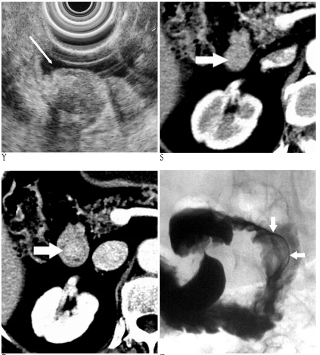
gram (EUS) revealed a mass occupying the mucosal and submucosal layers, and it showed mixed echogenecity with a central necrotic portion (Fig. 1A). Abdominal computed tomography (CT) was done. On the CT scan, a well-defined, polypoid mass was observed with intra- luminal protrusion into the second portion of the duode- num (Fig. 1B), but there were no signs of bowel obstruc-
tion. The mass was well and homogeneously enhanced in the arterial and venous phases, at 125-130 HU (Fig.
1C). On the barium study, about a 2.5×2 cm sized ovoid filling defect was noted in the second portion of the duodenum and this was sharply outlined with bari- um. The mass showed an abrupt and slightly obtuse an- gulation with the duodenal wall. The overlying mucosal
A B
C D
Fig. 1. A. The endoscopic ultrasonogram shows a well-defined mass occupying the mucosal and submucosal layers. The mass re- veals mixed echogenecity with a central necrotic portion.
B, C. On the axial CT images, the mass shows an intraluminal location in the second portion of the duodenum and it is well en- hanced (HU 125-130) with relative homogeneity (solid arrows).
D. The barium study shows an ovoid filling defect with smooth surface, sharp margination and an obtuse angulation with the duo- denal wall (solid arrows).
surface of the mass was intact and smoothly marginated (Fig. 1D). Here too, there was no evidence of passage disturbance. We suspected that the mass was a duode- nal polyp or a submucoal tumor such as gastrointestinal stromal tumor. The endoscopic biopsy showed chronic nonspecific inflammation and there were no tumor cells found. Duodenotomy with polypectomy was per- formed. At surgery, a pedunculated mass was removed from the posterior wall of the second portion of the duo- denum. The specimen consisted of a round mass of polypoid soft tissue that measured 1.8×1.5×1 cm. The
mucosal and submucosal layers were covering it. The cut section of the specimen showed a relatively defined, yellowish solid submucosal tumor that measured 1.8×
1.2×1 cm. Microscopically, the mass had infiltrated the mucosa and submucosa layers and it had extended to the proper muscle. The low magnification photomicro- graph demonstrated a solid islet cell tumor located in the more superficial portion as well as a heterotopic pancreas in deep muscular layer (Fig. 1E). The high magnification photomicrograph showed uniform round tumor cells in a trabecular or acinar arrangement with
E F
G H
Fig. 1. E. Low magnification photomicograph of the mass demonstrates a solid islet cell tumor located at the more superficial por- tion of the heterotopic pancreas in the deep muscular layer of the duodenum (solid arrows).
F. High magnification photomicrograph shows the uniform round tumor cells in a trabecular or acinar arrangement with amyloid deposition (asterisks).
G. The tumor cells are diffusely stained with anti-chromogranin A antibody with a strong intensity.
H. Electron micrograph shows the secretory granules in the tumor cell cytoplasm and the amyloid fibrils (asterisks).
amyloid deposition (Fig. 1F). The tumor cells were dif- fusely stained with anti-chromogranin A antibody with a strong intensity. This suggested that the tissue consist- ed of chromogranin A-positive neuroendocrine cell nests (Fig. 1G). Also, the sections showed amyloid depo- sition in the tumorous stroma. Amyloid was identified in the tumor focus where the cells were stained for anti- insulin antibody. Several studies have revealed that in- sulinomas commonly express islet amyloid polypeptide (IAPP) and in approximately 5% of these cases, IAPP may be precipitated as amyloid in the tumor stroma (1).
Electron microscopy was carried out and the further his- tologic examination of the resected tumor demonstrated many closely packed round to ovoid cells with abundant cytoplasmic organelles. Many membrane-bound neu- rosecretory granules and a disordered meshwork of nonbranching fibrils were noted in the cytoplasm.
Fibrillary depositions were also noted in the stroma (Fig.
1H). These features were all compatible to islet cell tu- mor with amyloid deposition arising from a heterotopic pancreas.
Discussion
Heterotopic pancreas is thought to arise at the time of embryonic development and during fusion of the pan- creatic buds. The alimentary tract is in close proximity to the developing pancreas; thus, tissue can become im- planted in the bowel wall and then it’s carried to its final location (5). The aberrant tissue consists of endoderm that is composed of all the cell types normally found in the pancreas, and it often includes islets of Langerhans.
Consequently, pancreatitis with fat necrosis, islet cell tu- mors with hyperinsulinism and pancreatic carcioma with metastases may occur in heterotopic pancreas (6).
Symptoms rarely come into existence with the presence of heterotopic pancreatic tissue alone. This is usually an asymptomatic condition that is found incidentally dur- ing laparotomy or at autopsy in the stomach, duodenum and small intestine, and it is even found in Meckel’s di- verticulum or in the biliary tract. Heterotopic pancreatic tissue is most commonly found in the gastric antrum along the greater curvature. In our patient, the tumor was located in the duodenum, a second most common location for heterotopic pancreas. The frequency of het- erotopic pancreas has been estimated to be 1 case per 500 explorations of the upper abdomen or 0.6-13.7% of autopsies (1). It is generally diagnosed with the develop- ment of complications such as hemorrhage, obstruction
and malignant transformation (7). However, malignant transformation in heterotopic pancreatic tissue is a rare finding, and a review of literature revealed only 15 well- documented cases of carcinoma arising in heterotopic pancreas (2). Islet cell tumor arising from heterotopic pancreas is also extremely rare. We found only one re- port of a case of an islet cell tumor arising from a hetero- topic pancreas in our review of literature. Tolentino et al (4) reported one case in 2003 and that was a pathologic report.
The radiologic diagnosis of a pancreatic tumor is usu- ally made on the basis of the typical location and ap- pearance, which is that of a small umblicated lesion in a submucosal mass (3). However, this appearance may be a nonspecific finding for diagnosis. That is, other sub- mucosal tumors such as gastrointestinal stromal tumor, lymphoma, carcinoid or adenomatous polyp can reveal as being small umblicated lesions such as an overlying mucosal ulceration. Besides, it is more difficult to distin- guish islet tumor originating from a heterotopic pan- creas tissue from the other submucosal tumors, and there are no reported specific findings about islet cell tu- mor arising from heterotopic pancreas. In our case, on the initial EUS and barium study, we suspected a sub- mucosal tumor such as gastrointestinal stromal cell tu- mor or duodenal polyp. Furthermore, CT was not help- ful for the diagnosis. The CT findings were merely a well defined, intraluminal mass in the duodenum. If there is one thing to be considered, it is that the mass was enhanced at 125-130 Housfield units (HU), and that was higher than the HUs of the normal enhancing pancreas, which showed an average of 100 HU in the venous phase of our case. Typically, but not always, islet tumor in a normal pancreas shows intense en- hancement in the arterial and venous phase.
Islet cell tumors are classified as functioning if they produce symptoms related to excessive hormone pro- duction; intractable hypoglycemia, low blood levels of glucose and high circulating plasma insulin (8).
Nonfunctioning tumors are those pancreatic tumors with endocrine differentiation in the absence of a clini- cal syndrome related to hormone production. In our case, GI symptoms such as vomiting and epigastric pain were presented, but the blood hormone levels were nor- mal. On the pathology, the uniform round tumor cells that were diffusely stained with anti-chromogranin A, which suggested that this tissue had differentiated into chromogranin A-positive neuroendocrine cells, and they were located in the more superficial portion of the mass
and in deep muscular layer where the heterotrophic pancreas was based. Also, the sections showed amyloid deposition in the tumorous stroma. Amyloid was identi- fied in the tumor focus where the cells were stained for anti-insulin antibody. So our case was a symptomatic nonfunctioning islet cell tumor arising from a hetero- topic pancreas in the duodenal wall.
Although malignant transformation of heterotopic pancreas, including islet cell tumor, is extremely rare, it remains an important consideration in the differential diagnosis for duodenal submucosal masses. We have demonstrated the radiologic appearance and clinical- pathologic findings of a rare case of islet cell tumor aris- ing from a heterotopic pancreas in the duodenal wall.
References
1. Barbosa JJ, Docketry MB, Waugh JM. Pancreatic heterotopia.
Review of the literature and report of 41 authenticated surgical
cases, of which 25 were clinically significant. Surg Gynecol Obstet 1946;82:527-542
2. Ura H, Denno R, Hirata K, Saeki A, Hirata K, Natori H. Carcinoma arising from ectopic pancreas in the stomach: endosonographic de- tection of malignant change. J Clinical Ultrasound 1998;26:265-268 3. Cho JS, Shin KS, Kwon ST, Kim JW, Song CJ, Noh SM, et al.
Heterotopic pancreas in the stomach: CT findings. Radiology 2000;
217:139-144
4. Tolentino LF, Lee H, Maung T, Stabile BE, Li K, French SW. Islet cell tumor arising from a heterotopic pancreas in the duodenal wall with ulceration. Exp Mol Pathol 2004;76:51-56
5. Harold KL, Sturdevant M, Matthews BD, Mishra G, Heniford BT.
Etopic pancreatic tissue presenting as submucosal gastric mass. J Laparoendosc Adv Surg Tech A 2002;12:333-338
6. Kilman WJ, Berk RN. The spectrum of radiologic features of aber- rant pancreatic rests involving the stomach. Radiology 1977;123:
291-296
7. Ozcan C, Celik A, Guclu C, Balik E. A rare cause of gastric outlet obstruction in the newborn: pyloric ectopic pancreas. J Pediatr Surg 2002; 37:119-120
8. Sheth S, Hruban RK, Fishman EK. Helical CT of islet cell tumors of the pancreas: typical and atypical manifestations. AJR Am J Roentgenol 2002;179:725-730
대한영상의학회지 2005;52:395-399
십이지장에 생긴 이소성췌장에서 발생한 도세포종양에 대한 증례보고1
1인제대학교 의과대학 일산백병원 진단방사선과
2인제대학교 의과대학 일산백병원 해부병리과
박주환・한윤희・김미영・차순주・주 미2
이소성 췌장에서 발생한 도세포종양을 다른 점막하 종양과 구별하기는 쉽지 않다. 비록 도세포종양을 포함하여 이소 성 췌장에서 발생하는 악성변환은 극히 드물지만 이것은 십이지장에서 발생하는 점막하 종양의 중요한 감별진단이 될 수 있다. 저자들은 십이지장에 생긴 이소성 췌장에서 기원한 도세포종양의 방사선학적 소견 및 해부병리 소견을 문헌 고찰과 함께 보고하고자 한다.