Korean J Hepatobiliary Pancreat Surg 2011;15:194-197
Case Report
Partial gastric outlet obstruction caused by a huge submucosal tumor originating in the heterotopic pancreas
Gum O Jung1, Dong Eun Park1, Ki Jung Yun2, and Kwon Mook Chae1
Departments of 1Surgery and 2Pathology, Wonkwang University Hospital, Wonkwang University of College of Medicine, Iksan, Korea
A 21-year-old woman presented gastrointestinal manifestation showing intermittent abdominal pain, nausea, and vomiting. An upper endoscopic examination showed round, elevated mucosa at the antrum of the stomach anterior wall. After CT scanning, a huge degenerated gastrointestinal stromal tumor was suspected. Subtotal gastrectomy with Billroth II anastomosis was performed. Histologically, pseudocystic degeneration of the heterotopic pancreas was confirmed. The patient showed eventful postoperative course except temporary dilated gastric emptying. The patient is doing well without any abnormal symptom at 8-month follow-up. This report is a rare case of gastric outlet obstruction caused by a pseudocyst originating from a heterotopic pancreas in the gastric antrum. (Korean J Hepatobiliary Pancreat Surg 2011;15:194-197)
Key Words: Heterotopic pancreas; Gastric outlet obstruction; Submucosal tumor
Received: July 28, 2011; Revised: August 11, 2011; Accepted: August 15, 2011 Corresponding author: Kwon Mook Chae
Department of Surgery, Wonkwang University Hospital, 344-2, Shinyoung-dong, Iksan 570-711, Korea Tel: +82-63-859-1491, Fax: +82-63-855-2389, E-mail: [email protected]
Copyright Ⓒ 2011 by The Korean Association of Hepato-Biliary-Pancreatic Surgery Korean Journal of Hepato-Biliary-Pancreatic Surgery ∙ pISSN: 1738-6349
INTRODUCTION
Heterotopic pancreas (HP) is a relatively uncommon condition that can occur at any age. It is ectopically lo- calized pancreatic tissue with no anatomic and vascular connections to the true pancreas, and is found in 2-15%
of all autopsies.1 Heterotopic pancreas is most frequently encountered in the duodenum (25-35%) and stomach (25-60%).2,3 The etiology of heterotopic pancreas is un- known; it is possible that during rotation of the foregut and fusion of the ventral and dorsal parts of the pancreas in early fetal life, small pieces of tissue become detached from the forming organ, leading to entrapment in a differ- ent location.4 Most patients with heterotopic pancreas are asymptomatic, but it can occasionally present as nausea, vomiting, and abdominal pain. Serious symptoms, such as peptic ulceration with severe gastrointestinal bleeding, malignant degeneration, pancreatitis, and pseudocyst, are rarely seen. This is a report on rare case of gastric outlet obstruction caused by a pseudocyst of a heterotopic pan- creas in the gastric antrum.
CASE
A 21-year-old-female was admitted to our hospital with 6 month history of chronic epigastric pain, postprandial dyspepsia, and recurrent vomiting after meals. The patient had an unremarkable medical history. She had never smoked or consumed alcoholic beverages. She showed with stable vital signs. The mass was palpated with mild direct tenderness in the epigastric area. However, the rest of the physical examination was normal. The white blood cell count was 1,100/ul in hematological examinations, and blood biochemical findings were normal. All tumor markers, including alpha-fetoprotein, carbohydrate antigen 19-9 (CA19-9) and carcinoembryonic antigen (CEA), were in the normal range.
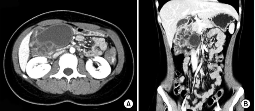
Gastroduodenoscopy showed chronic antritis and a sub- mucosal lesion in the antero-inferior wall of the gastric antrum. Multi-slice spiral computerized tomography in the portal venous phase showed a 7.5 cm diameter well-de- fined lesion in the antero-inferior wall of the stomach. It contained a large cystic mass with likely cystic degener- ation and multi-septated small cysts. The cystic mass
Gum O Jung, et al. Gastric outlet obstruction caused by heterotopic pancreas 195
Fig. 2. Magnetic resonance imaging study showing a definitive cystic mass in T2 weighted imaging. There was no connection between the cystic mass and the pancreatic duct or common bile duct.
Fig. 1. Computed tomography image shows a large cystic mass with multi-septated small cystic mass with enhancement in the delayed phase. Although the bo- undary between the cystic lesion and gastric antral wall was indis- tinct, there was no evidence of local invasion around solid or- gans and tissue.
showed no evidence of local invasion (Fig. 1). Magnetic retrograde cholangiopancreatography (MRCP) showed a large cystic lesion without connection to the common bile duct (CBD) or the pancreatic duct. The CBD showed mild dilatation due to compression of the cystic lesion (Fig. 2).
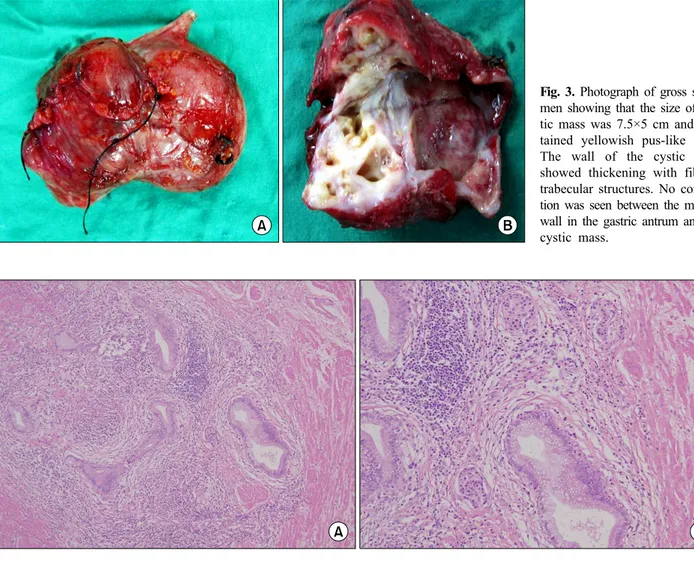
To rule out either gastrointestinal stromal tumor (GIST) or pancreatic pseudocyst, we decided to resect the cystic mass. During the operation, the cystic mass was not sepa- rated from the gastric antrum, but easily dissected from other surrounding tissues. We performed a wedge re- section of the antrum of the stomach, including the cystic lesion, and a Billroth II gastrojejunostomy due to narrow- ing of the gastric outlet. During operation, the cystic mass was seen to contain yellowish fluid, likely pus, and no connection between the cystic mass and the mucosa of the resected antral wall. The wall of the cystic mass showed thickening with trabecular structures (Fig. 3). The resected
specimen was 7.3 cm × 5.5 cm × 4 cm, and a histological examination showed benign glandular tissue within the muscle layer with the presence of islets of Langerhans and cystic changes of the ductular structure. There was no evi- dence of pancreatic acinar formation (Fig. 4). It was fi- nally decided that mass was formed from cystic degener- ation of heterotopic pancreas.
The patient was discharged 12 days after surgery.
However, she was re-admitted with gastric outlet ob- struction symptoms. Gastroscopy showed partial ob- struction at the gastrojejunostomy site. She was treated with conservative management for 1 week and discharged with relief of symptoms. The patient did not suffer from other postoperative complication or recurrence of disease for postoperative 8 month.
196 Korean J Hepatobiliary Pancreat Surg Vol. 15, No. 3, August 2011
Fig. 3. Photograph of gross speci- men showing that the size of cys- tic mass was 7.5×5 cm and con- tained yellowish pus-like fluid.
The wall of the cystic mass showed thickening with fibrous trabecular structures. No connec- tion was seen between the mucosa wall in the gastric antrum and the cystic mass.
Fig. 4. Microscopic photograph showed benign grandular tissue within the muscle layer with presence of islets of Langerhans and cystic changes in ductular structure. There was no evidence of pancreatic acinar formation. (A: H&E stain, ×100, B: H&E stain, ×200).
DISCUSSION
Heterotopic pancreas, also known as ectopic or aberrant pancreas, is defined as pancreatic tissue that lacks either anatomical or vascular communication with true pancreas and is reported in 0.6-13% of post-mortem examinations.4 Heterotopic pancreas can include all of the following: his- tological features, duct development, and islets of Langer- hans. Distribution of the heterotopic pancreas tissue varies throughout the gastrointestinal tract. The most common site is the stomach, accounting for 25-38.2% of all pa- tients with heterotopic pancreas.5 Most patients with het- erotopic pancreas are asymptomatic.6 Reported symptoms include non-specific abdominal pain (45.5%), epigastric discomfort (12.0%), nausea and vomiting (9.6%), bleeding (8.0%), and others (24.5%).5 Complications caused by
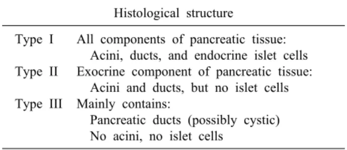
heterotopic pancreas are mechanical obstruction, cystic formation, acute inflammation, and malignant transforma- tion.3,7 Histologically, heterotopic pancreas has been clas- sified into three types by Heinrich (Table 1);8 the present case would be classified as type III.
Most patients with heterotopic pancreas had been diag- nosed post-laparotomy on histological examination on his- tological examination of the resected specimen, usually suspected preoperatively as a gastrointestinal stromal tu- mor, lymphoma, or duodenal submucosal tumor. As such, this disorder is difficult to diagnose preoperatively despite diagnostic procedures such as abdominal ultrasonography, gastroduodenoscopy, and computed tomography9,10 Accor- ding to one report,9 only 1 of 17 patients (6%) was con- sidered to have heterotopic pancreas preoperatively. Pre- sent case also was initially diagnosed as gastrointestinal
Gum O Jung, et al. Gastric outlet obstruction caused by heterotopic pancreas 197
Table 1. Heinrich’s classification of the heterotopic pancreas Histological structure
Type I Type II Type III
All components of pancreatic tissue:
Acini, ducts, and endocrine islet cells Exocrine component of pancreatic tissue:
Acini and ducts, but no islet cells Mainly contains:
Pancreatic ducts (possibly cystic) No acini, no islet cells
stromal tumor through preoperative imaging and endo- scopic study until the postoperative pathology showed otherwise.
If gastric outlet obstruction caused by heterotopic pan- creas occurs, as in present case, it can be treated by vari- ous operative procedures, including bypass gastro- enterostomy or antrectomy with gastroduodenal anastomo- sis. Lymphadenectomy is not considered necessary as a lymphatic spread rarely occurs from a heterotopic pan- creas or gastrointestinal stromal tumor.11 We performed antrectomy without lymph node dissection to resolve gas- tric outlet obstruction.
In conclusion, we herein present a rare case of gastric outlet obstruction caused by a pseudocyst originating from the heterotopic pancreas in gastric antrum. Although most of asymptomatic heterotopic pancreas usually is not clin- ical significance, it can be included in the differential di- agnoses of gastric outlet obstruction caused by a sub- mucosal gastric mass.
ACKNOWLEDGEMENTS
This case report was supported by Wonkwang Univer- sity in 2010.
REFERENCES
1. Jaffee R. The pancreas. In: Wigglesworth JS, Singer DB, eds.
Textbook of fetal and perinatal pathology. Vol 2nd ed. Boston, Massachusetts; Blackwell Scientific, 1991:1021-1055.
2. Mizuno Y, Sumi Y, Nachi S, et al. Acinar cell carcinoma arising from an ectopic pancreas. Surg Today 2007;37:704-707.
3. Mulholland KC, Wallace WD, Epanomeritakis E, Hall SR.
Pseudocyst formation in gastric ectopic pancreas. JOP 2004;5:
498-501.
4. Tolentino LF, Lee H, Maung T, Stabile BE, Li K, French SW.
Islet cell tumor arising from a heterotopic pancreas in the duode- nal wall with ulceration. Exp Mol Pathol 2004;76:51-56.
5. Kaneda M, Yano T, Yamamoto T, et al. Ectopic pancreas in the stomach presenting as an inflammatory abdominal mass. Am J Gastroenterol 1989;84:663-666.
6. Dolan RV, ReMine WH, Dockerty MB. The fate of heterotopic pancreatic tissue. A study of 212 cases. Arch Surg 1974;109:
762-765.
7. Song DE, Kwon Y, Kim KR, Oh ST, Kim JS. Adenocarcinoma arising in gastric heterotopic pancreas: a case report. J Korean Med Sci 2004;19:145-148.
8. Heinrich H. Ein beitrag zur histologie des sogen, akzessorichen pankreas Virchows Arch Path Anat 1909;198:392-401.
9. Hsia CY, Wu CW, Lui WY. Heterotopic pancreas: a difficult diagnosis. J Clin Gastroenterol 1999;28:144-147.
10. Cho JS, Shin KS, Kwon ST, et al. Heterotopic pancreas in the stomach: CT findings. Radiology 2000;217:139-144.
11. Cuschieri A. Laparoscopic gastric resection. Surg Clin North Am 2000;80:1269-1284.