Received April 9, 2010, Accepted June 17, 2010 Correspondence to: Sung Kim
Department of Surgery, Samsung Medical Center, Sungkyunkwan University School of Medicine, 50 Irwon-dong, Gangnam-gu, Seoul 135-710, Korea
Tel: +82-2-3410-0294, Fax: +82-2-3410-6981, E-mail: [email protected]
cc Journal of the Korean Surgical Society is an Open Access Journal. All articles are distributed under the terms of the Creative Commons Attribution Non-Commercial License (http://creativecommons.org/licenses/by-nc/3.0/) which permits unrestricted non-commercial use, distribution, and reproduction in any medium, provided the original work is properly cited.
Dae Hoon Kim, Kyoung Mee Kim , Seung Jong Oh, Jeong A Oh, Min Gew Choi, Jae Hyung Noh, Tae Sung Sohn, Jae Moon Bae, Sung Kim
Departments of Surgery and 1Pathology, Samsung Medical Center, Sungkyunkwan University School of Medicine, Seoul, Korea
The incidence of heterotopic gastric mucosa located in the submucosa in resected stomach specimens has been reported to be 3.0 to 20.1%. Heterotopic gastric mucosa is thought to be a benign disease, which rarely becomes malignant. Heterotopic gas- tric mucosa exists in the gastric submucosa, and gastric cancer rarely occurs in heterotopic gastric mucosa. Since tumors are located in the normal submucosa, they appear as submucosal tumors during endoscopy, and are diagnosed through endo- scopic biopsies with some difficulty. For such reasons, heterotopic gastric mucosa is mistaken as gastric submucosal tumor.
Recently, two cases of early gastric cancer arising from heterotopic gastric mucosa in the gastric submucosa were treated.
Both cases were diagnosed as submucosal tumors based on upper gastrointestinal endoscopy, endoscopic ultrasound, and computed tomography findings, and in both cases, laparoscopic wedge resections were performed, the surgical findings of which also suggested submucosal tumors. However, pathologic assessment of the surgical specimens led to the diagnosis of well-differentiated intramucosal adenocarcinoma arising from heterotopic gastric mucosa in the gastric submucosa.
Key Words: Early gastric cancer, Heterotopic gastric mucosa, Submucosal tumor
INTRODUCTION
The recent advancement of endoscopic diagnosis and increase of mass screening test in Korea results in the in- crease of the incidence of early gastric cancer and sub- mucosal tumor. Gastric cancer and submucosal tumor have different origins. The gastric adenocarcinoma origi- nates from the gastric epithelium, but submucosal tumor does not originate from gastric epithelium. So, their fea- tures are different, and the gastric cancer resembling sub- mucosal tumor is very rare. Heterotopic gastric mucosa in
the gastric submucosa has been reported of an incidence ranging 3.0 to 20.1% and were considered benign and rare- ly transformed malignant [1,2]. The gastric cancer arising from heterotopic gastric mucosa in the gastric submucosa is extremely rare, and its feature is similar to submucosal tumor [3,4]. We present two cases of early gastric cancer arising from heterotopic gastric mucosa in the gastric sub- mucosa mimicking submucosal tumor.
Fig. 1. (A) The endoscopic study revealed a 1.5 cm ovoid, elevated lesion with a central hyperemic depression on the anterior side of the high body. It was covered with normal-appearing mucosa, except for the central portion. (B) The endoscopic ultrasonographic study revealed a submucosal tumor that was 15.8 × 7.8 mm in size as a heterogenous hyperechoic lesion. (C) Abdominal computed tomography finding showed a 1.7 cm submucosal tumor in the gastric fundus.
Fig. 2. (A) Follow-up endoscopic findings 6 months later revealed a submucosal tumor that was slightly increased in size. (B) Endoscopic ultrasonographic finding showed slightly increased size (about 18.8 mm).
CASE REPORTS
Case 1
A 45-year-old male requested transfer for evaluation of a gastric submucosal tumor found on upper gastro- intestinal endoscopy performed during a regular health evaluation. Several months prior to the evaluation, no par- ticular findings existed except for epigastralgia. The medi- cal history, family history, and physical examination showed no particular findings. Upper gastrointestinal en- doscopy and endoscopic ultrasound (EUS) showed a 15.8
× 7.8 mm submucosal tumor on the high body of the stom- ach (Fig. 1A, B). An endoscopic biopsy was performed, and the pathologic diagnosis was chronic gastritis accom- panying infection with Helicobacter pylori. According to the
abdominal computed tomography (CT), a 17 mm tumor was located in the fundus of the stomach (Fig. 1C). After 6 months, an EUS was performed and the tumor had grown to 18.8 mm in size. The EUS findings suggested that it had malignant potential, such as a lymphoma, so the decision was made to perform a laparoscopic gastric wedge re- section for diagnosis and treatment (Fig. 2). At the time of hospitalization, the blood chemistry tests, including tu- mor markers, showed no abnormal findings. In May 2008, a laparoscopic gastric wedge resection was performed, and at the time of surgery a 2 cm submucosal tumor was found in the fundus of the stomach. A frozen biopsy was not performed because the gross appearance of the tumor appeared benign. No post-operative events occurred. The results of the post-operative pathologic findings were an
Fig. 3. (A) Low power view shows a 0.5 × 0.5 × 0.4 cm well-differentiated intramucosaladenocarcinoma arising from the heterotopic gastric mucosa in the gastric submucosa (H&E, ×40). (B) High power view shows a well-differentiated intramucosaladenocarioma (white arrow, H&E, ×400)
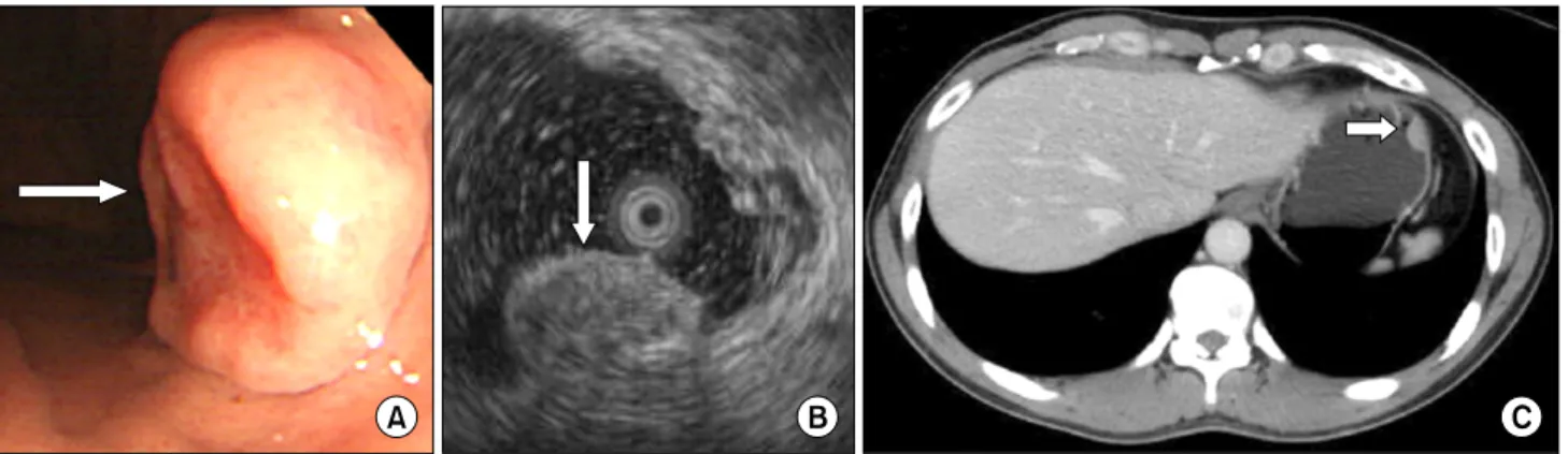
Fig. 4. (A) The endoscopic finding shows the submucosal tumor with normal mucosa on the greater curvature side of the proximal antrum, about 2 cm in size. A round, elevated lesion with normal covering mucosa, hard consistency, and central hyperemia was noted. (B) Abdominal computed tomography finding shows a 2 cm mass in the anterior wall of the gastric antrum.
early gastric cancer (0.5 × 0.5 × 0.4 cm well-differentiated adenocarcinoma) arising from heterotopic gastric mucosa in the gastric submucosa without lymphatic invasion, ves- sel invasion, or perineural invasion, and in the resection margin no infiltration of tumor cells was found (Fig. 3). We did not perform further treatment because we could not detect metastatic lymphadenopathy on CT and operative findings, and the pathologic finding was the same as the indication of endoscopic mucosal resection (EMR) that was suggested by Tsujitani et al [5]. Currently, the patient is on regular follow-up.
Case 2
A 50-year-old male requested transfer for further evalu- ation after detection of a submucosal tumor on upper gas-
trointestinal endoscopy performed during a regular health evaluation. Except for upper stomach brash symp- toms several months ago, the medical history, family his- tory, and physical examination showed no remarkable findings. On upper gastrointestinal endoscopic findings, a submucosal tumor, 20 mm in size and covered with hy- peremic mucosa on the greater curvature of the gastric an- trum, was found and a biopsy was performed (Fig. 4A).
The biopsy results indicated chronic gastritis accompany- ing an infection with H. pylori. An abdominal CT identified a 20 mm mass on the gastric antrum (Fig. 4B). The endo- scopic appearance and the findings on abdominal CT were highly suggestive of a gastrointestinal stromal tumor. We recommended a laparoscopic excision according to the in- dication of surgical excision of submucosal tumor that
suggested by Otani et al [6]. Blood chemistry tests at ad- mission, including tumor markers, showed no abnormal findings. In February 2009, a laparoscopic gastric wedge resection was performed. At the time of surgery, a 2 cm mass was detected on the greater curvature of the gastric antrum; upon visual inspection, the mass was shown to be a gastrointestinal stromal tumor, so we did not perform a frozen biopsy. No post-operative events occurred. The pathologic evaluation revealed a intramucosal adenocar- cinoma that was well-differentiated originating from a 1.5
× 1.5 × 1 cm heterotopic gastric mucosa in the gastric sub- mucosa, no tumor infiltration was found at the resection margin, and there was no detected lymphatic, vessel, and perineural invasion (Fig. 5). Because we did not detect metastatic lymphadenopathy on CT and operative find- ings, and the pathologic finding was the same as the in-
dication of EMR, no further treatment was performed.
Presently, the patient is on regular follow-up.
DISCUSSION
Heterotopic gastric mucosa in the gastric submucosa is reported to occur in 3.0 to 20.1% of resected stomach speci- mens [2,4,7]. Although the etiology of this disease is un- known, mucosal infoldings bulging in the submucosal tis- sue have been demonstrated, probably as a result of in- flammation or ulceration [2,7]. In addition, the histologic characteristics of heterotopic gastric mucosa with cystic expansion are very similar to gastritis cystic polyposa, but gastritis cystric polyposa is known to occur at anas- tromotic sites after gastrectomy [8]. Heterotopic gastric
Fig. 5. (A) Low power view shows a 1.5 × 1.5 × 1 cm well-diffe- rentiated adenocarcinoma that did not invade the muscularis mucosa arising from the heterotopic gastric mucosa in the gastric submucosa (H&E, ×40). (B) High power view shows a well-di- fferentiated adenocarcinoma (white arrow, H&E, ×400). (C) High power view shows heterotopic gastric mucosa in the gastric submucosa (black arrow, H&E, ×400).
gastric mucosa is due to congenital or acquired causes, heterotopic gastric mucosa is found in 20.1% of gastric specimens from adults, but not in gastric specimens from children, thus it is thought to occur by acquired causes [4,9]. Because heterotopic gastric mucosa is found primar- ily among adults in their 60s and is not found in autopsies of people <20 years of age, it is presumed to be due to re- petitive inflammation [8]. Indeed, both of our cases were accompanied by chronic gastritis associated with H. pylori infections.
Gastric cancers occurring in such heterotopic gastric mucosa are very rare [3,10] and the association with carci- nogenesis is controversial. Rubio and Mandai [4] argued that heterotopic gastric mucosa is related to carcino- genesis. Heterotopic gastric mucosa was found in 34 gas- tric resection samples, and among them, 3 cases were gas- tric cancer and 1 case was a stomach ulcer. The authors maintained that such findings are associated with the de- velopment of gastric cancer. However, other authors con- tend that heterotopic gastric mucosa is not associated with the development of cancer. Among 1,500 cases of gastric specimens, heterotopic gastric mucosa was found in 160 cases, and among heterotopic gastric mucosae, 15% were associated with stomach ulcers, 9.9% were associated with gastric cancer, 4% were associated with duodenal ulcers, and 11% were associated with chronic gastritis. With such findings, it was maintained that heterotopic gastric muco- sa is not associated with gastric cancer [2]. In other studies, heterotopic gastric mucosa resulted in no development of cancer; however, 11.7% of early gastric cancer with a single lesion were accompanied by heterotopic gastric mucosa, which was found in 28.6% of multicentric lesion gastric cancers. Multicentric lesions are frequently accompanied by heterotopic gastric mucosa, and causes include repeti-
the 11,100 patients who underwent gastrectomy for gas- tric cancer at Samsung Medical Center between November 1994 and March 2009, only 2 cases (0.02% of all cases) were diagnosed as gastric cancer of heterotopic gas- tric mucosa, and it is estimated that heterotopic gastric mucosa does not lead to a high risk of gastric cancer development.
Gastric cancer that originates from heterotopic gastric mucosa is difficult to diagnose, and on endoscopic find- ings, takes the form of submucosal tumors. In the case of gastric cancer arising from heterotopic gastric mucosa, cancer exists at the submucosa, and cancer components are not exposed on the surface, so biopsy-based diagnoses of gastric cancer are difficult, and based on upper gastro- intestinal barium studies and upper gastrointestinal en- doscopy, gastric cancer takes the form of submucosal tu- mors [10]. Since the findings on upper gastrointestinal en- doscopy, barium studies, or CT take the form of a sub- mucosal tumor, it was diagnosed as a submucosal tumor before surgery, and thus laparoscopic gastric wedge re- section was performed. After surgery, the pathologic eval- uation led to a diagnosis of gastric cancer in our cases.
Heterotopic gastric mucosa is considered to be a benign disease, and cancer rarely occurs within it. Since hetero- topic gastric mucosa takes the form of submucosal tumors, it is very hard to diagnose. When submucosal tumors are found, the possibility of being gastric adenocarcinoma arising from heterotopic gastric mucosa is very low, but should still be considered.
CONFLICTS OF INTEREST
No potential conflict of interest relevant to this article was reported.
REFERENCES
1. Matsumoto K, Shida S, Murakami T, Sugiyama Y, Yamagata N, Tsuchida H, et al. Multiple submucosal cysts of the stomach associated with IIc + III type early gastric cancer: report of a case. Gan No Rinsho 1987;33:848-53.
2. Yamagiwa H, Matsuzaki O, Ishihara A, Yoshimura H.
Heterotopic gastric glands in the submucosa of the stomach. Acta Pathol Jpn 1979;29:347-50.
3. Kosugi S, Kanda T, Hatakeyama K. Adenocarcinoma aris- ing from heterotopic gastric mucosa in the stomach. J Gastroenterol Hepatol 2006;21:483-4.
4. Rubio CA, Mandai K. Gastric adenocarcinomas in dis- placed mucosal glands. Anticancer Res 1999;19(3B):2381-5.
5. Tsujitani S, Oka S, Saito H, Kondo A, Ikeguchi M, Maeta M, et al. Less invasive surgery for early gastric cancer based on the low probability of lymph node metastasis. Surgery
1999;125:148-54.
6. Otani Y, Ohgami M, Igarashi N, Kimata M, Kubota T, Kumai K, et al. Laparoscopic wedge resection of gastric submucosal tumors. Surg Laparosc Endosc Percutan Tech 2000;10:19-23.
7. Iwanaga T, Koyama H, Takahashi Y, Taniguchi H, Wada A.
Diffuse submucosal cysts and carcinoma of the stomach.
Cancer 1975;36:606-14.
8. Littler ER, Gleibermann E. Gastritis cystica polyposa.
(Gastric mucosal prolapse at gastroenterostomy site, with cystic and infiltrative epithelial hyperplasia). Cancer 1972;29:205-9.
9. Rubio CA. Intramucosal gastric cysts simulating sub- mucosal cysts. Pathol Res Pract 1989;184:418-21.
10. Tomonori A, Toshihiro S, Akimichi I, Sei K, Takeshi H, Hitoshi N, et al. Gastric cancer arising from ectopic gastric mucosa glands, report of a case. Stomach Intestine 2003;38:1551-6.