고교 남자 역도 선수의 척측 측부인대 초음파 검사
대구가톨릭대학교병원 정형외과
장일웅∙김세식∙최창혁
서 론
야구와 같이 머리 위로 던지는 운동을 반복적으로 하는 선수들에게서 외반력 부하에 의한 주관절 내측 손상이 많이 발생한다는 것은 잘 알려져 있다2,7,10). 주관절에 대한 이러한 만성 외반력은 야구의 투구동
작, 테니스의 서브, 투창에서 창을 던지는 동작, 미 식축구의 패스, 하키의 슛, 배구의 스파이크 등과 같 은 동작에 의해 비롯되어 종종 내측 측부 인대 손상 등의 주관절 과사용증후군을 유발시킬 수 있다6,8). 이런 운동들은 비슷한 운동기전을 가지는데, 회외전 된 전완의 회내전 및 주관절 외반력과 동반된 빠르 고 강한 주관절 신전이 필요하다14). 역도 또한 이와 유사한 기전을 보이는 운동에 포함되며, 계속되는 훈련으로 인해 주관절에 지속 반복적으로 외반력 부 하가 가해질 경우 주관절 내측부의 반복적 미세 손 상을 유발시켜 척측 측부 인대 손상 및 주관절 내측
통신저자: 최 창 혁
대구광역시 남구 대명4동 대구가톨릭대학교병원 정형외과 Tel: 053-650-4276, Fax: 053-652-4272 E-mail: [email protected]
Ultrasonographic Assessment of the Ulnar Collateral Ligament in High School Male Weight Lifters
Il-Woong Jang, M.D., Se-Sik Kim, M.D., Chang-Hyuk Choi, M.D., Ph.D.
Department of Orthopaedic Surgery, College of Medicine, Catholic University of Daegu
Purpose: We evaluated the efficacy of ultrasonographic examination of the medial collateral ligament injury in elbow joints in the high school male weight lifting athletes.
Materials and Methods: The study group (group I) included 15 male weight lifting athletes (average age: 16.8) and the control group (group II) was demographically matched 9 male with no symptoms on their elbow. Both elbow joint was evaluated through physical examination, plain radiograph, valgus stress view and ultrasonography.
Results: On plain radiograph, there was no significant differences (3.6 mm, 2.7 mm; p>0.05) for the medial articular distances between both groups. The valgus stress view revealed the significant increase in group I (right, avr. 5.86 mm and 3.52 mm, p<0.01, left, avr. 5.33 mm and 3.64 mm, p<0.01).
On ultrasonography, medial joint space was increased in group I (right, avr. 4.66 mm and 3.29 mm, p<0.01, left elbow 4.28 mm and 3.38 mm, p<0.01). The lateral shifting of proximal ulna also increased in group I (right, avr. 0.73 mm and 1.43 mm, p<0.01, left, avr. 0.96 mm and 1.53 mm, p<0.05). The angular deformity were more prevalent in group I.
Conclusion: The medial joint space widening and angular deformity was prevalent in male weight lifting athletes. the ultrasonographic examination was useful in evaluating the degree and incidence of the medial collateral ligament injuries.
Key Words: High school male weight lifting athletes, Elbow joint, Ulnar collateral ligament, Ultrasonography
이완을 일으킬 것으로 생각된다. 저자들은 남자 고 교 역도 선수들에게 있어서 이러한 변화를 초음파 검사를 통해 확인해 봄으로써 적절한 운동 프로그램 을 만드는데 도움을 주고자 하였다.
대상 및 방법
고등학교 남자 역도 선수 15명을 대상으로 하였 으며(실험군), 평균연령은 16.8세(범위 16세~18 세)이었다. 체중은 평균 74 kg이었고, 신장은 평균 172 cm이었다. 운동경력은 평균 3.9년(2~6년)으 로, 운동 경력 1~2년이 3명, 3~4년이 6명, 5~6년 이 6명이었다. 대조군은 주관절부에 증상을 호소하 지 않는 일반 학생으로써 역기 등의 무거운 물건을 들거나 야구나 배구 등 머리위로 던지는 운동 선수 가 아닌 신장 및 몸무게가 조절된 일반 고교 남학생 9명을 대상으로 하였으며 평균 연령은 16.7세(범위 16~17세)이었다. 체중은 평균 68 kg이었고, 신장 은 평균 172 cm이었다.
주관절부 신체 검사와 주관절부 단순 방사선 촬영 과 외반 스트레스 촬영을 하였으며, 초음파검사는 Philips HD11 XE (Bothell, Washington)의 7.5 Hz linear array transducer를 사용하였으며, 중력 하 외반 상태에서 양측 주관절의 외반정도(A, B, C 형) (Fig. 1), 주관절의 간격 정도와 외측 이전 정도 그리고 주관절 주위 변화를 관찰하였다.
외반 스트레스 하에서 방사선 촬영은 환자를 편안
한 자세에서 앉도록 한 후 진행되었고, 주관절을 30 도 굴곡시킨 상태에서 한 술자에 의해 외반력을 주 어 촬영하였다.
초음파 검사는 환자를 앙와위 자세로 눕힌 상태에 서 검사하고자 하는 팔을 침대 밖으로 나오도록 하 고 견관절을 90도 외전시키고, 주관절을 70도 굴곡 시킨 후 전완부를 중립 위에 둔 상태에서 중력부하 를 전완부에 가해 주관절 내측부와 내측 측부 인대
Fig. 1. Three types of angular changes of the medial collateral ligament. (A) normal stable elbow joint. (B) increased medial elbow laxity, as manifested by widening of the medial joint space and lateral shift of the proximal part of the ulna. (C) increased lateral shift of the proximal part of the ulna (arrow) causing impinge- ment of the ulnar collateral ligament on the trochlea (arrowheads).
A B C
Fig. 2. Ultrasonographic examination. In supine posi- tion, the arm was stretched outside the bed.
After the shoulder joint was abducted 90 degrees and elbow flexed 70 degrees, and the forearm put in neutral position, ultrasonograph- ic examination were carried out with gravity bearing tension put on the forearm area.
가 긴장되도록 하였다(Fig. 2). 통계 분석은 SPSS 통계프로그램(SPSS for Windows Release 12.0;
SPSS, Chicago, Illinois)을 사용하였으며, stu- dent t-test와 chi-square test, Fisher’s test등 을 이용하여 통계적 유의성을 검정하였으며, 신뢰구 간은 95%로 하였다.
결 과 1. 신체검사
주관절 내측부에 압통이 있는 경우가 4명(5례)있 었는데 이중 양쪽 주관절 모두 압통이 있는 경우가 1명(2례)이었다. 외반 불안정성이 확인된 경우는 2 명(2례)이었다. 반면에 대조군에서 불안정성이나 압통이 있는 경우는 없었다.
2. 단순 방사선 촬영
내측 관절 간격은 실험군이 대조군에 비해 유의한 차이가 없었으나(3.6 mm, 2.7 mm; p>0.05), 외반 부하 방사선 사진상 실험군에서 내측 관절 간격이 유의하게 증가 되어 있었다[우측 실험군 5.86 mm (4.84~6.88 mm), 대조군 3.52 mm (2.8~4.24 mm); dependent t-test, p<0.01, 좌측 실험군 5.33 mm (4.61~6.05 mm), 대조군 3.64 mm (3.0~4.28 mm); dependent t-test, p<0.01]
(Table 1).
3. 초음파 검사
실험군에서 대조군에 비해 수평거리가 유의하게 증 가 되 어 있 었 으 며 [우 측 실 험 군 4.66 mm (3.36~5.96 mm), 대조군 3.29 mm (2.48~4.1 mm); dependent t-test, p<0.01, 좌측 실험군
Table 1. Comparison of medial elbow laxity between weight-lifter and control group
Numbers of Valgus stress Horizontal Vertical elbow view (mm)* distance (mm)* distance (mm)*
Right elbow
Weight-lifers 15 5.86±1.02 κ 4.66±1.30 φ 0.73±0.73 £ Control group 09 3.52±0.72 κ 3.29±0.81 φ 1.43±0.14 £ Left elbow
Weight-lifers 15 5.33±0.72 ψ 4.28±0.87 λ 0.96±0.89 Ω
Control group 09 3.64±0.64 ψ 3.38±0.81 λ 1.53±0.43 Ω
* The value are given as the mean and the standard deviation. There was a significant difference between the val- ues (p<0.01, dependent t test) of κ, φ, £, ψ, λ, and Ω.
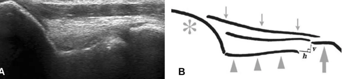
Fig. 3. Ultrasonographic image on normal elbow. The medial joint space is shown as a nonechoic space between the subchondral bone of the trochlea (arrowheads) and that of the coronoid process (large arrow). The medial collateral ligament (MCL) is identified as a band-like structure that attaches to the medial epicondyle and the tubercular portion of the coronoid process. The superficial surface of the ligament is seen outlined by a hyperechoic straight line (small arrows). h; horizontal distance of the medial joint space (with the assumption that the outline of the ulnar collateral ligament is a horizontal line), and v; vertical distance of the medial joint space. *; medial epicondyle.
A B
4.28 mm (3.41~5.15 mm), 대조군 3.38 mm (2.57~4.19 mm); dependent t-test, p<0.01], 수직거리 또한 유의하게 감소되어 있었다[우측 실 험군 0.73 mm (0~1.46 mm), 대조군 1.43 mm (1.29~1.57 mm); dependent t-test, p<0.01, 좌 측 실험군 0.96 mm (0.07~1.85 mm), 대조군 1.53 mm (1.1~1.96 mm); dependent t-test, p<0.05](Table 1) (Fig. 3). 주관절 각변형도 실험 군에서 대조군에 비해 유의하게 나타났다(우측 실 험군 A형 6례, B형 9례, 대조군 A형 9례; Fisher’s test, p<0.01, 좌측 실험군 A형 9례, B형 6례, 대조 군 A형 9례; Fisher’s test, p<0.05) (Fig. 4).
4. 실험군의 운동경력 따른 차이(1군: 1~2년, 2군:
3~4년, 3군: 5~6년)
외반 부하 방사선 사진상 내측 관절간격이 1군, 2 군 3군에서 각각 평균 우측 주관절 5.17 mm, 6.28 mm, 5.78 mm(ANOVA test, p>0.1), 좌측 주관절 5.03 mm, 5.57 mm, 5.25 mm (ANOVA test, p>0.1)로 유의한 차이가 없었다.
초음파 검사상 수평거리는 각각 평균 우측 주관 절 4.71 mm, 4.40 mm, 4.91 mm (ANOVA test, p>0.1), 좌측 주관절 4.67 mm, 4.14 mm, 4.22 mm (ANOVA test, p>0.1)으로 유의한 차이는 없 었다. 수직거리는 각각 평균 우측 주관절 0.33 mm, 0.87 mm, 0.80 mm (ANOVA test, p>0.1), 좌측 주관절 0.85 mm, 0.93 mm, 1.05 mm (ANOVA test, p>0.1)로 유의한 차이가 없었다. 각변형에서 도 우측 주관절의 경우 1군에서 B형 3례, 2군에서 A형 3례, B형 3례, 3군에서 A형 3례, B형 3례이었 으며(Chi-square test, p>0.1), 좌측 주관절에서 1군 A형 1례, B형 2례, 2군에서 A형 4례, B형 2례, 3군에서 A형 4례, B형 2례로써(Chi-square test, p>0.1)로 유의한 차이가 없었다.
5. 실험군의 우세팔 여부에 따른 차이
우측 우세팔이 12명, 좌측 우세팔이 3명으로서, 외반 스트레스 방사선 촬영상에서는 우세팔 평균 5.58 mm, 비우세팔 평균 5.61 mm로 우세팔 여부 에 따 른 차 이 가 없 었 다 (dependent t-test, p>0.1). 하지만 초음파 검사상에서 수평거리가 우 세팔 4.89 mm, 비우세팔 4.09 mm로 차이는 있었 으 나 유 의 하 지 않 았 고 (dependent t-test,
Table 2. Association between ultrasonographic results
Deformity of medial Number of Horizontal Vertical
collateral ligament elbows distance (mm)* distance (mm)*
Right elbow
Present 09 5.09±0.67 κ 0.21±0.39 φ
Absent 15 3.68±1.30 κ 1.41±0.17 φ
Left elbow
Present 06 4.94±0.72 ψ 0.03±0.0 λ
Absent 18 3.53±0.75 ψ 1.58±0.47 λ
* The value are given as the mean and the standard deviation. There was a significant difference between the val- ues (p<0.01, Mann-Whiney U test) of κ, φ, ψ, and λ.
Fig. 4. Ultrasonographic findings in ligament laxity.
The ultrasonographic image shows a large hor- izontal distance and a decreased vertical dis- tance. It also demonstrates type B angular deformity of the contour of the ulnar collateral ligament on the distal-medial corner of the trochlea.
p=0.056), 수직거리는 우세팔 0.6 mm, 비우세팔 1.09 mm로 차이를 보였으나 유의하지 않았다 (dependent t-test, p=0.01). 각변형은 우세팔 A 형 5례, B형 10례를, 비우세팔에서는 A형 10례, B 형 5례로 차이는 보였으나 통계적으로 유의하진 않 았다(Fisher’s test, p=0.072).
6. 실험군 및 대조군의 외반부하 방사선 소견 및 초음 파 검사간의 연관성
외반 부하 검사상 내측 관절 간격과 초음파 검사 상 수평거리 간에는 우측 주관절은 상관계수 0.01 수준에서 유의한 관계(chi-square test, p<0.01) 를 보이나 좌측 주관절은 유의한 관계를 보이지 않 았으며(chi-square test, p>0.1) 외반 부하 검사 상 척측 관절 간격과 초음파 검사상 수직거리 간에 는 좌, 우측 주관절 모두에서 유의한 관계를 보이지 않았다(chi-square test, p>0.05).
초음파 검사상 수평거리와 각변형은 서로 유의한 관계를 보였고(Mann-Whitney U test, p<0.01), 수직거리와 각변형에서도 서로 유의한 관계를 보였 다(Mann-Whitney U test, p<0.01) (Table 2).
고 찰
사체 조직을 통한 실험에서 주관절 내측 측부 인 대의 최대 신장력은 33 N/m정도가 된다고 하며, 투 구시 가속기에 약 64 N/m6,24), 테니스 서브시에 60 N/m이상의 주관절 외반력을 받게 되어 주관절 내 측 측부 인대 손상의 위험성이 높아지게 된다. 역도 선수들은 보통 50 kg에서 최대 200 kg까지의 역기 를 들게 되는데, 이때 전완의 위치는 회내전을 취하 게 되고 주관절의 외반각 및 역기를 잡을 때 어깨 넓 이보다 넓은 손의 위치로 인한 외반 자세로 인해 강 한 외반력을 받을 수 있으며, 반복적인 운동을 통하 여 만성적인 외반력을 받을 경우 내측 측부 인대의 손상 등으로 인해 내측 이완성이 증가될 수 있다.
주관절 내측부의 이완 및 손상은 정확한 병력확인 과 신체검사로 의심할 수 있으며, 단순 방사선 검사 및 부하방사선 검사, 관절조영술, 컴퓨터 단층촬영 (CT), MRI 그리고 동적 초음파 검사 등 영상 검사 로 확진할 수 있다.
투구 운동선수에 대한 방사선 부하 검사는 Goitz
등7)에 의하면 내측 측부 인대 손상의 진단에 있어서 94% 감수성과 100%의 특이성을 보인다고 하였다.
그러나 급성 내측 측부 인대 손상시 부하검사는 환 자의 방해(guarding)로 검사의 어려움이 있을 수 있다. Rijike 등18)은 부하 방사선 검사를 통하여, 완 전파열 내지는 거대 파열이 있는 경우 척상완 간격 이 손상이 없는 측에 비해 5 mm 이상 증가되었다 고 보고하였다. Ellenbecker 등5)은 우세수와 비우 세수의 주관절 내측 이완성을 비교하기 위해 주관절 증상이 없는 프로 야구선수 40명을 대상으로 부하 방사선 검사를 시행하였는데 주관절 내측 관절 간격 의 차이가 비우세수보다 우세수에서 5 mm정도의 차이를 보였다(p<0.01)고 보고하였다. 또한 Lee 등 은 정상 남녀 12명을 대상으로 주관절 외반력 부 하 단순 방사선 검사를 시행하였다. 중력부하시 내 측 척 상 완 골 간 격 이 유 의 하 게 증 가 되 었 고 (p<0.0001), 주관절 외반력을 중력부하와 25 N 부 하시 차이도 나타났지만(p<0.0001), 우세수와 비 우세수의 부하방사선상에서는 5 mm미만의 차이만 나타났다고 하였다13,18).
MRI는 급성 및 만성 손상을 포함한 인대 손상의 가장 정확한 진단이 가능하며9,16,22), 주변의 다른 구 조물들도 보여 줄 수 있는 이점이 있으나, 고가의 검 사로써 운동 선수의 screening 검사로 사용하기에 는 한계가 있다. Azar 등1)은 MRI 관절조영술이 내 측 측부인대의 하면 파열을 포함한 내측 측부 인대 손상 진단에서 97%의 민감도를 가진다고 하였다.
Timmerman 등22)에 의하면 MRI는 57%의 민감도 와 100%의 특이도를 가진다고 하였다. 하지만 Thompson 등23)에 의하면 내측 측부 인대 재건술을 시행한 선수의 79%에서 MRI 양성을 보였으나 21%에선 위 음성을 보였다고 하였다.
최근 주관절 내측 측부 인대 파열 및 변형에 대한 동적 초음파 검사의 유용성에 대한 관심이 높아지고 있으며3,15,17,20,21)
, Sasaki 등19)은 대학야구 수준의 남 자 야구선수 30명을 대상으로 한 검사에서 투구를 하는 주관절 부위가 비투구 주관절 부위에 비해 주 관절 내측 간격이 유의하게 증가되었고, 또한 투구 측 주관절의 내측 측부 인대의 각변형이 비투구측 보다 유의하게 증가되어 있었다고 보고하였다. 남자 역도 선수들을 대상으로 한 저자들의 연구에서는 비 운동선수에 비해 주관절 내측 관절 간격, 수직거리 및 각변형의 정도가 높았으며 이는 이는 성장기 고
교 남자 역도선수의 주관절이 주관절 외반 이완성이 증가되었으며, 내측 측부 인대 손상의 가능성이 높 다는 의미로 해석되었다. 그러나 우세수 여부에 따 른 차이를 보이지는 않았다.
가슴 부위에서 역기를 강한 힘으로 들어 올릴 때 힘의 감소가 보이는 지점이 있는데 이를 ”sticking region”이라 부른다. 초기 가속기에 힘의 감소는 주관절 내측부에 강한 부하를 주어 ”sticking region”에서 역기를 놓치게 만든다고 한다4). 따라 서 무리한 역도 훈련은 선수의 주관절 부위 특히 내 측 관절 부위를 손상 시킬 수 있으며, Lawton 등
11,12)
과 Wilardson 등25)에 의하면 계획된 횟수로 반 복하여 역기를 들어 올리고 그 사이에 적절한 휴식 을 동반하는 것이 계속 역기를 들어 올리는 것보다 근력 강화에 더욱 도움이 된다고 하였다. 특히 성장 기 운동 선수의 경우 이를 고려하여 훈련 프로그램 을 구성하여야 한다. 그리고 주관절 내측부의 증상 을 호소할 경우 초음파를 이용하여 조기에 관절이완 및 손상을 확인함으로써 부상을 방지하는데 도움을 줄 수 있을 것으로 생각되었다.
결 론
남자 고교 역도 선수들의 주관절 내측 부위는 만 성적인 과사용으로 인해 내측 측부 인대 손상 등 부 상의 위험성이 높다. 이를 동적 초음파를 이용하여 이완소견을 조기에 확인함으로써 부상을 방지하기 위한 운동 프로그램을 효과적으로 적용할 수 있을 것으로 생각된다.
참고문헌
01. Azar FM, Andrews JR, Wilk KE, Groh D:
Operative treatment of ulnar collateral ligament injuries of the elbow in athletes. Am J Sports Med, 28: 16-23, 2000.
02. Conway JE, Jobe FW, Glousman RE, Pink M: Medial instability of the elbow in throwing athletes. Treatment by repair or reconstruction of the ulnar collateral ligament. J Bone Joint Surg Am, 74: 67-83, 1992.
03. De Smet AA, Winter TC, Best TM, Bernhardt DT: Dynamic sonography with val- gus stress to assess elbow ulnar collateral liga-
ment injury in baseball pitchers. Skeletal Radiol, 31: 671-676, 2002.
04. Drinkwater EJ, Galna B, Pyne DB, Hunt PH, McKenna MJ: Validation of an optical encoder during free weight resistance movements and analysis of bench press sticking point power during fatique. J Strength Cond Res, 21: 510-517, 2007.
05. Ellenbecker TS, Mattaline AJ, Elam EA, Caplinger RA: Medial elbow joint laxity in pro- fessional baseball pitchers. A bilateral compari- son using stress radiography. Am J Sports Med, 26: 420-424, 1998.
06. Fleisig GS, Andrews JR, Dillman CJ and Escamilla RF: Kinetics of baseball pitching with implications about injury mechanisms, Am J Sports Med, 23: 233-239, 1995.
07. Goitz HT, Rijke AM and Andrews JP et al.:
Evaluation of elbow medial collateral ligament injury in the throwing athlete. 1994.
08. Hamilton CD, Glousman RE, Jobe FW, Brault J, Pink M, Perry J: Dynamic stability of the elbow: electromyographic analysis of the flex- or pronator group and the extensor group in pitchers with valgus instability. J Shoulder Elbow Surg, 5: 347-354, 1996.
09. Jobe FW and Kvitne RS: Elbow instability in the athlete. Instr Course Lect, 40: 17-23, 1991.
10. Jobe FW, Stark H, Lombardo SJ: Reconstruction of the ulnar collateral ligament in athletes. J Bone Joint Surg Am, 68: 1158-1163, 1986.
11. Lawton T, Cronin J, Drinkwater E, Lindsell R, Pyne D: The effect of continuous repetition training and intra-set rest training on bench press strength and power. J Sports Med Phys Fitness, 444: 361-367, 2004.
12. Lawton TW, Cronin JB, Lindsell RP: Effect of interrepetition rest intervals on weight train- ing repetition power output. J Strength Cond Res, 20: 172-176, 2006.
13. Lee GA, Katz SD, Lazarus MD: Elbow valgus stress radiography in an uninjured population.
Am J Sports Med, 26: 425-427, 1998.
14. Loftice J, Fleisig GS, Zheng N, Andrews JR:
Biomechanics of the elbow in sports. Clin Sports Med, 23: 519-530, 2004.
15. Miller TT, Adler RS, Friedman L: Sonography of injury of the ulnar collateral ligament of the
elbow-initial experience. Skeletal Radiol, 33: 386- 391, 2004.
16. Mirowitz SA and London SL: Ulnar collateral ligament injury in baseball pitchers: MR imaging evaluation. Radiology, 185: 573, 1992.
17. Nazarian LN, McShane JM, Ciccotti MG, O’’ Kane PL: Harwood MI Dynamic US of the ante- rior band of the ulnar collateral ligament of the elbow in asymptomatic major league baseball pitchers. Radiology, 227: 149-154, 2003.
18. Rijke AM, Goitz HT, McCue FC, Andrew JR, Berr SS: Stress radiography of the medial elbow ligaments. Radiology, 191: 213-216, 1994.
19. Sasaki J, Takahara M, Ogino T, Kashiwa H, Ishigaki D, Kanauchi Y: Ultrasonographic assessment of the ulnar collateral ligament and medial elbow laxity in college baseball players. J Bone Joint Surg Am, 84: 525-531, 2002.
20. Sugimoto K, Matui N, Taneda Y, Oyabu N, Nakano Y, Goto H: Ultrasonographic evalua- tion of the ulnar collateral ligament of the elbow joint. J Japanese Soc Orthop Ultrasonics, 6: 187-
188, 1994.
21. Sugimoto K, Ohta S: The natural course of the MCL injury of the elbow. J Japanese Soc Orthop Ultrasonics, 9: 15-19, 1997.
22. Timmerman LA, Schwartz ML, Andrews JR: Preoperative evaluation of the ulnar collat- eral ligament by magnetic resonance imaging and computed tomography arthrography:
Evaluation in 25 baseball players with surgical confirmation. Am J Sports Med, 22: 26-31, 1994.
23. Thompson WH, Jobe FW, Yocum LA, Pink MM: Ulnar collateral ligament reconstruction in athletes: Muscle splitting approach without transposition of the ulnar nerve. J Shoulder Elbow Surg, 10: 152-157, 2001.
24. Werner SL, Fleisig GS, Dillman CJ:
Biomechanics of the elbow during baseball pitch- ing. J Orthop Sports Phys Ther, 17: 274-278, 1993.
25. Wilardson JM: A brief review: Factors affect- ing the length of the rest interval between resis- tance exercise sets. J Strength Cond Res, 20: 978- 284, 2006.
목
목적적:: 남자 고교 역도 선수에게서 나타나는 주관절 부위의 변화와 내측 측부 인대 손상의 정도 및 빈도를 초음파 검사를 이용하여 확인하고자 하였다.
대
대상상 및및 방방법법:: 평균연령 16.8세(범위 16세-18세) 고교 남자 역도 선수 15명(실험군, Group I) 및 평균 연령 16.7세(범위 16-17세)인 대조군(Group II) 9명을 대상으로 하여 주관절 부위 병력 청취, 신체검사, 단순 주관절 방사선 촬영, 외반 부하 방사선 검사 및 초음파 검사를 통하여 관절 주위 변화를 관찰하였다.
결
결과과:: 단순 방사선 촬영상 내측 관절 간격은 양군 간에 유의한 차이가 없었으나(3.6 mm, 2.7 mm; p>0.05), 외반 부하 방사선 사진상 실험군이 대조군에 비해 유의하게 증가 되었다(우측 평균 5.86 mm 및 3.52 mm, p<0.01, 좌측 평균 5.33 mm 및 3.64 mm, p<0.01). 초음파 검사상 실험군에서 내측 관절 간격이 유의하게 증가 되어 있었고(우측 평균 4.66 mm 및 3.29 mm, p<0.01, 좌측 평균 8 mm 및 3.38 mm, p<0.01), 척골 근위부가 유의하게 외측 이동되어 있 었으며(우측 평균 0.73 mm 및 1.43 mm, p<0.01, 좌측 평균 0.96 mm 및 1.53 mm, p<0.05), 주관절 각변형 또한 실험군에서(우측 A형 6례, B형 9례, 좌측 A형 9례, B형 6례), 대조군(우측 A형 9례와 좌측 A형 9례)에 비해 유의하게 나타났다. 실험군에서 운동경력(p>0.1) 및 우세팔 여부(p>0.05)에 따른 유의한 차이를 보이지는 않았다.
결
결론론:: 남자 고교 역도 선수들의 주관절 내측 이완성의 빈도가 높았으며, 동적 초음파 검사를 이용시 보다 효과적인 평가 가 가능할 것으로 생각되었다.
색
색인인 단단어어:: 남자 고교 역도 선수, 주관절, 내측 측부 인대, 초음파 국문초록