Ultrasonographic Serial Evaluation
after Reconstruction of the Ulnar Collateral Ligament and the Common Flexor Tendon of the Elbow
in a High School Female Weight Lifter
Chang-Hyuk Choi, M.D. , Se-Sik Kim, M.D., Won-Jun Chang, M.D.*, and Hee-Soo Kim, M.D.
Department of Orthopaedic Surgery, Daegu Catholic University Medical Center, Daegu,
*Department of Orthopaedic Surgery, Gimcheon Medical Center, Gimcheon, Korea
A 16-year-old female weight-lifter with average records of 70 kg for the snatch event and 90 kg for the clean and jerk event suffered chronic ulnar collateral ligament injury, and underwent ulnar collateral ligament reconstruction. An ultrasonographic evaluation was performed at six weeks, three months, six months, nine months, and one year after the operation. The horizontal and vertical distances measured during the initial ultrasonographic examination as well as at six weeks, three months, six months, nine months, and one year after the operation were 8.4 mm, 2.0 mm, 2.6 mm, 2.8 mm, 2.7 mm, 2.7 mm, and -1.2 mm, 2.4 mm, 1.0 mm, 0.0 mm, 0.0 mm, 0.0 mm, respectively. The lifting records at one year after the operation were 65 kg for the snatch event and 90 kg for the clean and jerk event. The ultrasonographic method of serial examination was useful for evaluation of the rehabilitation program and for deciding on the time to return to competition.
Key words: weight-lifting athletes, ultrasonography, elbow, ulnar collateral ligament
Weight lifting usually involves lifting a weight heavier than one’s own body above the head level by using fast and strong elbow joint extensibility with a pronated forearm and a wide, weight-bearing grip, and this may lead to strong valgus force on the elbow joint.
Chronic repetitive valgus stress during exercise and abrupt resisted extension force on lifting could cause injuries at the medial side of the elbow joint. We experienced a case of chronic complete medial collateral ligament and common flexor tendon rupture in a 16-year- old female weight lifting athlete and she was treated with surgical reconstruction and she was evaluated with serial ultrasonographic examinations. We report here on the usefulness of an ultrasono- graphic examination for identifying the integrity of the reconstructed ligament and supporting the rehabilitation program.
CASE REPORT
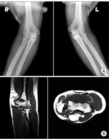
A 16-year-old high school female weight-lifter suffered a valgus overload injury with a popping sound during a weight lifting com- petition 6 months prior to visiting our outpatient clinic. She did not received specific treatment other than a few days rest regardless of the slight swelling and bruise on her left elbow joint area. She noticed progressive weakness of her elbow flexion power and she couldn’t elevate a barbell after experiencing a similar type of recur- rent injury during exercise 1 month ago. She was an elite weight lifter with a 3 years career and she averaged 70 kg for the snatch event and 90 kg for the clean and jerk event. On physical examina- tion, there were mild tenderness on the medial elbow joint and in- creased valgus laxity compared to the contralateral side. The medial joint width on the valgus stress view was 7.0 mm on the left elbow and 4.9 mm on the right side. Magnetic resonamce imaging (MRI) examination revealed a torn medial collateral ligament and a re- tracted common flexor tendon (Fig. 1). The initial ultrasonographic findings revealed a torn medial collateral ligament and common
Copyright © 2014 by The Korean Orthopaedic Association
“This is an Open Access article distributed under the terms of the Creative Commons Attribution Non-Commercial License (http://creativecommons.org/licenses/by-nc/3.0/) which permits unrestricted non-commercial use, distribution, and reproduction in any medium, provided the original work is properly cited.”
The Journal of the Korean Orthopaedic Association Volume 49 Number 2 2014 Received November 29, 2013 Revised February 19, 2014
Accepted February 22, 2014
Correspondence to: Chang-Hyuk Choi, M.D.
Department of Orthopaedic Surgery, Daegu Catholic University Medical Center, 33 Duryugongwon-ro 17-gil, Nam-gu, Daegu 705-718, Korea
TEL: +82-53-650-4276 FAX: +82-53-626-4272 E-mail: [email protected]
flexor tendon with hypoechogenic areas of discontinuity. The an- gular deformity on the Sasaki classification1) was type C and the gap of the elbow joint (horizontal distance) was 8.4 mm and the lateral movement of the elbow joint (vertical distance) was -1.2 mm (Fig. 2, 3).
She underwent ulnar collateral ligament reconstruction using the
palmaris longus tendon and common flexor tendon repair (Fig. 4).
After 6 weeks’ immobilization with a long arm cast, passive range of motion exercise and isometric exercise were begun. Active range of motion exercise and isotonic exercise were started after eight weeks within the range of the pain free motion arc and weight. An isokinetic exercise training program was started 3 months after the operation and weight lifting exercise permitted after 6 months. On the contrary to our rehabilitation program, she started lifting exer- cise 4 months after the operation to attend local competition. She complained of mild pain and tenderness on the medial joint space at 6 months’ follow-up. Based on 6 months’ ultrasonographic find- ings, we prohibited vigorous exercise program for another 1 month and restarted strengthening exercise seven months after operation.
The patient recovered 64% and 78% of the preinjury level records (45 kg for the snatch event and 70 kg for the clean and jerk event) at 9 months after operation and more than 90% of her pre-injury strength (65 kg for the snatch event and 90 kg for the clean and jerk event) at 1 year after operation. Mayo elbow performance score was 60 in initial and improved to 85 at 3 months and 6 months after operation, and 92 at one year follow-up examination. There were no limitation of elbow motion compared to nonoperated side and the medial joint width on the valgus stress view revealed an intact medial joint space compared to normal side (Fig. 5).
1. Ultrasonographic findings
The serial ultrasonographic evaluation was done at the initial ex- amination as well as at 6 weeks, 3 months, 6 months, 9 months, and 1 year after the operation to assess and evaluate the condition of the repaired ligament as well as its functional recovery. For the ultra- sonographic examination, the patient was positioned in the supine position, with the arm undergoing examination stretched across the bed. After the arm was 90o in external rotation, the elbow joint bent at 70o, and the forearm in a neutral position, gravity bearing stress Figure 1. (A) Stress radiograph of the elbow showed widening of the
medial joint space of the left elbow joint (right: 4.9 mm, left: 7.0 mm). (B) Magnetic resonance imaging (MRI) examination showing the rupture of the ulnar collateral ligament and common flexor tendon with retraction on the T2-weighted MRI examination and on the coronal and axial images.
Figure 2. Three types of angular changes of the medial collateral ligament. Type A: normal stable elbow joint. Type B: increased medial elbow laxity, as manifested by widening of the medial joint space and lateral shift of the proximal part of the ulna. Type C: increased lateral shift of the proximal part of the ulna (arrow) causing impingement of the ulnar collateral ligament on the trochlea (arrowheads).
Cited from the article of Sasaki et al.1) (J Bone Joint Surg Am. 2002;84:525-31).
was put on the forearm in order to cause tension in the medial por- tion of the elbow joint. The horizontal and vertical distances were 8.4 mm, 2.0 mm, 2.6 mm, 2.8 mm, 2.7 mm, 2.7 mm, and -1.2 mm,
2.4 mm, 1.0 mm, 0.0 mm, 0.0 mm, 0.0 mm, respectively (Table 1).
The angular deformity was type A at 6 weeks and 3 months after the operation, and type B at 6 months, 9 months and 1 year after the operation. The ultrasonographic findings of the 6 weeks after opera- tion revealed that the reconstructed ligament was maintained with a reduced distance of the vertical and horizontal spaces. The fibrillar pattern of the ligament was smooth and parallelized at 3 months follow-up, but increased inhomogeneity and swelling around the reconstructed ligament and a type B nature joint space were identi- fied at the 6 months evaluation, which might have been influenced by the increased activity and weight training. We restricted weight lifting exercise for 1 month and restarted lifting exercise 7 months after operation. A clearer hyperechogenic fibrillar pattern of the re- constructed ligament could be identified at the 9 months and 1 year follow-up in spite of the sustained hypoechogenecity around the reconstructed ligament and the hyperplasia of the surrounding soft tissue (Fig. 6).
Figure 4. Operative findings demonstrating the ruptured and retracted ulnar collateral ligament and common flexor tendon (A), which were reconstructed using the palmaris longus tendon (B).
Figure 5. The stress radiograph showed a joint width of 4.3 mm at one year after the operation (A) compared to a joint space width of 3.7 mm of the normal side medial joint space (B).
Figure 3. (A) Ultrasonographic image of the normal elbow. The medial joint space is shown as a nonechoic space between the subchondral bone of the trochlea (arrowheads) and that of the coronoid process (large arrow). The ulnar collateral ligament (UCL) is identified as a band-like structure that attaches to the medial epicondyle and the tubercular portion of the coronoid process. The superficial surface of the ligament is seen outlined by a hyperechoic straight line (thin arrows). (B) Ultrasonographic findings in ligament injury. The image showing type C angular changes with increased medial elbow laxity, as manifested by widening of the medial joint space (h=8.4 mm) and the lateral shift of the proximal part of the ulna (v=-1.2 mm).
UCL tear is shown as a nonechoic gap between the torn margin of the ligament (between x and +). h, horizontal distance of the medial joint space (with the assumption that the outline of the UCL is a horizontal line); v, vertical distance of the medial joint space.
DISCUSSION
Weight lifting needed strong muscular power for lifting as well as valgus stability for maintaining balance and control of the barbell.
During the concentric movement of the bench press, there is an ini- tial high-power push after chest contact, immediately followed by a characteristic area of low power, the so-called ‘sticking region’. The decline in power during the initial acceleration phase appears to be a factor in a failed lift attempt at the sticking point during high in- tensity lifting or fatigue repeat repetition training, and this increases the risk of injuries.2) Medial joint injuries can be identified with careful history taking and a thorough physical examination and they can be confirmed with radiologic imaging studies, including simple and stress radiography, arthrography, computerized tomography, MRI and dynamic ultrasonographic examination.1,3-8) MRI can be
a useful diagnostic study for medial collateral ligament injury and injury to the surrounding soft tissue structures in both the acute and chronic conditions, but its usefulness as a screening test for athletes might be limited due to MRI’s high cost. According to Timmer- man et al.,8) MRI showed 57% sensitivity and 100% specificity for diagnosing medial collateral ligament injury. However Thompson et al.9) reported 79% positive results and 21% false negative results for patients who received reconstructive surgery. The effectiveness of dynamic ultrasonography to confirm ulnar collateral ligament injury was recently reported by several authors.1,3,7) Sasaki et al.1) reported on performing dynamic ultrasonography for evaluating medial joint laxity and medial collateral ligament injury and classified three types of angular changes of the medial collateral ligament and measure- ment of the medial joint space. They revealed that the medial el- bow joint gap and angular deformity were more increased in the Figure 6. Ultrasonographic finding of both elbow medial joint spaces showing a type A angular deformity at 6 weeks and 3 months after operation (A, B), and type B at 6 months, 9 months, and 1 year after the operation with an intact graft ligament (C, D, E) compared to the type B normal-side elbow (F). Arrows indicated width of the reconstructed ligament.
Table 1.Ultrasonographic Measurement of the Medial Joint Space
Variable Preoperative Postoperative
6 weeks 3 months 6 months 9 months 1 year
Horizontal distance (mm) 8.4 2.0 2.6 2.8 2.7 2.7
Vertical distance (mm) -1.2 2.4 1.0 0.0 0.0 0.0
dominant arm of 30 males baseball pitchers. But there have been no reports on the usefulness of ultrasonographic study for identifying ligament injury and serial examination for evaluating the integrity of the reconstructed ligament to support a rehabilitation program.
Actually, presented patient started weight lifting exercise before 6 months after the operation to attend local competition, because general symptoms get improved and she had pressures to return to the pre-injury level of exercise. Serial ultrasonographic examina- tion could be used as an impressive imaging study identifying the condition of the reconstructed ligament to the athletes regardless of improved pain or strength after operation. Based on 6 month’s ultrasonographic findings, we delayed vigorous exercise program for 1 month. The patient recovered 64% and 78% of the preinjury level records (45 kg for the snatch event and 70 kg for the clean and jerk event) at 9 months after operation and more than 90% of her pre- injury strength (65 kg for the snatch event and 90 kg for the clean and jerk event) at 1 year after operation. Ultrasonographic examina- tion at 1 year revealed that the integrity and thickness of the recon- structed ligament were preserved with the type B angle of the elbow joint. But, there were hypoechogenesity around the reconstructed ligament and hyperplasia of the surrounding soft tissue, which required that the patient be aware of the risk of reinjury and that she should continue her rehabilitation exercise. Young athletes of growing age needed regular examination during training programs to identify injuries and so administer early treatment. An ultraso- nographic examination could be a useful diagnostic and follow-up study for evaluating musculoskeletal injuries of athletes because it is a noninvasive, well tolerated and quick study with immediate results.
REFERENCES
1. Sasaki J, Takahara M, Ogino T, Kashiwa H, Ishigaki D, Kanau- chi Y. Ultrasonographic assessment of the ulnar collateral
ligament and medial elbow laxity in college baseball players. J Bone Joint Surg Am. 2002;84:525-31.
2. Drinkwater EJ, Galna B, McKenna MJ, Hunt PH, Pyne DB.
Validation of an optical encoder during free weight resistance movements and analysis of bench press sticking point power during fatigue. J Strength Cond Res. 2007;21:510-7.
3. De Smet AA, Winter TC, Best TM, Bernhardt DT. Dynamic sonography with valgus stress to assess elbow ulnar col- lateral ligament injury in baseball pitchers. Skeletal Radiol.
2002;31:671-6.
4. Ellenbecker TS, Mattalino AJ, Elam EA, Caplinger RA. Medial elbow joint laxity in professional baseball pitchers. A bilat- eral comparison using stress radiography. Am J Sports Med.
1998;26:420-4.
5. Lee GA, Katz SD, Lazarus MD. Elbow valgus stress radi- ography in an uninjured population. Am J Sports Med.
1998;26:425-7.
6. Miller TT, Adler RS, Friedman L. Sonography of injury of the ulnar collateral ligament of the elbow-initial experience. Skel- etal Radiol. 2004;33:386-91.
7. Nazarian LN, McShane JM, Ciccotti MG, O'Kane PL, Har- wood MI. Dynamic US of the anterior band of the ulnar col- lateral ligament of the elbow in asymptomatic major league baseball pitchers. Radiology. 2003;227:149-54.
8. Timmerman LA, Schwartz ML, Andrews JR. Preoperative evaluation of the ulnar collateral ligament by magnetic reso- nance imaging and computed tomography arthrography.
Evaluation in 25 baseball players with surgical confirmation.
Am J Sports Med. 1994;22:26-31.
9. Thompson WH, Jobe FW, Yocum LA, Pink MM. Ulnar col- lateral ligament reconstruction in athletes: muscle-splitting approach without transposition of the ulnar nerve. J Shoulder Elbow Surg. 2001;10:152-7.
고교 역도선수의 내측 측부인대와 총굴곡건의 재건술 후 연속적 초음파 검사를 이용한 추시 관찰
최창혁 • 김세식 • 장원준* • 김희수
대구가톨릭대학교병원 정형외과, *김천의료원 정형외과
인상 70 kg, 용상 90 kg의 평균기록을 지닌 16세 여자 역도선수가 주관절의 만성 내측 측부인대 손상으로 진단되어 인대 재건술을 시행하였다. 술 전 및 술 후 6주, 3개월, 6개월, 9개월과 1년에 초음파를 이용하여 인대 재건상태를 확인하였으며, 초음파 검사상 수 평거리는 각각 8.4 mm, 2.0 mm, 2.6 mm, 2.8 mm, 2.7 mm, 2.7 mm였고, 수직거리는 -1.2 mm, 2.4 mm, 1.0 mm, 0.0 mm, 0.0 mm, 0.0 mm로 측정되었다. 수술 후 1년째 역도 기록은 인상 65 kg, 용상 90 kg으로 회복되었다. 초음파를 이용한 내측 측부인대의 연속적인 검사는 재활 상태를 평가하며 시합 복귀시기를 결정하는 데 유용하게 이용될 수 있을 것으로 생각되었다.
색인단어: 역도선수, 초음파, 주관절, 내측 측부인대
접수일 2013년 11월 29일 수정일 2014년 2월 19일 게재확정일 2014년 2월 22일 책임저자 최창혁
대구시 남구 두류공원로 17길 33, 대구가톨릭대학교병원 정형외과 TEL 053-650-4276, FAX 053-626-4272, E-mail [email protected]
Copyright © 2014 by The Korean Orthopaedic Association
“This is an Open Access article distributed under the terms of the Creative Commons Attribution Non-Commercial License (http://creativecommons.org/licenses/by-nc/3.0/) which permits unrestricted non-commercial use, distribution, and reproduction in any medium, provided the original work is properly cited.”
대한정형외과학회지:제 49권 제 2호 2014