Kim SH and Kim HY: Congenital Esophageal Stenosis in Children
Journal of Korean Association of Pediatric Surgeons 1
pISSN 2383-5036 eISSN 2383-5508
J Korean Assoc Pediatr Surg Vol. 24, No. 1, June 2018
https://doi.org/10.13029/jkaps.2018.24.1.1
Review Article
소아의 선천성 식도 협착증
김수홍1, 김현영2
1부산대학교어린이병원 소아외과, 2서울대학교어린이병원 소아외과
Congenital Esophageal Stenosis in Children: From Etiology to Prognosis
Soo-Hong Kim1, Hyun-Young Kim2
1Department of Pediatric Surgery, Pusan National University Children’s Hospital, Yangsan, 2Department of Pediatric Surgery, Seoul National University Children’s Hospital, Seoul, Korea
Congenital esophageal stenosis (CES) is a rare disease that has been reported to occur once in every 25,000 to 50,000 births. According to its etiology, CES is divided into 3 subtypes, tracheobronchial remnants (TBR), fibromuscular hypertrophy (FMH) and membranous diaphragm (MD). Symptoms begin at the weaning period and the introduction of solid food around 6 months with dysphagia and vomiting. Esophagography is first screening test and endoscopic ultrasonography plays important roles to diagnose subtypes deciding therapeutic plan. TBRs were generally treated with surgical resection and end-to-end anasotomosis, whereas FMH and MD had good response rate to endoscopic or radiologic guided dilatation. This article reviews the literature on the etiology, clinical course, diagnosis and management of CES including recent opinion.
Keywords: Congenital esophageal stenosis, Esophagus, Tracheobronchial remnants, Child
Received: March 5, 2018, Accepted: April 13, 2018 Correspondence: Hyun-Young Kim
Department of Pediatric Surgery, Seoul National University Children’s Hospital, 101 Daehak-ro, Jongno-gu, Seoul 03080, Korea. Tel: +82-2- 2072-2478, Fax: +82-2-747-5130, E-mail: spkhy02@snu.ac.kr
Copyright © 2018 Korean Association of Pediatric Surgeons. All right reserved.
This is an Open Access article distributed under the terms of the Creative Commons Attribution Non-Commercial License (http://creativecommons.org/licenses/by-nc/4.0) which permits unrestricted non-commercial use, distribution, and reproduction in any medium, provided the original work is properly cited.
선천성 식도 협착증(congenital esophageal stenosis)은 식도 벽 구조의 선천적인 기형으로 인한 식도 내강의 협착을 말하는 매우 드문 질환으로, 특히 위 식도 역류 등에 의한 이 차적인 식도 협착과 혼동이 되는 경우가 많다[1,2].
협착의 원인이 되는 병리조직학적 소견에 따라 세 가지로 분류된다. 첫 번째는 기관 기관지 잔류물(tracheobronchial remnants)로 이소성의 기관 기관지 조직(연골, 호흡 상피 및 점액선, 원주 상피 등)이 존재하는 경우이며(Fig. 1), 두 번째 는 섬유 근육성 협착(fibromuscular hypertrophy)으로 전 반적인 섬유화를 동반하는 근육층, 점막하층의 부분적인 증 식을 보이는 경우, 그리고 세 번째는 막성 가로막(membranous diaphragm)이 존재하는 경우이다[1]. 이 중 기관 기관지 잔 류물이 대부분을 차지하는 것으로 알려져 있으며, 막성 가로 막이 가장 적은 것으로 알려져 있다[3].
기관 기관지 잔류물은 재태 25일에 발생하는 호흡기와 식 도의 원기의 분리 과정에서 이상이 발생하여 식도에 기관의
조직이 일부 남으면서 발생하는 것으로 여겨지고 있다. 임신 1개월에 발생한 자궁 내 스트레스나 저산소증이 이에 영향 을 주는 것으로 추정된다[3]. 섬유 근성 협착 및 막성 가로막 에 대해서는 그 원인을 정확히 모르는 상태이다. 섬유 근성 협착 및 막성 가로막이 재태기간 중에 발생한 위 식도 역류의 흔적으로, 출생 시에는 위 식도 역류에 의한 다른 흔적은 호 전되고 협착만 남아 있을 것이라는 추정이 있으며, 막성 가로 막은 gross type A 식도폐쇄증의 변형된 형태가 아닐까라는 의견이 있지만, 그 어느 것도 확실하지 않다[1].
25,000명에서 50,000명의 출생아 중 1명 정도로 보고되고 있지만, 협착의 정도가 경미할 경우 진단이 안 되는 경우가 많을 것으로 보이기 때문에 실제 빈도는 더 높을 것으로 생각 된다[1,2,4]. 성별에 따른 발생 빈도의 차이는 없거나[3], 남 아에서 조금 더 빈도가 높은 것으로 알려져 있다[5]. 다른 기 형이 동반되는 경우가 논문에 따라 17%-33%로 보고되고 있 다. 가장 흔하게 동반되는 기형은 기관-식도루가 있거나 없
J Korean Assoc Pediatr Surg 2018;24(1):1-4
2 Journal of Korean Association of Pediatric Surgeons
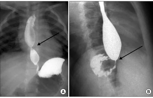
Fig. 2. Upper gastrointestinal contrast study revealed a dilated esophagus proximal to a distal esophageal stenosis (arrows). (A) Anteroposterior view. (B) Lateral view.
Fig. 1. Histologic findings of the resected congenital esophageal stenosis segment revealed respiratory epithelium (arrows) and cartilage (arrowheads) in the esophageal wall (H&E, ×40).
는 선천성 식도 폐쇄이고, 심장 기형, 선천성 장 폐쇄, 직장항 문 기형, 염색체 이상, 요도하열, 두경부 기형 등이 동반된다 [1,6].
선천성 식도 협착증의 가장 흔한 증상은 고형식을 시작할 무렵인 생후 6개월 전후부터 발생하는 삼킴 장애와 구토이 다. 이외에 반복되는 구토에 의한 흡인성 폐렴, 상부 호흡기 질환과 음식물 섭취 부족에 의한 성장 저하 등도 보고되었다 [7]. 협착의 정도에 따라 증상 발생 시기가 다르고 다양한 정 도의 증상이 나타나기 때문에 진단 시기가 매우 다양하고, 지 연되는 경우도 많다[8]. 대부분 소아에서 진단되지만, 협착 의 정도가 경미할 경우 식이 습관의 조절로 극복하는 경우도
있어 드물게 성인기에 진단되기도 한다[9]. 75세 성인에서 기관 기관지 잔류물에 의한 식도 협착이 보고된 바도 있어 [10], 청소년기나 성인기에서도 원인을 찾을 수 없는 구토나 삼킴 장애가 지속될 때는 감별 진단으로 고려해보아야 한다.
상부 식도에 협착이 있는 경우 반복적 호흡기 감염이나, 흡인 성 폐렴 등 호흡기계 증상이 많이 나타나지만, 하부 식도에 협착이 있는 경우 구토 및 음식이나 이물 걸림을 주소로 호소 하는 경우가 많다[8,11].
선천성 식도 협착증은 발생이 매우 드물고, 식도 이완 불 능증 및 위 식도 역류로 의한 역류성 식도염에 의한 후천적인 협착 등과 감별이 어려워 진단이 늦어지는 경우가 많은 것으 로 알려져 있다. 특히 다른 원인으로 발생하는 후천적인 식도 협착질환과의 구분이 중요하다[12,13]. 선천성 식도 협착증 의 주된 진단 방법이며 가장 먼저 시행하는 선별 검사는 식도 조영술로, 특징적인 식도의 협착 소견과 협착 근위부 식도의 확장소견은 진단에 도움이 된다(Fig. 2). 또한 협착부의 위치 와 형태, 협착의 정도를 알 수 있다[3,8,12,14]. 그러나 식도 조영술에는 몇 가지 제한점이 있다. 근위부에 다른 병변이 위 치할 경우, 특히 협착부와 매우 가까운 근위부에 선천성 식도 폐쇄증 수술의 문합부가 있는 경우 근위부의 병변에 의해 선 천성 식도 협착증이 숨겨질 수 있다[15]. 또한, 병리 소견에 따른 분류가 치료 계획 수립에 매우 중요한데, 식도조영술로 는 기관 기관지 잔류물, 섬유근성 협착, 막성 가로막의 구분 이 불가능하다[16]. 식도 내시경 또한 널리 이용되는 진단 방 법으로, 협착 부위 위치와 형태를 직접 확인할 수 있다. 식도 염 여부를 확인함으로써 역류성 식도염이나 식도이완불능 증에 의한 이차적인 식도 협착증과 감별할 수 있지만, 안 되
Kim SH and Kim HY: Congenital Esophageal Stenosis in Children
Journal of Korean Association of Pediatric Surgeons 3
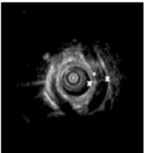
Fig. 3. Endoscopic ultrasound revealed hypoechoic structure in the esophageal wall. Data from the article of Quiros et al. (J Pediatr Gastroenterol Nutr 2013;56:e14) [18].
는 경우도 많다. 식도 내시경 역시 병리 소견에 따른 형태의 분류는 불가능하다[12]. 최근에는 이런 문제점을 극복하기 위하여 내시경 초음파가 이용되고 있다(Fig. 3). 내시경 초음 파는 병변 부위에 있는 연골 등 기관 잔류물을 확인할 수 있 기 때문에, 기관 기관지 잔류물과 다른 형태의 구분이 가능하 여, 치료 계획 수립에 큰 도움을 주고 있으며 그 중요성이 점 차 강조되고 있다[17,18]. 이차적 식도 협착증과 감별이 어려 운 경우 식도 내압 검사와 24시간 식도 산도 검사가 도움이 되는 것으로 알려져 있다[12].
선천성 식도 협착증의 치료는 수술적 치료와 보존적 치료 가 있으며, 원인 질환에 따라 초기 치료방침이 달라진다 [16,17]. 연구에 따라 추천하는 방법이 다르기는 하나, 기관 기관지 잔류형의 경우 풍선 확장술은 크게 도움이 되지 않으 며, 협착 부위의 절제 및 식도 단단문합술을 시행하는 것이, 수술 후 합병증이 적고, 장기적인 결과도 좋은 것으로 알려져 있다[19]. 반면 섬유 근육성 협착의 경우에는 풍선 확장술과 같은 보존적 치료에 반응하는 경우가 많아 이를 일차적으로 시도하는 경우가 많다. 그러나, 보존적 치료가 효과가 없는 경우에는 역시 협착 부위 절제 및 식도 단단문합술을 시행해 야 한다. 일부 연구에서는 식도 확장술 만으로도 기관 기관지 잔류형을 포함한 모든 형태의 식도 협착증에서 완전한 증상 해소를 보였다고 주장하고 있지만[20], 풍선 확장술의 경우 일반적으로 기관 기관지 잔류형에는 증상완화를 보여주지 않으며, 오히려 합병증이 많이 발생한 것으로 알려져 있다 [8]. 특히, 시술 후 식도 천공 및 종격동염이 발생하는 경우가 보고되고 있어 시술의 결정 및 시행에 특별한 주의가 필요할
것으로 생각된다[15].
수술을 계획할 때 흉부와 복부 접근, 두 가지 방법이 있다.
병변의 위-식도 접합부와의 거리에 따라 더욱 편리한 방법 을 선택하면 될 것이다. 흉부 접근법을 시행할 경우에 이전에 선천성 식도폐쇄증 및 기관-식도루 등으로 개흉술을 받은 병력이 있을 경우 문제가 된다. 이전 수술로 인하여 흉막의 유착이나 변형 등이 있을 수 있기 때문에 이에 대한 주의가 필요하다[8]. 복부 접근을 할 경우 병변이 특히 위 식도 접합 부와 가까이 있을 때, 미주 신경의 손상에 주의해야 하며, 경 우에 따라 위 유문 성형술을 해야 할 수 있다[21]. 최근에는 흉강경 수술 등 최소 침습 수술이 성공한 사례도 보고되고 있 다[13,22]. 선천성 식도 협착증 환자는 식도 운동 장애나, 위 식도 역류증이 흔히 동반된다[23]. 이 때문에 구토 등의 증상 은 선천성 식도 협착증에 대한 수술 이후에도 지속될 수 있 어, Nissen 위저성형술 등 역류 방지수술이 필요할 수도 있 다[5,12,24].
치료 후 증상 호전을 보이는 경우가 대부분이기는 하나 연 구에 따라 비율이 다르며, 호전을 보이지 않는 경우도 상당수 있는 것으로 보인다. 절제 후 단단문합술을 시행한 경우, 문 합부 누출과 이에 따른 영양 실조가 보고되어 있으며[13], 수 술 후에도 상당수의 환자에서 삼킴 장애와 같은 증상이 지속 될 수 있다. 문합부 협착이나 식도의 운동 장애가 원인으로 알려져 있다. 문합부 협착이 발생하면 내시경 등을 통한 식도 확장술을 시행한다. 수술 후 식도 운동 장애에 대해서는 아직 까지 연구가 부족한 실정으로 장기 예후를 알아보기 위해서 는 추가적으로 연구가 진행되어야 한다[13,25]. 내시경 식도 확장술을 시행받은 경우 내시경 초음파로 사전에 아형을 알 고 시행한 경우는 89%에서 증상이 호전되는 좋은 경과를 보 이지만, 확인하지 않은 경우는 30% 미만에서만 호전을 보인 다고 알려져 있다[26].
요 약
선천성 식도 협착증은 기관 기관지 잔류물, 섬유 근육성 협착, 막성 가로막의 3가지 원인이 있다. 식도조영술이 가장 흔하게 쓰이며, 진단을 위한 선별 검사이며, 내시경 초음파 검사가 원인 감별에 유용하기 때문에 큰 도움이 된다. 섬유 근육성 협착이나 막성 가로막의 경우 내시경 등을 이용한 식 도 확장술에 반응하여 효과가 있지만, 기관 기관지 잔류물형 의 경우 식도 확장술에 반응하지 않고, 합병증 발생률이 높아 주로 절제 후 단-단 문합 수술을 치료 방침으로 한다.
J Korean Assoc Pediatr Surg 2018;24(1):1-4
4 Journal of Korean Association of Pediatric Surgeons
CONFLICTS OF INTEREST
No potential conflict of interest relevant to this article was reported.
REFERENCES
1. Nihoul-Fékété C, De Backer A, Lortat-Jacob S, Pellerin D. Congenital esophageal stenosis. Pediatr Surg Int 1987;2:86-92.
2. Ramesh JC, Ramanujam TM, Jayaram G. Congenital esophageal stenosis: report of three cases, literature review, and a proposed classification. Pediatr Surg Int 2001;17:188-92.
3. Nemolato S, De Hertogh G, Van Eyken P, Faa G, Geboes K.
Oesophageal tracheobronchial remnants. Gastroenterol Clin Biol 2008;32:779-81.
4. Valerio D, Jones PF, Stewart AM. Congenital oesophageal stenosis.
Arch Dis Child 1977;52:414-6.
5. Zhao LL, Hsieh WS, Hsu WM. Congenital esophageal stenosis ow- ing to ectopic tracheobronchial remnants. J Pediatr Surg 2004;39:
1183-7.
6. Nishina T, Tsuchida Y, Saito S. Congenital esophageal stenosis due to tracheobronchial remnants and its associated anomalies. J Pediatr Surg 1981;16:190-3.
7. Harmon CM, Coran AG. Congenital anomalies of the esophagus.
In: Coran AG, Adzick NS, Krummel TM, Laberge J, Shamberger RC, Caldmone AA, eds. Pediatric surgery. 7th ed. Philadelphia:
Elsevier Saunders; 2012. p.915-6.
8. Nam SH, Kim DY, Kim SC, Kim IK. The diagnosis and treatment of congenital esophageal stenosis. J Korean Surg Soc 2009;76:383-7.
9. Oh CH, Levine MS, Katzka DA, Rubesin SE, Pinheiro LW, Amygdalos MA, et al. Congenital esophageal stenosis in adults:
clinical and radiographic findings in seven patients. AJR Am J Roentgenol 2001;176:1179-82.
10. Younes Z, Johnson DA. Congenital esophageal stenosis: clinical and endoscopic features in adults. Dig Dis 1999;17:172-7.
11. Kim SH, Kim HY, Jung SE, Lee SC, Park KW. Clinical study of con- genital esophageal stenosis: comparison according to association of esophageal atresia and tracheoesophageal fistula. Pediatr Gastroenterol Hepatol Nutr 2017;20:79-86.
12. Amae S, Nio M, Kamiyama T, Ishii T, Yoshida S, Hayashi Y, et al.
Clinical characteristics and management of congenital esophageal stenosis: a report on 14 cases. J Pediatr Surg 2003;38:565-70.
13. Trappey AF 3rd, Hirose S. Esophageal duplication and congenital esophageal stenosis. Semin Pediatr Surg 2017;26:78-86.
14. Murphy SG, Yazbeck S, Russo P. Isolated congenital esophageal stenosis. J Pediatr Surg 1995;30:1238-41.
15. Yoo HJ, Kim WS, Cheon JE, Yoo SY, Park KW, Jung SE, et al.
Congenital esophageal stenosis associated with esophageal atre- sia/tracheoesophageal fistula: clinical and radiologic features.
Pediatr Radiol 2010;40:1353-9.
16. Lee KS. Preoperative diagnosis of congenital esophageal stenosis caused by tracheobronchial remnants using miniprobe endo- scopic ultrasonography in a child. Pediatr Gastroenterol Hepatol Nutr 2012;15:52-6.
17. Takamizawa S, Tsugawa C, Mouri N, Satoh S, Kanegawa K, Nishijima E, et al. Congenital esophageal stenosis: therapeutic strategy based on etiology. J Pediatr Surg 2002;37:197-201.
18. Quiros JA, Hirose S, Patino M, Lee H. Esophageal tracheobron- chial remnant, endoscopic ultrasound diagnosis, and surgical management. J Pediatr Gastroenterol Nutr 2013;56:e14.
19. Suzuhigashi M, Kaji T, Noguchi H, Muto M, Goto M, Mukai M, et al. Current characteristics and management of congenital esoph- ageal stenosis: 40 consecutive cases from a multicenter study in the Kyushu area of Japan. Pediatr Surg Int 2017;33:1035-40.
20. Romeo E, Foschia F, de Angelis P, Caldaro T, Federici di Abriola G, Gambitta R, et al. Endoscopic management of congenital esoph- ageal stenosis. J Pediatr Surg 2011;46:838-41.
21. Vasudevan SA, Kerendi F, Lee H, Ricketts RR. Management of con- genital esophageal stenosis. J Pediatr Surg 2002;37:1024-6.
22. Saka R, Okuyama H, Sasaki T, Nose S, Oue T. Thoracoscopic re- section of congenital esophageal stenosis. Asian J Endosc Surg 2017;10:321-4.
23. Kawahara H, Oue T, Okuyama H, Kubota A, Okada A. Esophageal motor function in congenital esophageal stenosis. J Pediatr Surg 2003;38:1716-9.
24. Elhalaby EA, Elbarbary MM, Hashish AA, Kaddah SN, Hamza AF.
Congenital esophageal stenosis: to dilate or to resect. Ann Pediatr Surg 2006;2:2-9.
25. Michaud L, Coutenier F, Podevin G, Bonnard A, Becmeur F, Khen-Dunlop N, et al. Characteristics and management of con- genital esophageal stenosis: findings from a multicenter study.
Orphanet J Rare Dis 2013;8:186.
26. Terui K, Saito T, Mitsunaga T, Nakata M, Yoshida H. Endoscopic management for congenital esophageal stenosis: a systematic review. World J Gastrointest Endosc 2015;7:183-91.