J Korean Soc Radiol 2017;77(6):367-371 https://doi.org/10.3348/jksr.2017.77.6.367
INTRODUCTION
Localized Stanford type A dissection at the sinus of Valsalva is a condition with difficult early diagnosis, and requires emer- gency surgical treatment (1). Coronary computed tomography angiography (CCTA) with electrocardiography (ECG) gating can be a useful imaging modality for diagnosis of aortic dissec- tion (2). Herein, we report a case of aortic dissection limited to the sinus of Valsalva, focusing on CCTA findings.
CASE REPORT
A 69-year-old man was transferred to our hospital because of bilateral acute pyelonephritis detected by abdominal CT scan.
The patient had complaints of fever, chilliness, and bilateral
flank pain for 7 days prior to visiting the local clinic, and did not have any specific hospital history. Methicillin-sensitive Staphylo- coccus aureus was isolated in blood cultures at the time of ad- mission at the local clinic. Based on suspicion of acute pyelone- phritis and bacteremia, the patient was medicated with ceftriaxone and nafcillin for 5 days, however symptoms persisted.
At transfer, his vital signs were as follows: blood pressure 100/60 mm Hg, pulse 86/min, respiratory rate 20/min, and body tem- perature 36.4°C. Five days after transfer, vital signs were still unsta- ble: blood pressure 96/44 mm Hg, pulse 92/min, respiratory rate 17/min, and body temperature 38.0°C. Because a grade III sys- tolic murmur, splinter hemorrhage, and petechiae were present in the physical examination, the attending physician suspected infective endocarditis.
Chest radiography revealed cardiomegaly and blunting of both
Evaluation of Localized Aortic Dissection at Sinus of Valsalva by Coronary CT Angiography with Multiplanar Reformation:
A Case Report
다절편 전산화단층촬영을 이용한 발살바동에 국한된 대동맥 박리의 평가: 증례 보고
Ki Wook Lee, MD
1, Dong Hun Kim, MD
1*, Eun Ju Yoon, MD
1, Hong-Joo Seo, MD
2, Sang-Wan Ryu, MD
2,3, Dong-Hyun Choi, MD
4Departments of 1Radiology, 2Thoracic and Cardiovascular Surgery, 4Internal Medicine, Chosun University Hospital, Gwangju, Korea
3Department of Thoracic and Cardiovascular Surgery, Saint Carollo General Hospital, Suncheon, Korea
Localized aortic dissection at the sinus of Valsalva is a rare finding in patients with Stanford type A dissections. This condition can be fatal and difficult to diagnose. In this study, we report a case of a 69-year-old man with aortic dissection at the sinus of Val- salva, which was accurately diagnosed with multiplanar reformatted images of coro- nary computed tomography angiography, and later confirmed at surgery.
Index terms
Multidetector Computed Tomography Aorta
Sinus of Valsalva Dissection, Blood Vessel
Received January 31, 2017 Revised May 22, 2017 Accepted July 20, 2017
*Corresponding author: Dong Hun Kim, MD Department of Radiology, Chosun University Hospital, 365 Pilmun-daero, Dong-gu, Gwangju 61453, Korea.
Tel. 82-62-220-3543 Fax. 82-62-228-9061 E-mail: [email protected]
This is an Open Access article distributed under the terms of the Creative Commons Attribution Non-Commercial License (http://creativecommons.org/licenses/by-nc/4.0) which permits unrestricted non-commercial use, distri- bution, and reproduction in any medium, provided the original work is properly cited.
costophrenic angles, indicating bilateral pleural effusion (Fig.
1A). Cardiac enzymes, such as creatine kinase MB fraction and hs troponin T were increased, with values of 35.02 ng/mL (nor- mal range: 0–6.22 ng/mL) and 0.161 ng/mL (normal range:
0.000–0.013 ng/mL), respectively. Other laboratory tests indicat- ed inflammation: white blood cell 13.650/µL, neutrophils 83%, erythrocyte sedimentation rate 59 mm/h (normal range: 0–20
mm/h), C-reactive protein 29.3 mg/dL (normal range: 0.0–0.3 mg/dL), procalcitonin 67.9 ng/mL (normal range: 0–0.5 ng/
mL). ECG revealed wide Q waves in leads II, III, and aVF with- out ST segment elevation, indicating a chronic inferior wall in- farction. Portable echocardiography was performed, and dem- onstrated decreased wall motion with less contractility in the inferior wall of left ventricle (LV). Concerned with the possibili-
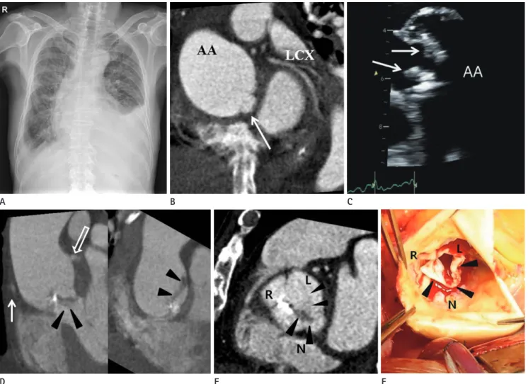
Fig. 1. Localized aortic dissection at the sinus of Valsalva with vegetations and dystrophic calcification in a 69-year-old man.
A. Chest radiograph shows cardiomegaly, pulmonary congestion, and bilateral pleural effusion.
B. Short axis view of initial CCTA represents dilated AA without evidence of dissection. There is a small contrast filled outpouching which communi- cates with the non-coronary Valsalva sinus (arrow).
C. Trans-esophageal echocardiogram demonstrates dilatation of AA and vegetations of aortic valve (arrows).
D. Coronal oblique MPR image in the follow-up CCTA (right panel) shows the dissection flap involving the non-coronary sinus of Valsalva (arrow- heads). Ascending thoracic aorta is dilated. Solid and empty arrows indicate right coronary artery and left main coronary artery, respectively. An- other coronal oblique MPR image (left panel), obtained posteriorly to the right panel, demonstrates localized aortic dissection at the sinus of Val- salva (arrowheads).
E. Axial oblique MPR image in the follow up CCTA, which is reconstructed to correspond with the intra-operative picture (F), presents a dissection flap (arrowheads) that starts from the N and extends to the L.
F. Intra-operative picture shows the extensive dissection flap (arrowheads), extending from the N to the L..
AA = ascending thoracic aorta, CCTA = coronary computed tomography angiography, L = left coronary sinus, LCX = left circumflex coronary artery, MPR = multiplanar reformatted, N = non-coronary sinus, R = right coronary sinus
D E F
A B C
ty of ischemic heart disease, CCTA was performed. A 320 slice CT scanner (Aquilion ONE, Toshiba Medical Systems, Nasu, Ja- pan) with three-dimensional volumetric cardiac imaging during the diastole of R-R interval (Gantry rotation time of 350 msec, scan time of 1.224 sec, 0.5 mm slice thickness with 0.25 recon- struction interval) was done and short axis view, 2 chamber view, 4 chamber view, and volume rendering were acquired. In CCTA, 80 mL of contrast agent, Tomoray 320 (Ioversol; Dong- guk Pharm, Seoul, Korea), followed by 40 mL of a 50%/50% sa- line/contrast medium mixture was infused at 4.5 mL/sec by a dual injection system (Stellant, Medrad Inc., Indianola, PA, USA). CT showed suspicious aortic regurgitation displaying an enlarged LV with poor coaptation of the right and non coronary cusps in dias- tole, as well as mild dilatation of the noncoronary sinus of Val- salva. There was 50% luminal narrowing at the ramus interme- dius. We could not find abnormal valve thickening or vegetation, but small aneurysmal dilatation at the non-coronary Valsalva si- nus was discovered (Fig. 1B). Trans-esophageal echocardiogram (TEE) could not be performed immediately due to patient’s non-cooperation. Therefore, the clinician decided to start con- servative therapy on suspicion of ischemic heart disease. Nine- teen days later, the first TEE was performed and echocardiogra- phy demonstrated the presence of infective endocarditis with severe aortic regurgitation and dilatation of the ascending aorta, vegetations, and decreased contractility in the anteroseptal wall of the LV (Fig. 1C). Follow-up CCTA following TEE showed suspicious infective endocarditis with valvular vegetations, focal calcification, and a dissection flap involving the non-coronary sinus of Valsalva. The dissection flap only involved the sinus of Valsalva (Fig. 1D).
The patient was submitted to surgery 6 days later due to de- layed patient’s consent. During the procedure, it was found that the annulus of the aortic valve was destroyed by infective endo- carditis and there was an aortic dissection present from the non- coronary cusp to the left coronary sinus. The reconstructed image corresponding to the intra-operative picture revealed a dissec- tion flap between the non-coronary cusp and the left coronary sinus (Fig. 1E, F). It was decided to obliterate the dissection site and insert a grafted aorta with a prosthetic aortic valve. Histo- logic evaluation of the aortic valve showed acute inflammation with fibrinoid necrosis, focal abscess formation, and dystrophic calcification. After surgery, the patient’s symptoms vanished,
and he was discharged on the 63rd day.
DISCUSSION
Aortic dissection can occur when the intimomedial layer of the aortic wall is torn by trauma, iatrogenic manipulation, or in- flammation. Dissection of aortic wall layers can extend to the proximal and distal aortic parts. The Stanford classification sys- tem classifies aortic dissection into two types based on its loca- tion. All aortic dissections involving the ascending aorta are de- fined as “type A” and the rest as “type B.” Type A aortic dissection requires urgent surgical treatment, with dissections limited at the sinus of Valsalva being treated by coronary stenting. Patients with type A aortic dissection have a high risk of aortic wall rup- ture, causing secondary cardiac tamponade, aortic regurgita- tion, and visceral malperfusion (3).
Unlike other common type A dissections, detecting localized dissection flaps at the sinus of Valsalva require more attention (1). Radiologists should try to detect these small flaps, as pul- sating artifacts often occur at the same site, and small dissection flaps mimic normal coronary cusps (1). Therefore, in this case, multiplanar reformatted (MPR) images (Fig. 1D, E) of the aor- tic valve were used to diagnose aortic dissection. Particularly, these reformatted images assist in understanding three-dimen- sional relationships between normal anatomy and pathologic lesions.
Conventional aortography, transesophageal echocardiogra- phy, CT, or magnetic resonance imaging can be used to diagnose aortic dissection (4). Many studies have been performed con- cerning diagnostic accuracy of aortic dissection by echocardiog- raphy; recently developed three-dimensional echocardiography provides more helpful information to understand the anatomi- cal structure of dissection than two-dimensional echocardiog- raphy (5). However, several limitations exist: the need of a well- trained user, limited availability in hospitals, obscured view of distal ascending aorta and proximal arch trapping air in the tra- chea and left main bronchus, and an obscured dissection flap under severe aortic valve calcification (6). Furthermore, detected lesions by echocardiography are difficult to differentiate between vegetation, calcification and tumor (7). The selection of initial diagnostic tools for suspected aortic dissection is controversial (4, 8). MPR images of CCTA, which show very high sensitivity,
specificity and contains various reconstruction techniques, can also be used as a valuable method to detect aortic dissection (9).
In this case, aortic dissection at the sinus of Valsalva with val- vular vegetations and focal valvular calcification was confirmed by follow-up CCTA. The abscess of the Valsalva sinus was sus- pected to be the origin site of the dissection. Although mixed lo- calized dissection and abscess were not differentiated in CCTA, MPR images helped to understand lesion extent and direction, as well as evaluate other combined lesions. After being diagnosed with aortic dissection at the sinus of Valsalva by CCTA, the pa- tient was submitted to emergency surgery and recovered com- pletely.
Although type A aortic dissection is a well-known pathology, aortic dissection which is limited at the sinus of Valsalva needs more attention for detection and can be easily misdiagnosed (1, 2, 7). CCTA with multiplanar reformation may be a useful re- construction technique for localized aortic dissection diagnosis.
Acknowledgments
This study was supported by research fund from Chosun Uni- versity, 2015.
REfERENCES
1. Batra P, Bigoni B, Manning J, Aberle DR, Brown K, Hart E, et al. Pitfalls in the diagnosis of thoracic aortic dissection at CT angiography. Radiographics 2000;20:309-320
2. Roos JE, Willmann JK, Weishaupt D, Lachat M, Marincek B, Hilfiker PR. Thoracic aorta: motion artifact reduction with retrospective and prospective electrocardiography-assisted multi-detector row CT. Radiology 2002;222:271-277
3. Hagan PG, Nienaber CA, Isselbacher EM, Bruckman D, Kara- vite DJ, Russman PL, et al. The International Registry of Acute Aortic Dissection (IRAD): new insights into an old dis- ease. JAMA 2000;283:897-903
4. Sommer T, Fehske W, Holzknecht N, Smekal AV, Keller E, Lu- tterbey G, et al. Aortic dissection: a comparative study of diagnosis with spiral CT, multiplanar transesophageal echo- cardiography, and MR imaging. Radiology 1996;199:347- 352
5. Zhao H, He B, Shen X, Qiao Z, Xu T, Lian F, et al. Aortic root dissection with left valsalva sinus perforation detected by transesophageal 3D echocardiography in a patient with Be- hcet’s disease. J Clin Ultrasound 2014;42:59-62
6. Manghat NE, Morgan-Hughes GJ, Roobottom CA. Multi- detector row computed tomography: imaging in acute aor- tic syndrome. Clin Radiol 2005;60:1256-1267
7. Bae JM, Choe YH, Hwang HW, Kim JS, Kim WS, Peck KR, et al. Tumor-mimicking large vegetation attached to the tri- cuspid valve without predisposing factors: a case report on CT and echocardiographic findings. J Koren Soc Radiol 2015;73:269-273
8. Cigarroa JE, Isselbacher EM, DeSanctis RW, Eagle KA. Diag- nostic imaging in the evaluation of suspected aortic dissec- tion. Old standards and new directions. N Engl J Med 1993;
328:35-43
9. Zeman RK, Berman PM, Silverman PM, Davros WJ, Cooper C, Kladakis AO, et al. Diagnosis of aortic dissection: value of he- lical CT with multiplanar reformation and three-dimensional rendering. AJR Am J Roentgenol 1995;164:1375-1380
다절편 전산화단층촬영을 이용한 발살바동에 국한된 대동맥 박리의 평가: 증례 보고
이기욱
1· 김동훈
1* · 윤은주
1· 서홍주
2· 류상완
2,3· 최동현
4발살바동에 국한된 대동맥 박리증은 스탠포드 A형 박리의 드문 소견이다. 이 병변은 생명을 위협하며 진단이 매우 어렵다.
저자들은 관상동맥 CT 혈관 조영술과 다방면 재구성 영상을 이용해 발살바동 박리증으로 진단한 69세 남자 증례를 보고 하고자 한다.
조선대학교병원 1영상의학과, 2흉부외과, 4내과, 3성가롤로병원 흉부외과