Clinical Significance of Persistent Tumor in Bone Marrow during Treatment of High-risk Neuroblastoma
The records of 63 high-risk neuroblastoma patients with bone marrow (BM) tumors at diagnosis were retrospectively reviewed. All patients received nine cycles of induction chemotherapy followed by tandem high-dose chemotherapy and autologous stem cell transplantation (HDCT/auto-SCT). Follow-up BM examination was performed every three cycles during induction chemotherapy and every three months for one year after the second HDCT/auto-SCT. BM tumor cells persisted in 48.4%, 37.7%, 23.3%, and 20.4% of patients after three, six, and nine cycles of induction chemotherapy and three months after the second HDCT/auto-SCT, respectively. There was no difference in progression-free survival (PFS) rate between patients with persistent BM tumor and those without during the induction treatment. However, after tandem HDCT/auto-SCT, the PFS rate was worse in patients with persistent BM tumor than in those without (probability of 5-yr PFS 14.7% ± 13.4% vs. 64.2% ± 8.3%, P = 0.009). Persistent BM tumor during induction treatment is not associated with a worse prognosis when intensive tandem HDCT/auto-SCT is given as consolidation treatment. However, persistent BM tumor after tandem HDCT/
auto-SCT is associated with a worse prognosis. Therefore, further treatment might be needed in patients with persistent BM tumor after tandem HDCT/auto-SCT.
Keywords: Neuroblastoma; Bone Marrow Tumors; Prognosis; Treatment Young Bae Choi,1* Go Eun Bae,2*
Na Hee Lee,1 Jung-Sun Kim,2 Soo Hyun Lee,1 Keon Hee Yoo,1 Ki Woong Sung,1 and Hong Hoe Koo1 Departments of 1Pediatrics and 2Pathology, Samsung Medical Center, Sungkyunkwan University School of Medicine, Seoul, Korea
* Young Bae Choi and Go Eun Bae equally contributed to this work.
Received: 27 January 2015 Accepted: 30 April 2015 Address for Correspondence:
Ki Woong Sung, MD
Department of Pediatrics, Samsung Medical Center, Sungkyunkwan University School of Medicine, 81 Irwon-ro, Gangnam-gu, Seoul 135-710, Korea
Tel: +82.2-3410-3529, Fax: +82.2-3410-0043 E-mail: [email protected]
Funding: This study was supported by a grant from the National R&D Program for Cancer Control, Ministry of Health and Welfare, Republic of Korea (No. HI13C1521).
http://dx.doi.org/10.3346/jkms.2015.30.8.1062 • J Korean Med Sci 2015; 30: 1062-1067
INTRODUCTION
Neuroblastoma is the most common extracranial solid tumor of children. Many patients have metastatic disease at diagnosis.
The bone marrow (BM) is a common metastatic site, and many high-risk patients have BM tumor at diagnosis. The current treat- ment for high-risk neuroblastoma consists of induction treat- ment, high-dose chemotherapy and autologous stem cell trans- plantation (HDCT/auto-SCT) as consolidation treatment, and 13-cis-retinoid acid treatment to reduce relapse from possible residual disease. Regular evaluation of tumor response during and after treatment is needed to assess treatment efficacy.
Several researchers have reported that persistent BM tumor after a certain period of induction treatment is associated with poor outcomes (1-3). However, these studies differed in the tim- ing of BM examination and used strategies with various treat- ment intensities and durations. Therefore, it remains controver- sial whether persistent BM tumor is really a significant risk fac- tor. In our previous retrospective reports, a longer induction treat- ment followed by tandem HDCT/auto-SCT to increase treat- ment intensity was associated with a better outcome in patients with high-risk neuroblastoma (4). We have prospectively used tandem HDCT/auto-SCT following nine cycles of induction che- motherapy since January 2004. Interestingly, many patients with
persistent BM tumor during induction treatment were found to remain progression free. This finding suggests that the signifi- cance of persistent BM tumor might differ from what was previ- ously reported when treatment intensity and duration are in- creased. For this reason, we retrospectively evaluated the sig- nificance of persistent BM tumor in patients with high-risk neu- roblastoma treated at our center.
MATERIALS AND METHODS Patients
Among 197 patients who were newly diagnosed with neuro- blastoma at Samsung Medical Center from January 2004 to De- cember 2012, 88 had high-risk tumors. In the present study, 63 high-risk neuroblastoma patients with BM tumor at diagnosis were retrospectively reviewed. Patients were staged according to the International Neuroblastoma Staging System (5). MYCN amplification was determined using competitive PCR, quanti- tative RT-PCR (qRT-PCR), or fluorescence in situ hybridization.
Tumors were classified as histologically favorable or unfavor- able according to the International Neuroblastoma Pathology Classification (6). Serum ferritin, neuron-specific enolase (NSE), lactic acid dehydrogenase (LDH), and 24-hr urine vanillylman- delic acid (VMA) were measured at diagnosis. Stage 4 tumors
in patients older than 1 yr or any MYCN-amplified tumors were stratified as high-risk tumors.
Treatment of patients
For induction chemotherapy, CEDC (cisplatin + etoposide + doxorubicin + cyclophosphamide) and ICE (ifosfamide + car- boplatin + etoposide) regimens were used in an alternating man- ner (Table 1). Overall, nine cycles of induction chemotherapy were administered prior to HDCT/auto-SCT. Excisional biopsy of the primary tumor was performed at diagnosis if the tumor was resectable. Otherwise, incisional or percutaneous needle biopsy was performed, and definitive surgery was deferred un- til after six cycles of chemotherapy. Peripheral blood stem cells (PBSCs) were collected during the recovery phase after the sev- enth chemotherapy cycle. After induction treatment, tandem HDCT/auto-SCT was administered. The CEC (carboplatin + et- oposide + cyclophosphamide) regimen was used for the first HDCT. As the second HDCT regimen, a TM (thiotepa + melph- alan)-total body irradiation (TBI) regimen was used for patients who were diagnosed up until December 2008. For patients di- agnosed from January 2009 onward, TBI was substituted with high-dose 131I-metaiodobenzylguanidine (MIBG) treatment to reduce late adverse effects (Table 1). Local radiotherapy was ap- plied to the primary site in all patients about 6 weeks after the second HDCT/auto-SCT. Differentiation therapy with 13-cis- retinoic acid (125 mg/m2/day for 14 days every 4 weeks) along with immunotherapy using interleukin-2 (2 × 10⁶ U/m2/day for 5 days every 4 weeks) was administered until one year after HD-
CT/auto-SCT to reduce relapse from possible residual tumor cells (7).
BM examination
BM examination (bilateral aspiration and biopsies) was per- formed at diagnosis, after every three cycles of induction che- motherapy, and then every three months for the first year after tandem HDCT/auto-SCT. Hematoxylin and eosin (H&E) sec- tions were prepared from paraffin embedded biopsy specimens fixed in 10% buffered formalin. Tumor cells in the BM biopsy were confirmed by immunohistochemical staining with anti- bodies against NSE (1:100, polyclonal, CAT 18-0042, Invitrogen, Carlsbad, CA, USA) and CD56 (1:200, CD564, NCL-56-5041, No- vocastra, Newcastle upon Tyne, UK) (8, 9). The degree of BM tumor differentiation (undifferentiated neuroblasts, differenti- ating neuroblasts, and ganglion cells), stroma composition (neu- ropil and Schwannian stroma), and presence of rosettes, necro- sis, fibrosis, hemorrhage, calcification, and foam cells was eval- uated. The percent tumor area was calculated as the proportion of the area occupied by tumor cells in the total BM area.
Detection of tyrosine hydroxylase (TH) transcripts The levels of TH transcripts in PBSCs were measured. PBSC samples were collected in EDTA tubes and mononuclear cells were separated. The mononuclear cells were lysed in Trizol re- agent (Invitrogen, Carlsbad, CA, USA), and RNA was extracted according to the manufacturer’s instructions. When RNA could not be extracted on collection day, the specimen was immedi- ately stored below -70°C and RNA was extracted later (Ambion, Austin, TX, USA). Complementary DNA was generated using an RNA PCR kit (Applied Biosystems, Foster City, CA, USA).
Real-time qRT-PCR was performed using the Real-Q TH Quan- tification Kit (BioSewoom, Seoul, Korea) on a LightCycler (Roche Diagnostics, Mannheim, Germany). qRT-PCR was performed under the following cycling conditions: denaturation at 95°C for 10 min, 45 cycles at 95°C for 10 sec, 60°C for 10 sec, and 72°C for 30 sec, followed by a cooling step at 40°C for 30 sec. Samples were regarded as TH transcript positive if a cycle threshold val- ue was 42 or less. The levels of TH transcripts in biopsied BM samples were also measured according to the methods which we previously reported (10). In brief, fresh biopsied BM tissues were directly lysed in Trizol agent immediately after biopsy, and the following RNA isolation steps were identical to those used for PBSCs.
Statistical analysis
The Mann-Whitney U-test and Kruskal Wallis test were used to compare continuous variables between groups. The chi-square test was used to compare frequencies between groups. The pro- gression-free survival (PFS) rate and 95% confidence interval were determined using the Kaplan-Meier method. Differences Table 1. Induction and high-dose chemotherapy regimens
Regimen/drugs Dose Schedule
Induction regimens CEDC
Cisplatin Etoposide Doxorubicin Cyclophosphamide
60 mg/m2/dose 100 mg/m2/dose 30 mg/m2/dose 30 mg/kg/dose
Day 0 Days 2, 5 Day 2 Days 3, 4 ICE
Ifosfamide Carboplatin Etoposide
1,200 mg/m2/dose 400 mg/m2/dose 100 mg/m2/dose
Days 0-4 Days 0-1 Days 0-4 First HDCT regimen
Carboplatin Etoposide Cyclophosphamide
650 mg/m2/dose 650 mg/m2/dose 1,800 mg/m2/dose
Days -7, -6, -5 Days -7, -6, -5 Days -4, -3, -2 Second HDCT regimen
2004-2008 Thiotepa Melphalan TBI
200 mg/m2/dose 60 mg/m2/dose 3.33 Gy/dose
Days -8, -7, -6 Days -5, -4 Days -3, -,2, -1 2009-2012
Thiotepa Melphalan 131I-MIBG
200 mg/m2/dose 60 mg/m2/dose 12 or 18 mCi/kg
Days -6, -5, -4 Days -3, -2 Day -21 HDCT, high-dose chemotherapy; TBI, total body irradiation; 131I-MIBG, 131I-metaio do- benzylguanidine.
in the PFS rates between groups were compared using the log- rank test. P value < 0.05 were considered significant.
Ethics statement
The institutional review board (IRB) of Samsung Medical Cen- ter approved this study and waived the requirement for inform- ed consent (IRB No. 2014-12-123).
RESULTS
Patient characteristics
A total of 63 high-risk patients (39 boys and 24 girls) had BM tu- mors at diagnosis during the study period. Median age at diag- nosis was 39.5 months (range 1-231). Twenty-two patients had MYCN-amplified tumors and 47 patients had histologically un- favorable tumors. Three patients experienced progression dur- ing induction treatment. The remaining 60 patients underwent the first HDCT/auto-SCT, and three patients died from toxici- ties during the first HDCT/auto-SCT. Therefore, 57 patients un- derwent the second HDCT/auto-SCT. Twenty patients experi- enced relapse/progression after the second HDCT/auto-SCT (nine patients within one year after the second HDCT/auto- SCT).
Result of BM examination
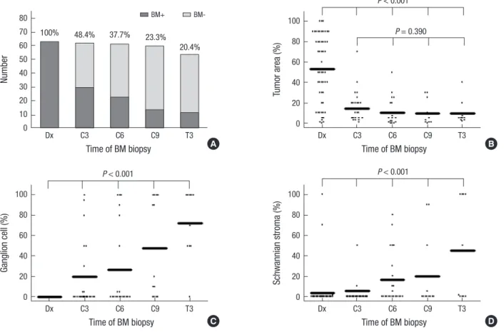
A total of 484 BM biopsies from 63 patients were reviewed. The proportion of patients with persistent BM tumor gradually de- creased during induction treatment and follow-up after tandem HDCT/auto-SCT (Fig. 1A). BM tumor cells were persistent in 14 (23.3%) of 60 biopsies at the end of induction and 11 (20.4%) of 54 biopsies at three months after the second HDCT/auto-SCT.
In patients with persistent BM tumor, the BM tumor area de- creased significantly during the first three cycles of chemother- apy; however, it did not change during further follow-up (Fig.
1B). The evidence of tumor maturation (proportions of gangli- on cells and Schwannian stroma) gradually increased during treatment and follow-up (Fig. 1C and D).
Detection of TH mRNA transcripts
TH transcripts in PBSCs were positive in 1 of 22 patients with persistent BM tumor and 1 of 37 patients without BM tumor af- ter six cycles of induction chemotherapy (P = 1.000). While the former patient remains progression free, the latter patient expe- rienced progression. When the presence of BM tumor was eval- uated with qRT-PCR for TH, the results were correlated with those with histologic examination (sensitivity 71.6% and speci- ficity 82.8%). TH transcripts were positive in 25 (17.2%) of 145
Fig. 1. Results of bone marrow examination. (A) Proportion of patients with persistent BM tumor gradually decreases during follow-up. (B) In patients with persistent BM tumor, tumor area in BM decreases significantly during the first three cycles of chemotherapy (Dx-C3); however, it does not change during further follow-up (C3-T3). Proportions of ganglion cell (C) and Schwannian stroma (D) gradually increase during treatment and follow-up.
Number
Time of BM biopsy
Dx C3 C6 C9 T3 80
70 60 50 40 30 20 10 0
100% 48.4% 37.7% 23.3%
20.4%
BM+ BM-
Tumor area (%)
Time of BM biopsy
Dx C3 C6 C9 T3 100
80 60 40 20 0
P < 0.001 P = 0.390
A B
Schwannian stroma (%)
Time of BM biopsy
Dx C3 C6 C9 T3 100
80 60 40 20 0
P < 0.001
Ganglion cell (%)
Time of BM biopsy
Dx C3 C6 C9 T3 100
80 60 40 20 0
P < 0.001
C D
BM samples without tumor in histological examination.
PFS according to presence/absence of BM tumors
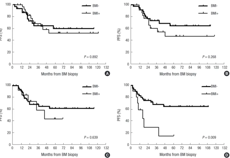
There was no difference in the PFS rates between patients with persistent BM tumor and those without at each time point dur- ing induction treatment (Fig. 2A-C). However, the PFS rate in patients with BM tumor was worse than those without at three months after tandem HDCT/auto-SCT (probability of 5-yr PFS 14.7 ± 13.4% vs. 64.2 ± 8.3%, P = 0.009, Fig. 2D). When TH tran- script positive BM samples without histological evidence were regarded as having persistent BM tumors, the prognostic signif- icance of persistent BM tumors was not changed during induc- tion treatment. However, after tandem HDCT/auto-SCT, the prognostic significance of persistent BM tumors disappeared (probability of 5-yr PFS 60.7 ± 9.3% vs. 42.9 ± 13.8%, P = 0.359).
While seven of 11 patients with histologically persistent BM tu- mors experienced relapse/progression, only one of seven TH transcript positive patients without histological evidence expe- rienced relapse/progression (P = 0.014).
Factors determining early BM response
Various clinical and pathologic parameters associated with BM response were analyzed after the first three cycles of chemother- apy (Table 2). High NSE level (> 100 ng/mL) and low 24 hr urine VMA level (< 15 mg/day) at diagnosis were associated with low- er frequency of persistent BM tumor after the first three cycles of chemotherapy. Younger age (< 18 months) and MYCN am- plification were also associated with lower frequency of persis- tent BM tumor, but with borderline significance.
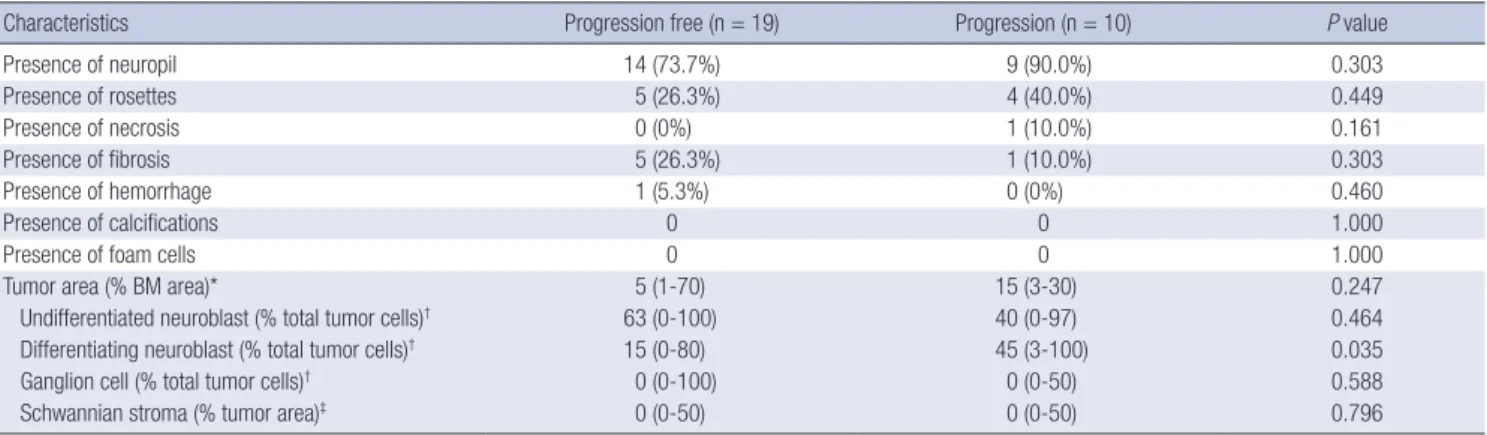
Pathologic parameters associated with poor outcome BM specimens were analyzed in 29 of 30 patients with persis- tent BM tumor after the first three cycles of induction chemo- therapy to evaluate a possible association between pathologic findings after three chemotherapy cycles and subsequent prog- nosis (Table 3). A higher proportion of differentiating neuroblasts in the BM tumor was associated with a higher probability of sub- sequent progression/relapse. However, there was no difference in the presence of rosettes, necrosis, fibrosis, hemorrhage, cal- cifications, and foam cells between patients who experienced
Fig. 2. PFS according to presence/absence of persistent BM tumor. There was no difference in progression free survival (PFS) rates between patients with persistent BM in- volvement (BMI) of tumor and those without at three (A), six (B), and nine (C) cycles of induction chemotherapy. (D) However, the PFS rate in patients with BMI of tumor was worse than those without at three months after tandem HDCT/auto-SCT.
PFS (%)
Months from BM biopsy
0 12 24 36 48 60 72 84 96 108 120 132 100
80
60
40
20
0
BMI- BMI+
P = 0.892
PFS (%)
Months from BM biopsy
0 12 24 36 48 60 72 84 96 108 120 132 100
80
60
40
20
0
BMI- BMI+
P = 0.268
PFS (%)
Months from BM biopsy
0 12 24 36 48 60 72 84 96 108 120 132 100
80
60
40
20
0
BMI- BMI+
P = 0.639
PFS (%)
Months from BM biopsy
0 12 24 36 48 60 72 84 96 108 120 132 100
80
60
40
20
0
BMI- BMI+
P = 0.009
A B
C D
subsequent relapse/progression and those who did not. There was also no difference in percent tumor area between the two patient groups.
DISCUSSION
According to previous studies, persistent BM tumor during in- duction treatment is a poor prognostic factor (1-3). However,
Table 2. Clinical factors determining BM response after three cycles of chemotherapy
Parameters Persistent BM tumor
(n = 30) P value
Age at diagnosis < 18 months (n = 7)
≥ 18 months (n = 55) 1 (14.3%)
29 (52.7%)
0.055
MYCN amplification Absent (n = 41)
Present (n = 21) 23 (56.1%)
7 (33.3%)
0.090
Pathology (INPC) Favorable (n = 11) Unfavorable (n = 46) Unknown (n = 5)
6 (54.5%) 21 (45.7%) 3 (60.0%)
0.596
Differentiation
Undifferentiated (n = 14) Poorly differentiated (n = 25) Differentiating (n = 12) Ganglioneuroblastoma (n = 6) Unknown (n = 5)
3 (21.4%) 12 (48.0%) 8 (66.7%) 3 (50.0%) 4 (80.0%)
0.136
LDH (U/L) < 1,500 (n = 29) ≥ 1,500 (n = 27) Unknown (n = 6)
16 (55.2%) 10 (37.0%) 4 (66.7%)
0.174
Ferritin (ng/mL) < 300 (n = 29) ≥ 300 (n = 28) Unknown (n = 5)
13 (44.8%) 14 (50.0%) 3 (60.0%)
0.696
NSE (ng/mL) < 100 (n = 26) ≥ 100 (n = 33) Unknown (n = 3)
16 (61.5%) 11 (33.3%) 3 (100%)
0.031
24-hr urine VMA (mg/day) < 15 (n = 26) ≥ 15 (n = 32) Unknown (n = 4)
8 (30.8%) 19 (59.4%) 3 (75.0%)
0.030
BM, bone marrow; INPC, International Neuroblastoma Pathology Classification; LDH, lactic acid dehydrogenase; NSE, neuron-specific enolase; VMA, vanillylmandelic acid.
Table 3. Pathologic characteristics according to outcome in patients with persisting tumor cells in BM after three cycles of chemotherapy
Characteristics Progression free (n = 19) Progression (n = 10) P value
Presence of neuropil 14 (73.7%) 9 (90.0%) 0.303
Presence of rosettes 5 (26.3%) 4 (40.0%) 0.449
Presence of necrosis 0 (0%) 1 (10.0%) 0.161
Presence of fibrosis 5 (26.3%) 1 (10.0%) 0.303
Presence of hemorrhage 1 (5.3%) 0 (0%) 0.460
Presence of calcifications 0 0 1.000
Presence of foam cells 0 0 1.000
Tumor area (% BM area)* 5 (1-70) 15 (3-30) 0.247
Undifferentiated neuroblast (% total tumor cells)† 63 (0-100) 40 (0-97) 0.464
Differentiating neuroblast (% total tumor cells)† 15 (0-80) 45 (3-100) 0.035
Ganglion cell (% total tumor cells)† 0 (0-100) 0 (0-50) 0.588
Schwannian stroma (% tumor area)‡ 0 (0-50) 0 (0-50) 0.796
*Tumor area ÷ BM area × 100; †Percent of neuroblasts or ganglion cells among total tumor cells; ‡Stroma area ÷ tumor area × 100. BM, bone marrow.
these studies differed in the timing of BM examination and used various regimens with different treatment intensity and dura- tion. Some studies used single HDCT/auto-SCT after a relative- ly short induction treatment (1, 2) while others used conven- tional chemotherapy (3). In our previous reports for high-risk neuroblastoma, we showed that longer induction treatment and intensive consolidation using tandem HDCT/auto-SCT is associated with better outcomes (4, 7). We hypothesized that the significance of persistent BM tumor might differ from what has been previously reported when intensive tandem HDCT/
auto-SCT is administered as a consolidation treatment. The re- sults indicate that persistent BM tumor during induction treat- ment was not associated with worse prognosis. This finding sug- gests that the prognostic significance of persistent BM tumor during induction treatment may be irrelevant if intensive con- solidation treatment is given. However, histologically persistent BM tumor even after tandem HDCT/auto-SCT was associated with worse prognosis. This finding suggests that further treat- ment with a novel strategy is needed in patients with persistent BM tumor after tandem HDCT/auto-SCT.
Various clinical and pathologic parameters associated with early BM response after the first three cycles of chemotherapy were analyzed. Higher NSE level and lower urine VMA level at diagnosis were associated with lower frequency of persistent BM tumors after the first three cycles of chemotherapy. Young- er age and MYCN amplification were also associated with lower frequency of persistent BM tumors, but with borderline signifi- cance. Higher urine VMA level (i.e., more mature tumor) at di- agnosis and higher proportion of differentiating (not undiffer- entiated) neuroblasts in BM tumor after three cycles of chemo- therapy were associated with lower frequency of persistent BM tumors after the first three cycles of chemotherapy and higher probability of subsequent progression, respectively. These find- ings suggest that favorable tumor biology might not indicate a favorable prognostic factor in high-risk patients who receive in- tensive treatment. We previously reported that the degree of tu- mor volume reduction during the early phase of induction che-
motherapy was higher in undifferentiated and MYCN-ampli- fied tumors (11). Similarly, the current findings suggest that tu- mors with unfavorable biology in high-risk neuroblastoma pa- tients show better treatment response when treated with longer and more intensive protocols.
The detection of minimal residual disease (MRD) status in PBSCs might be crucial in high-risk neuroblastoma because PBSCs contaminated with tumor cells are thought to contribute to relapse (12, 13). Detection of TH transcripts by RT-PCR is one way to assess whether PBSCs are contaminated with tumor cells.
The present study evaluated whether persistent BM tumor at time of PBSC collection is related to tumor cell contamination in PBSCs. There was no difference in TH positivity of PBSCs be- tween patients with persistent BM tumor and those without.
These findings suggest that it might not be necessary to defer PBSC collection even when BM tumors were persistent after 6 cycles of chemotherapy. However, it is not clear whether PBSC could be collected without tumor cell contamination in the ear- lier treatment period when BM tumors were persistent.
The significance of minimal residual BM tumors detected only with qRT-PCR after tandem HDCT/auto-SCT was differ- ent from that of histologically persistent BM tumors. There was no difference in the PFS between patients with MRD in the BM and those without after tandem HDCT/auto-SCT. These find- ings suggest that minimal residual BM tumors which can be de- tected only with qRT-PCR might be controlled by subsequent differentiation treatment and immunologic treatment.
In conclusion, persistent BM tumor during induction treat- ment is not associated with a worse prognosis when intensive tandem HDCT/auto-SCT is given as consolidation treatment.
However, persistent BM lesions after tandem HDCT/auto-SCT are associated with a worse prognosis. Therefore, further treat- ment with a novel strategy might be required in patients with persistent BM tumor after tandem HDCT/auto-SCT.
DISCLOSURE
The authors have no competing financial conflicts of interest to declare.
AUTHOR CONTRIBUTION
Conception and coordination of the study: Sung KW. Design of ethical issues: Choi YB, Lee NH. Acquisition of clinicopatholog- ical data: Bae GE, Kim JS, Sung KW. Analysis and interpretation of data: Choi YB, Sung KW. Manuscript preparation: Choi YB, Sung KW. Critical review of manuscript: Lee SH, Yoo KH, Koo HH. Manuscript approval: all authors.
ORCID
Young Bae Choi http://orcid.org/0000-0001-7016-8827 Ki Woong Sung http://orcid.org/0000-0001-5989-4772 REFERENCES
1. Seeger RC, Reynolds CP, Gallego R, Stram DO, Gerbing RB, Matthay KK. Quantitative tumor cell content of bone marrow and blood as a pre- dictor of outcome in stage IV neuroblastoma: a Children’s Cancer Group Study. J Clin Oncol 2000; 18: 4067-76.
2. Fukuda M, Miyajima Y, Miyashita Y, Horibe K. Disease outcome may be predicted by molecular detection of minimal residual disease in bone marrow in advanced neuroblastoma: a pilot study. J Pediatr Hematol Oncol 2001; 23: 10-3.
3. Cai JY, Pan C, Tang YJ, Chen J, Ye QD, Zhou M, Xue H, Tang JY. Minimal residual disease is a prognostic marker for neuroblastoma with bone marrow infiltration. Am J Clin Oncol 2012; 35: 275-8.
4. Sung KW, Lee SH, Yoo KH, Jung HL, Cho EJ, Koo HH, Lee SK, Kim J, Lim DH, Suh YL, et al. Tandem high-dose chemotherapy and autolo- gous stem cell rescue in patients over 1 year of age with stage 4 neuro- blastoma. Bone Marrow Transplant 2007; 40: 37-45.
5. Brodeur GM, Pritchard J, Berthold F, Carlsen NL, Castel V, Castelberry RP, De Bernardi B, Evans AE, Favrot M, Hedborg F, et al. Revisions of the international criteria for neuroblastoma diagnosis, staging, and response to treatment. J Clin Oncol 1993; 11: 1466-77.
6. Peuchmaur M, d’Amore ES, Joshi VV, Hata J, Roald B, Dehner LP, Gerb- ing RB, Stram DO, Lukens JN, Matthay KK, et al. Revision of the Interna- tional Neuroblastoma Pathology Classification: confirmation of favor- able and unfavorable prognostic subsets in ganglioneuroblastoma, nod- ular. Cancer 2003; 98: 2274-81.
7. Sung KW, Son MH, Lee SH, Yoo KH, Koo HH, Kim JY, Cho EJ, Lee SK, Choi YS, Lim DH, et al. Tandem high-dose chemotherapy and autolo- gous stem cell transplantation in patients with high-risk neuroblastoma:
results of SMC NB-2004 study. Bone Marrow Transplant 2013; 48: 68-73.
8. Ahn S, Lee JJ, Ha SY, Sung CO, Kim J, Han J. Clinicopathological analysis of 21 thymic neuroendocrine tumors. Korean J Pathol 2012; 46: 221-5.
9. Lee SS, Kang M, Ha SY, An J, Roh MS, Ha CW, Han J. Morphologic analysis of pulmonary neuroendoscrine tumors. Korean J Pathol 2013; 47: 16-20.
10. Lee ST, Suh YL, Ko YH, Ki CS, Sung KW, Kim HJ, Kim JW, Kim SH, Chueh H, Lee SH, et al. Measurement of tyrosine hydroxylase transcripts in bone marrow using biopsied tissue instead of aspirates for neuroblastoma. Pe- diatr Blood Cancer 2010; 55: 273-8.
11. Yoo SY, Kim JS, Sung KW, Jeon TY, Choi JY, Moon SH, Son MH, Lee SH, Yoo KH, Koo HH. The degree of tumor volume reduction during the ear- ly phase of induction chemotherapy is an independent prognostic factor in patients with high-risk neuroblastoma. Cancer 2013; 119: 656-64.
12. Burchill SA, Kinsey SE, Picton S, Roberts P, Pinkerton CR, Selby P, Lewis IJ. Minimal residual disease at the time of peripheral blood stem cell har- vest in patients with advanced neuroblastoma. Med Pediatr Oncol 2001;
36: 213-9.
13. Moss TJ, Cairo M, Santana VM, Weinthal J, Hurvitz C, Bostrom B. Clo- nogenicity of circulating neuroblastoma cells: implications regarding peripheral blood stem cell transplantation. Blood 1994; 83: 3085-9.