www.jpis.org
pISSN 2093-2278 eISSN 2093-2286 Copyright © 2010 Korean Academy of PeriodontologyThis is an Open Access article distributed under the terms of the Creative Commons Attribution Non-Commercial License (http://creativecommons.org/licenses/by-nc/3.0/).
A radiographical study on the changes in height of grafting materials after sinus lift: a comparison between two types of xenogenic materials
Pham-Duong Hieu, Jin-Hyung Chung, Sung-Bin Yim, Ki-Seok Hong* Department of Periodontology, Dankook University College of Dentistry, Cheonan, Korea
Purpose: The performance of implant surgery in the posterior maxilla often poses a challenge due to insufficient available bone. Sinus floor elevation was developed to increase the needed vertical height to overcome this problem. However, grafting materials used for the sinus lift technique eventually show resorption. The present study radiographically compared and eval- uated the changes in height of the grafting materials after carrying out maxillary sinus elevation with a window opening pro- cedure. This study also evaluated the difference between two xenogenic bone materials when being used for the sinus lifting procedure.
Methods: Twenty-one patients were recruited for this study and underwent a sinus lift procedure. All sites were treated with either bovine bone (Bio-Oss®) with platelet-rich plasma (PRP) or bovine bone (OCS-B®)/PRP. A total of 69 implants were placed equally 6-8 months after the sinus lift. All sites were clinically and radiographically evaluated right after the implant surgery, 7-12 months, 13-24 months, and 25-48 months after their prosthetic loading.
Results: Changes of implant length/bone length with time showed a statistically significant decreasing tendency (P < 0.05).
There was no significant change in the Bio-Oss® group (P > 0.05). In contrast, the OCS-B® group showed a significant decrease with time (P < 0.05). However, no significant difference was observed between the two groups (P > 0.05).
Conclusions: The results showed that there was significant reduction in comparison with data right after placement, after 7 to 12 months, 13 to 24 months, and over 25 months; however, reduction rates between each period have shown to be without significance. No significant difference in height change was observed between the Bio-Oss® and the OCS-B® groups.
Keywords: Bone substitutes, Maxillary sinus.
INTRODUCTION
The goal modern dentistry should be to restore the patient to normal contour, function, comfort, esthetics, speech, and health. A dentist provides this restoration for a living, wheth- er removing caries from a tooth or replacing several teeth.
What makes implant dentistry unique is the ability to achieve this ideal goal regardless of the atrophy, disease, or injury of the stomatognathic system. That is why the demands for den-
tal implants have recently been increasing.
Long-term success in implant dentistry requires the evalu- ation of more than 50 dental criteria, many of which are unique to this discipline. However, the amount and density of avail- able bone in the edentulous site of the patient are arguably the primary determining factors in predicting individual pa- tient success [1,2]. The amount of available bone for implant, however, is difficult to evaluate exactly since the bone resorp- tion process occurs soon after tooth extraction, particularly in
Received: Dec. 7, 2009; Accepted: Feb. 1, 2010
*Correspondence: Ki-Seok Hong
Department of Periodontology, Dankook University College of Dentistry, Sinbu-dong, Dongnam-gu, Cheonan 330-716, Korea E-mail: [email protected], Tel: +82-41-550-1983, Fax: +82-41-555-7364
loses volume faster than any other region. Tooth loss in the posterior maxilla results in a rapid resorption of both hori- zontal and vertical alveolar bone due to lack of intraosseous stimulation by periodontal ligament fibers. The absence of upper molars leads to increased osteoclast activity in Sch- neider’s membrane, causing pneumatization of the sinus by resorbing bone within a few months [3-5]. Moreover, peri- odontal disease causes aggressive alveolar bone resorption.
The base of the maxillary sinus tends to expand inferiorly to make the alveolar bone height shorter in patients who are edentulous for long periods [6]. According to Lekholom and Zarb’s index [7], severely atrophic maxillas usually have unfa- vorable bone conditions such as type IV bone quality and D or E bone quantity.
These problems have been solved by various surgical meth- ods performed on the maxillary sinus. Maxillary grafting al- ways involves an intraoral maxillary sinusotomy, elevating the mucoperiosteum lining of the maxillary sinus and the Schneiderian membrane upward into the maxillary sinus, and placing a graft — consisting of autogenous bone, allograft, al- loplast, or a combination of these materials — into the surgi- cally created submucoperiosteal pocket to augment the alve- olar bone and implant the recipient site. Boyne and James [8]
carried out maxillary sinus lift for the first time and Tatum [5]
first developed two methods which approach from the alveo- lar crest and lateral wall. Jensen et al. [9] and Adell et al. [10]
introduced an onlay graft technique for alveolar ridge aug- mentation, while Isaksson [11] and Kahnberg et al. [12] intro- duced a bone graft technique with LeFort I.
Summers first performed a sinus elevation technique using an osteotome to recover the vertically insufficient bone mass for implantation on the posterior region of the maxilla [13] As a modified form of this technique, bone added osteotome si- nus floor elevation (BAOSFE), which adds bone graft material to the site of the osteotome procedure was introduced [14].
In the window opening procedure, a primary incision is made in the crestal bone and the flap is reflected bucally to reveal the lateral bony wall of the sinus. In the region of im- plant placement, the bony wall is carefully perforated, taking care not to damage the sinus mucosa. The caudal border of the window should be about 5 mm above the crest of the al- veolar ridge. The sinus mucosa is slowly separated from the bone, creating room for the bone graft. The created space is now filled with the augmentation material, and then covered completely by the mucoperiosteal flap, which must be secure- ly sutured. High success rates have been shown to be achieved using this procedure. Therefore, it is a highly recommended procedure among the optional treatments for the posterior region of the maxilla where the alveolar bone is severely re-
could be used for sinus lift: autogenous bone, allogenic bone, xenogenous bone, and alloplastic bone.
Autogenous bone, as a bone graft material, was first used by Nabers and O’Leary [16]. Dragoo and Sullivan [17] reported that autogenous bone had the highest regeneration ability and Nishibori et al. [18] said that autogenous bone provided appropriate bone quality when used for sinus elevation. How- ever, it is difficult to apply in common clinical situations be- cause of the necessity for additional surgeries, the risk of com- plications, limitations of bone quantity and additional costs.
Allogenous bone also has some issues, including immune problems, potential for infection, and high cost.
Xenogenous bone that originates from bovine bone is be- ing developed to provide solutions to those problems. The xenograft material has osteoconductive abilities, and it is more resistant to resorption than autogenous bone [19]. Both Bio- Oss® (Geistlich Sons Ltd., Wolhusen, Switzerland) and OCS-B® (Nibec, Seoul, Korea) bovine xenograft materials are sold in Korea now.
Stability of graft material grafted into the sinus and chang- es in the height of graft material over time have been impor- tant issues. Wanschitz et al. [20] found a 10-13.9% resorption rate of graft material after a bone graft in the sinus. Hatano et al. [21] tracked vertical changes of mixed bone graft mate- rial (autogenous bone : xenogenous bone = 2 : 1) for up to 10 years. Until 2-3 years after grafting, it showed a statistically significant resorption rate with time, but after that, the bone resorption rate was not considerable. Cho and Kim [22] re- ported that a significant decrease in bone height occurred when either autogenous bone or alloplastic bone was used in a study on the changes in sinus base after graft. In contrast, Keller et al. [23], Blomqvist et al. [24] and Hallman et al. [25]
found that the height and volume of bone graft material re- mained steady.
The present study radiographically compared and evaluat- ed the changes in height of the grafting materials after carry- ing out maxillary sinus elevation with a window opening pro- cedure. This study also evaluated the difference between two xenogenous bone materials, Bio-Oss® and OCS-B®, when be- ing used for the sinus lifting procedure.
MATERIALS AND METHODS
Selection of experimental groups
The study population was comprised of patients who had been treated with implant surgery using the sinus elevation technique on the maxillary posterior of the edentulous region.
Place: Department of Periodontics, Dankook University.
•
Time: between 2004 and 2006.
•
months, 13 to 24 months, and 25 to 48 months after their prosthetic loading
Cases were limited to patients whose charts and radiograph- ic records were trackable. Therefore, only 69 implants in 29 patients met the study’s criteria for inclusion. Patients’ ages varied from 19 to 61 years (average, 51.7 years). This study was exempted from the approval of the institutional review board because the time zone is not applicable to the process.
Methods
Patients received detailed explanations of the difficulties and complications that could take place during the surgery and all patients agreed before the surgery. All the consenting patients were examined to determine whether there were any signs or symptoms of oral disease before conducting si- nus elevation. Patients who had absolute contraindications for implant surgery, such as uncontrolled diabetes, cardio- vascular disease, and blood-related disease, were not select- ed. Two stages of surgery were performed (delayed implan- tation) for all the patients.
This study restricted patients only to those who had at least 3 panoramic radiographs during the study. All the patients in- cluded underwent panorama radiographs at 7 to 12 months and 13 to 24 months after implantation, but only 26 implants in 13 patients had panorama radiography at 25 months after implantation.
For the sinus lift surgery, Bio-Oss® bone graft was used for 40 implants in 15 patients and OCS-B® bone graft was used for 29 implants in 11 patients.
For the implant surgery, two types of implant system were chosen equally: Paragon® (Zimmer Dental Inc, Carlsbad, USA) and Spider II® (BioTIS, Seoul, Korea).
Sinus lifting technique
Local anesthesia was conducted with 2% lidocaine contain- ing 1:100,000 epinephrine. A horizontal incision was made along on the crestal bone in the edentulous area and then buc- cal vertical incisions were made to elevate the muco-periosteal flap.
After elevation of a full-thickness mucoperiosteal flap, ac- cess was gained to the anterior bony wall of the sinus. The lateral bony wall of the sinus was cut by using a small diamond bur under a high speed handpiece. All the cortical bone was removed up to the sinus membrane. Once the membrane was exposed, it was elevated with instruments. The sinus was never lifted more than 2 cm to avoid occluding the sinusal ostium and was never lifted less than 12 mm to allow place- ment of implants of sufficient size to guarantee adequate
sinus cavity was then packed with mixtures of bovine xeno- graft material and PRP. An absorbable collagen membrane (Bio-Gide®, Geistlich Pharma AG, Wolhusen, Switzerland) was then placed on the vestibular wall of the sinus to avoid migration of the graft and its invasion by soft tissues. A com- plete wound closure was performed.
The bovine xenograft materials Bio-Oss® and OCS-B® were used for sinus elevation for the comparison. After the surgery, patients were prescribed 875 mg of augmentins twice a day for a week, and advised to rinse their mouths daily with chlo- rhexidine (0.2%) for 10 days. The patients were examined 1 week post-surgery when the sutures were removed. All pa- tients were checked regularly to verify healing. After a period of 6 to 8 months, the implants were placed by the traditional method. The choice of the implant length was based on the postpanorama after the sinus lift surgery. Two types of im- plant system were chosen equally: the Paragon Implant from Zimmer Dental Inc, United State and the Spider II Implant from BioTIS, Korea.
Measurement of graft material’s height
Height of graft material was measured at the following points.
1st measurement: right after the implantation (baseline).
•
2nd measurement: after the prosthetic restoration (7 to 12
•
months later).
3rd measurement: a year later from the 2nd measurement
•
(13 to 24 months later).
4th measurement: last visit after the 3rd measurement (25
•
to 48 months later).
Three to four panoramic radiographs were taken between right after the implantation to the last observation period.
Not all patients were tested for a 4th time. If the border of the graft material was not clear, then it was not measured.
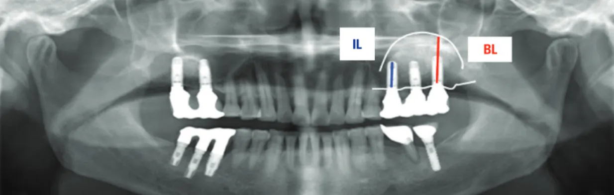
The implant length, alveolar crest, the original base line of the sinus and the base line of the sinus were traced on trac- ing paper. Two measurement points were measured with a digital caliper to the nearest 1/10 mm according to the meth- od suggested by Hatano et al. [21] (Fig. 1).
To evaluate changes in the height of graft material, these values were measured:
Implant length (IL): the distance from implant platform
•
to the apex.
Bone length (BL): the distance from implant platform to
•
the base of the maxillary sinus, which was elevated with graft material.
BL ratio (BL/IL ratio): this value used to evaluate changes
•
in mass of the graft material under the implant.
Statistical analysis
All the data were classified according to those values mea- sured from the radiographs and treatment records. Their means and standard deviations were calculated. Statistical program (SPSSTM, SPSS Inc., Chicago, USA) was used to eval- uate the resorption rate of graft material with time and the differences between graft materials. Changes in the BL/IL ra- tio with time were evaluated through ANOVA and simple linear regression analysis, while comparisons between graft materials were assessed through repeated measures of the general linear model. We considered it statistically significant when P < 0.05.
RESULTS
Changes of BL/IL with time
Sixty-nine dental implants with sinus grafts were placed in 26 patients. There were 26 fixtures with radiographs acquired after 25 months from implant placements. The mean BL was 23.50 mm right after placement, 21.97 mm after 7 to 12 months, 20.63 mm after 13 to 24 months and 20 mm after at least 25 months. The value of BL/IL was 1.54 right after placements, 1.44 after 7 to 12 months, 1.35 after 13 to 24 months, and 1.31 over 25 months (Table 1).
Changes of BL/IL in the Bio-Oss® group with time
There were 21 fixtures with radiographs acquired at least 25 months after implant placement. In patients with Bio-Oss®, the mean BL right after placement was 22.70 mm, 21.23 mm
after 7 to 12 months, 21.21 mm after 13 to 24 months, and 20.41 mm after 25 months. The mean IL was consistently 15.37 mm.
The mean value of BL/IL was 1.47 directly after the placement, 1.38 after 7 to 12 months, 1.38 after 13 to 24 months, and 1.33 after 25 months (Table 2).
Changes in BL/IL in the OCS-B® group with time
There were 5 fixtures with radiographs acquired at least 25 months after implant placement. The mean BL was 24.50 mm right after placement in patients with OCS-B®, 22.90 mm af- ter 7 to 12 months, 22.68 mm after 13 to 24 months, and 18.53 mm after 25 months. The mean IL was consistently 15.21 mm.
The mean value of BL/IL was 1.61 right after placement, 1.50 after 7 to 12 months, 1.49 after 13 to 24 months, and 1.22 at 25 months or more (Table 3).
Results with statistical analysis
Changes in BL/IL showed a statistically significant decreas- ing tendency over time (P< 0.05) (Fig. 2). There was no signif- icant change in the Bio-Oss® group (P> 0.05) (Fig. 1). In con- trast, the OCS-B® group showed a significant decrease with Figure 1. Diagram for the measurement of bone level and height. IL: implant length, BL: bone level.
IL BL
Table 1. Distribution of BL/IL changes with time.
Baseline 7-12 months 13-24 months 25-48 months
BL (mm) 23.50 21.97 20.63 20.00
IL (mm) 15.29 15.29 15.29 15.29
BL/IL 1.54 1.44 1.35 1.31
BL: bone level, IL: implant length.
Table 2. Distribution of BL/IL changes with time in the Bio-Oss® group.
Baseline 7-12 months 13-24 months 25-48 months
BL (mm) 22.70 21.23 21.21 20.41
IL (mm) 15.37 15.37 15.37 15.37
BL/IL 1.48 1.38 1.38 1.33
BL: bone level, IL: implant length.
Table 3. Distribution of BL/IL changes with time in the OCS-B® group.
Baseline 7-12 months 13-24 months 25-48 months
BL (mm) 24.50 22.90 22.68 18.53
IL (mm) 15.21 15.21 15.21 15.21
BL/IL 1.61 1.50 1.49 1.22
BL: bone level, IL: implant length.
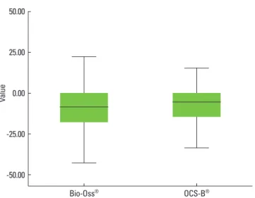
time (P< 0.05) (Fig. 3). However, no significant difference was observed between the 2 groups (P> 0.05) (Fig. 4).
DISCUSSION
It is difficult to place dental implants where maxillary pos- terior teeth are lost due to insufficient bone quantity and com- promised bone quality caused by pneumatization [26-30]. It is difficult to gain a large enough amount of bone for implant placement when there are alveolar bone loss after extractions of maxillary posterior teeth and severe pneumatization. In these situations where alveolar bone has poor quality and short height, the sinus lift technique could be the first option for treatment.
Air pressure from respiration may bring about pneumatiza- tion in the maxillary sinus [3], and this could accelerate resorp- tion of graft material in the maxillary sinus [31]. It is known that the stability of the graft material is one of the major fac- tors influencing further implant stability, and many studies have been done on this issue.
One in vivo experiment used autogenous bone graft mate- rials. Graft height reduced continuously and finally the apex of the implant was exposed to the maxillary sinus [32]. Anoth- er study reported that it was impossible to place dental im- plants after the bone graft technique was usedbecause of rap- id bone resorption [33].
Jensen et al. [34] reported on different resorption patterns with various kinds of graft material. Every case showed a re- sorption tendency regardless of the type of graft materials used. Block et al. [35] performed sinus lift procedures with dif- ferent types of graft material and evaluated the changes in height of the graft materials by computed tomography. At ob- servations 5 to 10 years after the procedure, all the kinds of Figure 4. Statical analysis between groups as time passed.
Value
25.00
0.00
-25.00
-50.00
Bio-Oss® OCS-B®
Figure 3. Statically analysis of each group as time passed.
Bio-Oss®
Value
2.50
2.00
1.50
1.00
baseline 7-12 13-24 25-
Time
OCS-B®
Value
2.50
2.00
1.50
1.00
baseline 7-12 13-24 25-
Time
Figure 2. Statical analysis of BL/IL as time passed. BL: bone level, IL: implant length.
Value
2.00
1.50
1.00
baseline 7-12 13-24 25-
Time
of cases, the graft materials were positioned superior to the apex of the implant.
Hatano et al. [21] reported that graft materials were reduced a statistically significant amount during 2 to 3 years after a si- nus lift. On the other hand, Nystrom et al. [36], Listrom and Symington [37] observed that the force loading to dental im- plants caused graft height to be sustained at a consistent level.
Jensen et al. [34] reported that the resorption rate of bone is in- fluenced by the types of graft materials, and that the amount of resorption was 1.8 mm in an autograft, 2.1 mm in an de- mineralized allograft, 0.9 mm in an alloplast and 0.8 mm in an autograft mixed with alloplast.
In the present study, as in most other studies, graft materi- als were resorbed over time with statistical significance. The value of BL/IL was 1.54 at the time of placement, 1.44 after 7 to 12 months, 1.35 after 13 to 24 months and 1.31 after at least 25 months. There was a significant decrease in comparison with data directly after placement, after 7 to 12 months, after 13 to 24 months and after at least 25 months; however, reduc- tion rates from one period to the next were not statistically significant. However, bone grafts were resorbed with time, as mean BL/IL was observed to be more than 1 in every peri- od. Grafted bones remained in the superior position of the implant apex.
In the Bio-Oss® group, the mean value of BL/IL was 1.47 right after the placement, 1.38 after 7 to 12 months, 1.38 after 13 to 24 months and 1.33 after at least 25 months, but there was no statistical significance.
In the OCS-B® group, the mean value of BL/IL was 1.61 right after placement, 1.50 after 7 to 12 months, 1.49 after 13 to 24 months, and 1.22 over at least 25 months, and it showed a re- duction with statistical significance. However, no significant changes occurred until 24 months. Therefore, it is thought that a significant decrease occurred since only 5 out of 29 fix- tures that had been observed for more than 25 months were investigated. also In addition, there was no significant differ- ence in resorption between the two graft materials.
In conclusion, graft materials are resorbed with time, but without resorption to the apex of the implant, the graft sup- ported the implant structure consistently. Furthermore, no significant difference in height change was observed between the two graft materials.
In the current study, radiographs taken directly after place- ment, after 7 to 12 months, and 13 to 24 months were from the same patients. However, the number of radiographs tak- en after at least 25 months was insufficient; there were limi- tations in achieving accurate follow-up on the changes in grafts with time. We faced limitations in analysis because the number of cases was not large enough and only 2 types of
In the current study, we selected 2D panoramic images.
However, if we took radiographs through 3D images like computed tomography or MRI, we could observe changes in the graft height and volume in maxillary sinus more accu- rately. Since the present study has been done retrospectively without consideration of the type of prosthesis, implant sur- face modifications, diameter and length of implant, and length of available bone, further studies with controlled variables should be done. It is thought that prospective studies with 3D images are needed.
CONFLICT OF INTEREST
No potential conflict of interest relevant to this article was reported.
ACKNOWLEDGMENTS
The present research was conducted by the research fund of Dankook University in 2008.
REFERENCES
Adell R, Eriksson B, Lekholm U, Branemark PI, Jemt T. Long- 1.
term follow-up study of osseointegrated implants in the treatment of totally edentulous jaws. Int J Oral Maxillofac Implants 1990;5:347-59.
Albrektsson T, Dahl E, Enbom L, Engevall S, Engquist B, 2.
Eriksson AR, et al. Osseointegrated oral implants: a Swed- ish multicenter study of 8139 consecutively inserted No- belpharma implants. J Periodontol 1988;59:287-96.
Chanavaz M. Maxillary sinus: anatomy, physiology, sur- 3.
gery, and bone grafting related to implantology: eleven years of surgical experience (1979-1990). J Oral Implantol 1990;16:199-209.
Rosen MD, Sarnat BG. Change of volume of the maxillary 4.
sinus of the dog after extraction of adjacent teeth. Oral Surg Oral Med Oral Pathol 1955;8:420-9.
Tatum H Jr. Maxillary and sinus implant reconstructions.
5.
Dent Clin North Am 1986;30:207-29.
Misch CE. Bone character: second vital implant criterion.
6.
Dent Today 1988;7:39-40.
Lekholom U, Zarb GA. Patient selection and preparation.
7.
In: Branemark PI, Zarb GA, Albrektsson T, editors. Tissue- integrated prostheses: osseointegration in clinical dentist- ry. Chicago: Quintessence; 1985. p.199-220.
Boyne PJ, James RA. Grafting of the maxillary sinus floor 8.
with autogenous marrow and bone. J Oral Surg 1980;38:
613-6.
of the severely resorbed maxilla with bone grafting and os- seointegrated implants: a preliminary report. J Oral Max- illofac Surg 1990;48:27-32.
Adell R, Lekholm U, Grondahl K, Branemark PI, Lindstrom 10.
J, Jacobsson M. Reconstruction of severely resorbed eden- tulous maxillae using osseointegrated fixtures in immedi- ate autogenous bone grafts. Int J Oral Maxillofac Implants 1990;5:233-46.
Isaksson S. Evaluation of three bone grafting techniques 11.
for severly resorbed maxillae in conjunction with imme- diate endosseous implants. Int J Oral Maxillofac Implants 1994;9:679-88.
Kahnberg KE, Nystrom E, Bartholdsson L. Combined use 12.
of bone grafts and Branemark fixtures in the treatment of severely resorbed maxillae. Int J Oral Maxillofac Implants 1989;4:297-304.
Summers RB. A new concept in maxillary implant surgery:
13.
the osteotome technique. Compendium 1994;15:152-8.
Summers RB. The osteotome technique. Part 3: Less inva- 14.
sive methods of elevating the sinus floor. Compendium 1994;15:698-704.
Buchmann R, Khoury F, Faust C, Lange DE. Peri-implant 15.
conditions in periodontally compromised patients follow- ing maxillary sinus augmentation. A long-term post-ther- apy trial. Clin Oral Implants Res 1999;10:103-10.
Nabers CL, O’Leary TJ. Autogenous bone transplants in the 16.
treatment of osseous defects. J Periodontol 1965;36:5-14.
Dragoo MR, Sullivan HC. A clinical and histological eval- 17.
uation of autogenous iliac bone grafts in humans. I: Wound healing 2 to 8 months. J Periodontol 1973;44:599-613.
Nishibori M, Betts NJ, Salama H, Listgarten MA. Short-term 18.
healing of autogenous and allogeneic bone grafts after si- nus augmentation: a report of 2 cases. J Periodontol 1994;
65:958-66.
McAllister BS, Margolin MD, Cogan AG, Buck D, Hollinger 19.
JO, Lynch SE. Eighteen-month radiographic and histolog- ic evaluation of sinus grafting with anorganic bovine bone in the chimpanzee. Int J Oral Maxillofac Implants 1999;14:
361-8.
Wanschitz F, Figl M, Wagner A, Rolf E. Measurement of 20.
volume changes after sinus floor augmentation with a phy- cogenic hydroxyapatite. Int J Oral Maxillofac Implants 2006;
21:433-8.
Hatano N, Shimizu Y, Ooya K. A clinical long-term radio- 21.
graphic evaluation of graft height changes after maxillary sinus floor augmentation with a 2:1 autogenous bone/xe- nograft mixture and simultaneous placement of dental im- plants. Clin Oral Implants Res 2004;15:339-45.
Cho SH, Kim OS. Radiographic change of grafted sinus 22.
of dental implant. J Korean Acad Periodontol 2006;36:345- 59.
Keller EE, Eckert SE, Tolman DE. Maxillary antral and na- 23.
sal one-stage inlay composite bone graft: preliminary re- port on 30 recipient sites. J Oral Maxillofac Surg 1994;52:
438-447.
Blomqvist JE, Alberius P, Isaksson S. Retrospective analysis 24.
of one-stage maxillary sinus augmentation with endosseous implants. Int J Oral Maxillofac Implants 1996;11:512-21.
Hallman M, Hedin M, Sennerby L, Lundgren S. A pro- 25.
spective 1-year clinical and radiographic study of implants placed after maxillary sinus floor augmentation with bo- vine hydroxyapatite and autogenous bone. J Oral Maxillo- fac Surg 2002;60:277-84.
Jaffin RA, Berman CL. The excessive loss of Branemark fix- 26.
tures in type IV bone: a 5-year analysis. J Periodontol 1991;
62:2-4.
Friberg B, Jemt T, Lekholm U. Early failures in 4,641 con- 27.
secutively placed Branemark dental implants: a study from stage 1 surgery to the connection of completed prostheses.
Int J Oral Maxillofac Implants 1991;6:142-6.
Jemt T, Lekholm U. Implant treatment in edentulous max- 28.
illae: a 5-year follow-up report on patients with different degrees of jaw resorption. Int J Oral Maxillofac Implants 1995;10:303-11.
Bahat O. Treatment planning and placement of implants 29.
in the posterior maxillae: report of 732 consecutive Nobel- pharma implants. Int J Oral Maxillofac Implants 1993;8:
151-61.
Adell R, Lekholm U, Rockler B, Branemark PI. A 15-year 30.
study of osseointegrated implants in the treatment of the edentulous jaw. Int J Oral Surg 1981;10:387-416.
Hurzeler MB, Kirsch A, Ackermann KL, Quinones CR. Re- 31.
construction of the severely resorbed maxilla with dental implants in the augmented maxillary sinus: a 5-year clini- cal investigation. Int J Oral Maxillofac Implants 1996;11:
466-75.
Coombs CJ, Mutimer KL, Holmes AD, Levant BA, Courte- 32.
manche DJ, Clement JG. Osseointegration in sinus-form- ing bone. Plast Reconstr Surg 1995;95:866-75.
Johansson B, Grepe A, Wannfors K, Aberg P. CT-scan in 33.
assessing volumes of bone grafts to the heavily resorbed maxilla. J Craniomaxillofac Surg 1998;26 Suppl 1:85.
Jensen OT, Shulman LB, Block MS, Iacono VJ. Report of 34.
the Sinus Consensus Conference of 1996. Int J Oral Maxil- lofac Implants 1998;13 Suppl:11-45.
Block MS, Kent JN, Kallukaran FU, Thunthy K, Weinberg 35.
R. Bone maintenance 5 to 10 years after sinus grafting. J Oral Maxillofac Surg 1998;56:706-714.
severely resorbed maxillae with bone graft and titanium implants: histologic review of autopsy specimens. Int J Oral Maxillofac Implants 1993;8:167-72.
plants in conjunction with bone grafts. Int J Oral Maxillo- fac Surg 1988;17:116-8.