1
1)이화여자대학교 임상치의학대학원 임상치주보철학과,
2)이화여자대학교부속 목동병원 치주과,
3)이화여자대학교 의과대학 치과학교실 치주과 이 지 은1), 박 소 민1), 이 종 빈1, 2), 방 은 경1, 2, 3)*
상악동저 거상술에서 이식재 양에 따른 이식골 높이 변화에 대한 방사선학적 평가
A change of sinus floor level related to the amount of grafted material after bone added osteotome sinus floor elevation (BAOSFE) technique:
A radiographic retrospective study
1)Department of Periodontology and Prosthodontics, Graduated School of Clinical Dentistry, Ewha Womans University,
2)Department of Periodontology, Mokdong Hospital, Ewha Womans University,
3)Department of Periodontology, School of Medicine, Ewha Womans University Ji-Eun Lee1), So-Min Park1), Jong-Bin Lee1, 2), Eun-Kyoung Pang1, 2, 3)*
Purpose: The purpose of this article is to evaluate a change o bone level on the sinus floor by a bone added osteotome sinus floor elevation (BAOSFE) technique, according to the amount of deproteinized bovine bone mineral (DBBM). And Changes in augmented bone height after BAOSFE procedure were also assessed for 6 months after the implant procedure.
Materials and Methods: Forty eight single implants were placed in the posterior maxilla using BAOSFE technique. The implantation sites were classified into two groups according to the amount of grafted DBBM, 0.25 group (0.25g) and 0.5 group (0.5 g). Panoramic views or cone-beam computed tomography (CBCT) were taken at the time of implant placement with BAOSFE and after at least 6 months to assess the bone level changes in the elevated sites with DBBM.
Results: Alveolar bone level around all implants was stable clinically and radiographically during the follow-up. Mean augmented bone height was 5.21 0.94 mm in 0.25 group and 6.92 1.19 mm in 0.5 group. Statistically significant difference in augmented bone height was found in the comparison between the 0.25 group and 0.5 group at the time of surgery. There was a positive correlation between the length of the implant protruding into the maxillary sinus and the augmented bone height. After 6 months, mean reduction of augmented bone height was 0.50 0.34 mm in 0.25 group and 0.41 0.30 mm in 0.5group. There was no specific correlation between the reduction of augmented bone height and amount of grafted DBBM.
Conclusion: Within the limit of this study, the amount of grafting materials and the protrusion length of implant into the maxillary sinus affect the amount of the augmented bone height.
Key words : Sinus floor augmentation, Cone-Beam Computed Tomography, Graft material, Dental implants ABSTRACT
Corresponding Author Eun-Kyoung Pang, DDS, PhD Associate Professor
Department of Periodontology, School of Medicine, Ewha Womans University, 1071 Anyangcheon-ro, Yangcheon-gu, Seoul 07985, Korea.
Tel : +82-2-2650-2725, Fax : +82-2-2650-5764, E-mail : [email protected]
이식재양에따른이식골높이변화에대한방사선학적평가
Ⅰ. INTRODUCTION
Posterior maxilla commonly poses a challenge for successful dental implants due to a poor bone quality and insufficient bone quantity1). Sinus pneumatization causes the poor bone quantity. In some patients the loss of alveolar bone coupled with increased antral pneumatization may result in only a 2-3mm thickness of alveolar bone height. To overcome this anatomic limitation, sinus floor elevation has been performed as a common surgical procedure in oral implant treatment. Until today elevation of the maxillary sinus in conjunction with sinus grafting have already been carried out for nearly 40years2, 3).
Recently, there have been many studies on the maxillary sinus lift using the non-invasive technique to reduce the complications of the maxillary sinus lift. A representative method of the non-invasive technique is the crestal approach, the osteotome sinus floor elevation (OSFE) technique or bone added osteotome sinus floor elevation(BAOSFE) technique, which has been used so far, and the results of studies on the residual bone height suitable for this method vary. Jensen(1996) suggested two stage procedures when the residual bone height remains less than 4mm, according to the height of residual bone. He also recommended one stage procedure with OSFE technique when the residual bone height is more than 7mm.
Additionally, Summer et al.(1994) reported that osteotome technique is possible when the residual bone height is 5-6 mm4, 5).
In general, OSFE technique can be used when
residual bone height is more than 7mm6). In BAOSFE technique, grafting material can be added with osteotome technique in cases remained less than 7mm of residual bone height.
Compared to the lateral approach, BAOSFE is less invasive and less traumatic. However, it shows relatively limited amount of sinus elevation because it is a blind technique. Several studies have reported that the increase in bone height available through BAOSFE is about 2-
4mm1, 7, 8). Since the amount of this augmented
bone height may vary depending on the amount of graft material, it is important to objectify the maxillary sinus elevation according to the amount of graft material.
In order to establish the quantitative criteria for the bone graft material to be augmented, the amount of loss should also be considered. In previous study, augmented bone loss was greatest during the first 6months because of the consolidation of the graft, and thereafter only minimal changes were occurred. They noted that if the mechanical loading of the implant caused a microstrain that exceeded the remodeling threshold, bone resorption and formation tend to be same, and then augmented bone height remain stable9). Therefore, the purpose of this article is to evaluate a change of bone level on the sinus floor by a bone added osteotome sinus floor elevation (BAOSFE) technique, according to the amount of deproteinized bovine bone mineral(DBBM). And changes in augmented bone height after BAOSFE procedure were also assessed using radiographs for 6 months after the implant procedure.
Ⅱ. MATERIALS AND METHODS
Subjects
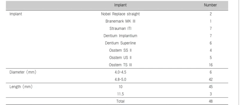
Forty eight patients(22 males and 26 females, mean age of 53.60 10.91) were selected in Ewha Womans University Mokdong Hospital department of periodontology from 2005 through 2016. The study was in accordance with the Declaration of Helsinki and was approved by the Instituitional Review Board of Ewha Womans Mokdong Hospital(IRB No. EUMC 2017-04- 009-001). All patients had single implant in the posterior maxilla(first molar or second molar) with sinus augmentation using BAOSFE technique and most of them were implanted with 5.0 mm diameter and 10 mm length implant. The type, diameter, length of implants are shown (Table. 1).
All surgeries were performed by three
specialists who trained periodontics. Implants were placed in various residual bone height of the posterior maxilla(Table. 1). The range of residual bone height was 4-9mm. Most of implants were placed at 4-8 mm of residual bone height. The implantation sites were classified into two groups according to the amount of grafted DBBM(Bio- Oss, Geistlich, Wolhusen, Switzerland), 0.25 group(0.25 g) and 0.5 group(0.5 g).
Surgical procedures
Antibiotics and steroids were given orally 1 hour before surgery. Patients rinsed their mouth with 0.1% chlorhexidine for 30 seconds, and their face were disinfected with povidone and saline gauze. A local anesthesia of 2% lidocaine with 1:100,000 epinephrine was administered to the edentulous area to be treated. A mid-crestal incision was made and a full-thickness mucoperiosteal flap was reflected. Under a
Implant Nobel Replace straight 2
Branemark MK III 1
Strauman ITI 7
Dentium Implantium 7
Dentium Superline 6
Osstem SS II 4
Osstem US II 5
Osstem TS III 16
Diameter (mm) 4.0-4.5 6
4.8-5.0 42
Length (mm) 10 45
11.5 3
Total 48
Table 1. Characteristics of implants
Implant Number
이식재양에따른이식골높이변화에대한방사선학적평가 surgical stent, the implant position was marked
on the alveolar crest with a guide drill. Twist and pilot drills of each implant system were used in sequence for preparing the implant site to a distance of approximately 1mm below the sinus floor. The first osteotome(ACE Surgical Supply Co, Inc, USA) with diameter of 1.6mm was used.
With light tapping, the osteotome was pushed toward the compact bone of the sinus floor. After reaching the sinus floor, the osteotome was pushed about 1mm further with light tapping in order to create a ‘greenstick’ fracture on the compact bone of the sinus floor. The second osteotome with a larger diameter(2.0mm) increased the fracture area of the sinus floor. The third osteotome was with a diameter of 2.8 mm or about 1mm smaller than that of the implant to be placed. Before placement of any grafting material, the Valsalva maneuver was done in order to check for any perforation of sinus membrane. If the sinus membrane was judged to be intact, the preparation was filled with DBBM.
The grafting material was then slowly pushed into the sinus cavity with the osteotome. This procedure was repeated several times. After the application of the grafting material, implant is installed in the prepared site.
Post-surgical care
In addition to the standard oral home care, antiseptic rinsing with 0.2% chlorhexidine twice daily for the first 2 weeks after surgery was recommended. The patients were placed on antibiotic prophylaxis(Ceclor 750 mg three times daily) for a period of 3-5 days.
Prosthetic procedures
The implants were allowed to heal for 6 months for grafted cases. A panoramic view, periapical radiograph and CBCT image were taken to examine whether there was continued radiolucency around the implant body and evaluate marginal bone level at the implant shoulder. Additionally, the ISQ value was recorded by OsstellTM Mentor(Integration Diagnostics AB, Goteborg, Sweden). When the ISQ value of the implant was measured as 70 or more, it was regarded as stable and prostheses was made10).
Radiographic evaluation
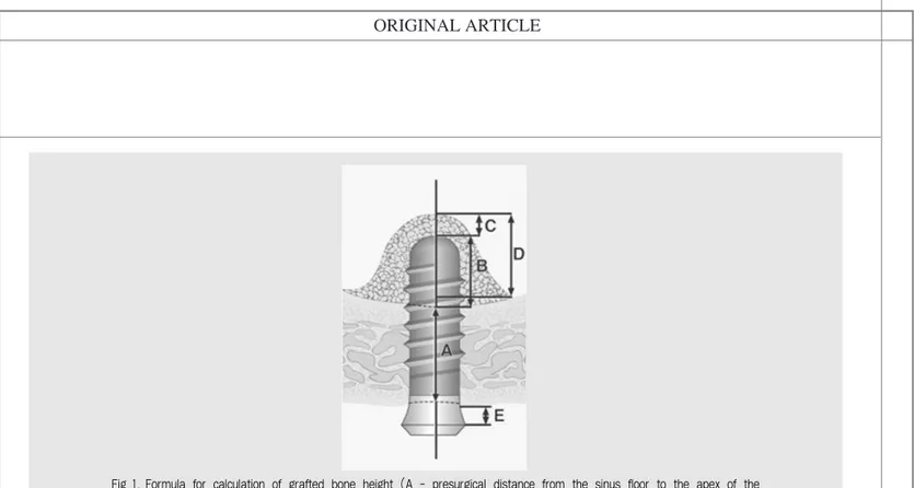
Panoramic view or CBCT were taken at the time of implant placement with BAOSFE and after at least 6 months to assess the bone level changes in the elevated sites with DBBM. To evaluate the change of height in augmented sinus floor, defined as the distance from the intra-sinus marginal bone to the grafted sinus floor directly above the lowest point of the original sinus height, B+C(Fig. 1), and has been measured11). And the protrusion length of implants in the maxillary sinus, B(Fig. 1), was measured to evaluate the effect of the implant length in sinus on the augmented bone height. In the panorama, magnification was corrected using the actual length of the placed implants in consideration of the enlargement ratio(True implant length/
implant length on radiograph)12). These evaluations were made by one examiners who majored in periodontology.
Statistical analysis
Radiographic data of augmented bone height were used to calculate group mean SD values.
Changes of grafted bone level according to the amount of DBBM were evaluated using student t- test through SPSS ver.20.0(SPSS Inc., Chicago, IL, USA). A P-value less than 0.05 was considered statistically significant. The relationship between amount of grafted DBBM and the changes of grafted bone level after 6 months and the effect of implant length in the maxillary sinus on augmented bone height were evaluated by Pearson correlation analysis.
Ⅲ. RESULTS
Clinical findings
All implants were stable clinically and radiographically during the follow-up. Of the 48 patients, only 37patients were followed up. At
least 6 months after surgery, there was no implant showed radiographically marked marginal bone resorption(more than 1mm), and clinically deep probing depth(more than 3 mm)(Fig. 2).
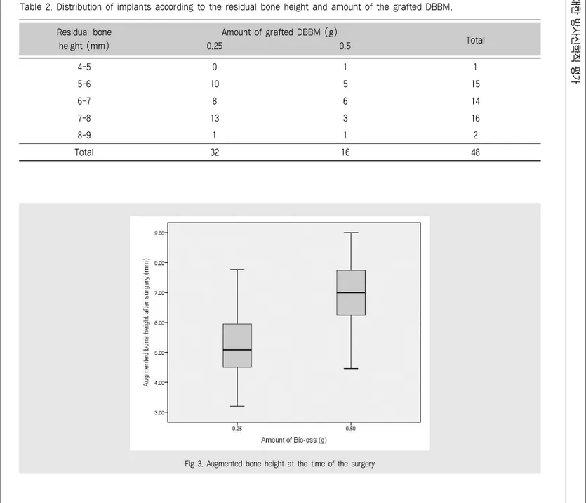
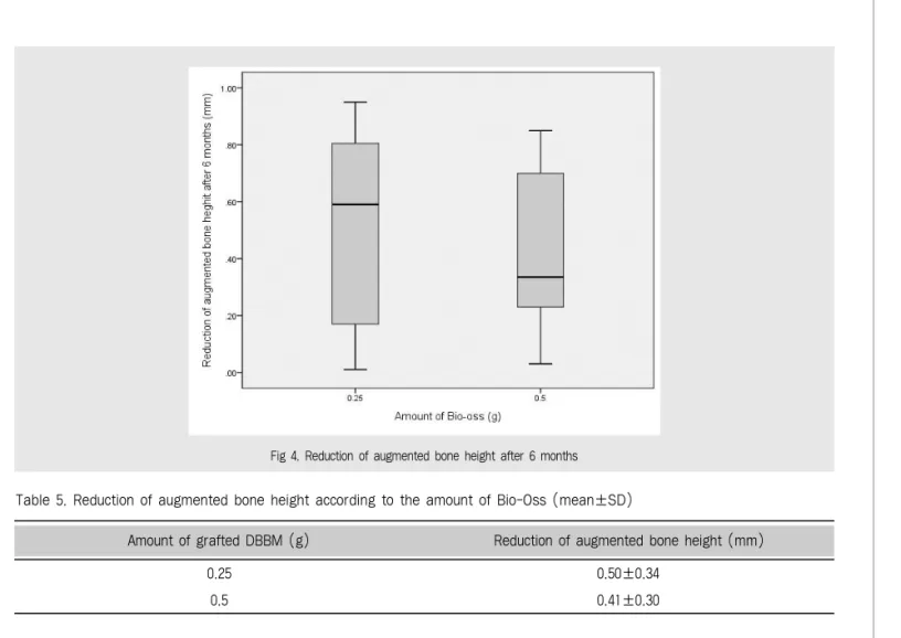
The Mean augmented bone height was 5.21 0.94mm in 0.25g, 6.92 1.19mm in 0.5g DBBM group. Statistically significant difference in augmented bone height was found in the comparison between the group of 0.25 g and 0.5 g at the time of surgery(Fig. 3, Table. 3). The mean protrusion length of implants into the sinus was 3.61 0.78mm ranging from 2.0mm to 5.44mm. There was a positive correlation between the length of the implant protruding into the maxillary sinus and the augmented bone height(r = 0.49; p < 0.01)(Table 4). The graft apical to the implants(C) also measured and the mean value of them is 2.19 1.21 mm. After 6 months, mean reduction of augmented bone height was 0.50 0.34mm in 0.25g and 0.41 0.30mm in 0.5g DBBM group(Table 5, Fig. 4).
Fig 1. Formula for calculation of grafted bone height (A - presurgical distance from the sinus floor to the apex of the implant; B - apical portion of the implant surpassing the boundary of the sinus; C - apical graft height; D - total graft height (B+C); E distance from the implant shoulder to the bone crest.)
이식재양에따른이식골높이변화에대한방사선학적평가
Fig 2. Panoramic radiograph was taken before surgery (a), immediately after surgery (b), 6 months after surgery (c).
Fig 3. Augmented bone height at the time of the surgery
4-5 0 1 1
5-6 10 5 15
6-7 8 6 14
7-8 13 3 16
8-9 1 1 2
Total 32 16 48
Table 2. Distribution of implants according to the residual bone height and amount of the grafted DBBM.
Residual bone Amount of grafted DBBM (g)
Total
height (mm) 0.25 0.5
There was no specific correlation between the reduction of augmented bone height and amount
of grafted DBBM.(r = - 0.05; p = 0.79)
0.25 5.21±0.94
0.5 6.92±1.19
Table 3. Mean augmented bone height according to the amount of Bio-Oss (mean±SD)
Amount of grafted DBBM (g) Augmented bone height after surgery (mm)
*Statistically significant difference in augmented bone height between the group of 0.25 g Bio-Oss and 0.5 g Bio-Oss (P<0.05).
Fig 4. Reduction of augmented bone height after 6 months
5.78±1.30 3.61±0.78 0.49* < 0.01
Table 4. Correlation between implants protrusion length and the amount of augmented bone height (mean±SD)
Augmented bone height (mm) Protrusion length (mm) r p
*Statistically significant correlation between implants protrusion length and the amount of augmented bone height (P<0.01).
0.25 0.50±0.34
0.5 0.41±0.30
Table 5. Reduction of augmented bone height according to the amount of Bio-Oss (mean±SD)
Amount of grafted DBBM (g) Reduction of augmented bone height (mm)
이식재양에따른이식골높이변화에대한방사선학적평가
Ⅳ. DISCUSSION
In this study, we evaluated the change of bone level on the sinus floor by a bone added osteotome sinus floor elevation(BAOSFE) technique, according to the amount of deproteinized bovine bone mineral(DBBM). And Changes in augmented bone height after BAOSFE procedure were also assessed using radiographs for 6months after the implant procedure. All implants were stable regardless of the amount of grafted DBBM during the 6 months follow-up periods. This result is consistent with the results of other studies that showed a predictable survival rate ranging from 95-100%
when implant were placement with BAOSFE simultaneously. However, long-term follow up is needed because the 6-month follow up period is too short to predict survival rate1, 13, 14).
Within the limit of this study, amount of augmented height was significantly increased according to the amount of grafting material.
However, this study did not include the use of larger amounts of graft materials(0.75g, 1g), so further research is needed. And there was a positive correlation between the protrusion length of implant in the maxillary sinus and augmented bone height. This implies that a longer protruding length of the implant may lead to a better tenting effect and may provide more space for grafted space.
According to Nkenke et al. (2002), the maxillary sinus membrane was perforated due to tension of the sinus membrane more than 3.0 0.8mm(range 2 to 5mm) when it was elevated
through the osteotome technique alone15). In our study, perforation did not occur even though the augmented height was higher than the height mentioned above. Long-term follow up is needed because tension may act to collapse the augmented portion supported by the graft material.
In general, fewer native bones and more grafted material are expected to lead to more absorption of augmented bone height, but in this study less absorption was observed in the 0.5 group. However, there was no specific correlation between the reduction of augmented bone height and amount of grafted DBBM.
Moreover, there was a controversial opinions whether graft material is necessary or not. In a retroprospective study assessing the pattern of tissue remodeling after maxillary sinus floor elevation using the transalveolar osteotome technique with or without grafting materials, no grafting material group showed some dense structure at apical to the implant. However, when grafting material was used, a cloudy dome structure with a hazy demarcation was usually visible after implant placement16).
The height of bone graft decreased during the first 2-3years after augmentation. Jung et al.
(2010) performed the BAOSFE procedure with 7 non-submerged SLA implants on 4 patients. The average gain in the augmented bone height of the implants was 8.6mm(range, 6.9-9.9mm). The height of grafted bone was reduced markedly by an overall mean of 1.6mm during the first 2 years. During long-term healing periods, over 5 years, the height of grafted bone was reduced by
an overall mean of 1.9mm17). Bragger et al.
(2004) reported the patterns of tissue remodeling after placement of 25 implants in 19 patients using the transalveolar technique with composite xenografts and autografts. The mean height of the new structure reaching apical and mesial to the implants was 1.52mm at surgery, but was reduced significantly to 1.24mm at 3 months and 0.29mm after 12months. The authors concluded that the grafted area apical to the implants underwent shrinkage and remodeling11). In this study, augmented bone height after 6months was measured and a slight amount of absorptions was observed. However, previous studies have shown that absorption is more frequent in the first 1-2
years, therefore longer-term follow up study is needed.
Ⅴ. CONCLUSION
BAOSFE is a predictable treatment option for atrophic posterior maxilla. Within the limit of this study, amount of augmented bone height was significantly increased according to the amount of grafting material and no specific correlation between the reduction of augmented bone height and amount of grafted DBBM. However, because of the short follow up period of this study, further long-term follow up studies are needed.
이식재양에따른이식골높이변화에대한방사선학적평가 1. Zitzmann NU, Scharer P. Sinus elevation procedures
in the resorbed posterior maxilla. Comparison of the crestal and lateral approaches. Oral Surg Oral Med Oral Pathol Oral Radiol Endod 1998;85(1):8-17 2. Boyne PJ, James RA. Grafting of the maxillary sinus
floor with autogenous marrow and bone. J Oral Surg 1980;38(8):613-616
3. Tatum H, Jr. Maxillary and sinus implant reconstructions. Dent Clin North Am 1986;30(2):207- 229
4. Summers RB. A new concept in maxillary implant surgery: the osteotome technique. Compendium 1994;15(2):152, 154-156, 158 passim; quiz 162 5. Summers RB. The osteotome technique: Part 3--
Less invasive methods of elevating the sinus floor.
Compendium 1994;15(6):698, 700, 702-694 passim;
quiz 710
6. Jensen OT, Shulman LB, Block MS, Iacono VJ.
Report of the Sinus Consensus Conference of 1996.
Int J Oral Maxillofac Implants 1998;13 Suppl:11-45 7. Cavicchia F, Bravi F, Petrelli G. Localized
augmentation of the maxillary sinus floor through a coronal approach for the placement of implants. Int J Periodontics Restorative Dent 2001;21(5):475-485 8. Winter AA, Pollack AS, Odrich RB. Placement of
implants in the severely atrophic posterior maxilla using localized management of the sinus floor: a preliminary study. Int J Oral Maxillofac Implants 2002;17(5):687-695
9. Kim SM, Park JW, Suh JY, et al. Bone-added osteotome technique versus lateral approach for sinus floor elevation: a comparative radiographic study. Implant Dent 2011;20(6):465-470
10. Bornstein MM, Hart CN, Halbritter SA, et al. Early loading of nonsubmerged titanium implants with a chemically modified sand-blasted and acid-etched surface: 6-month results of a prospective case
series study in the posterior mandible focusing on peri-implant crestal bone changes and implant stability quotient (ISQ) values. Clin Implant Dent Relat Res 2009;11(4):338-347
11. Bragger U, Gerber C, Joss A, et al. Patterns of tissue remodeling after placement of ITI dental implants using an osteotome technique: a longitudinal radiographic case cohort study. Clin Oral Implants Res 2004;15(2):158-166
12. Si MS, Shou YW, Shi YT, et al. Long-term outcomes of osteotome sinus floor elevation without bone grafts: a clinical retrospective study of 4-9 years. Clin Oral Implants Res 2016;27(11):1392- 1400
13. Fugazzotto PA, Vlassis J. Long-term success of sinus augmentation using various surgical approaches and grafting materials. Int J Oral Maxillofac Implants 1998;13(1):52-58
14. Rosen PS, Summers R, Mellado JR, et al. The bone-added osteotome sinus floor elevation technique: multicenter retrospective report of consecutively treated patients. Int J Oral Maxillofac Implants 1999;14(6):853-858
15. Nkenke E, Schlegel A, Schultze-Mosgau S, et al.
The endoscopically controlled osteotome sinus floor elevation: a preliminary prospective study. Int J Oral Maxillofac Implants 2002;17(4):557-566 16. Pjetursson BE, Ignjatovic D, Matuliene G, et al.
Transalveolar maxillary sinus floor elevation using osteotomes with or without grafting material. Part II: Radiographic tissue remodeling. Clin Oral Implants Res 2009;20(7):677-683
17. Jung JH, Choi SH, Cho KS, Kim CS. Bone-added osteotome sinus floor elevation with simultaneous placement of non-submerged sand blasted with large grit and acid etched implants: a 5-year radiographic evaluation. J Periodontal Implant Sci 2010;40(2):69-75
참 고 문 헌